Dear Editor,
The blocking of D antigen sites in neonate by maternal IgG anti-D in severe cases of haemolytic disease of the fetus and new born (HDFN) is not a new phenomenon [1]. The coating anti-D prevents the agglutination of the D positive red cells (RBC) by the IgM anti-D typing reagent. Blocking tests were actually the first one used to prove the existence of non-agglutinating IgG antibodies [1]. The antigen blocking phenomenon is not uncommon in examples of human anti-D [2]. However the total blocking of D antigen sites by IgG resulting in false negative D typing when a monoclonal IgM anti-D was used are rare. Chloroquine diphosphate (CDP) is a chemical used in immunohematology laboratories to dissociate IgG from red blood cells without compromising the integrity of the red cell membrane antigens by neutralization of the charged groups on amino acids that govern the tertiary structure of antibody molecules [3]. However, chloroquine diphosphate may not totally remove the coating of antibody from red cells and it is not able to remove complement component coating. Here we report a case of blocking phenomenon occurs in a neonate with a positive direct antiglobulin test due to maternal anti-D, anti-C antibodies and the serological challenge was resolved by chloroquine diphosphate treatment of neonatal red cells.
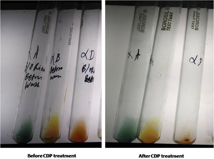
A 23 years old female (G2P1+0) with 36 weeks of gestation admitted in a tertiary care teaching institute of eastern India where she delivered a female child of weight 2.3 kg with hyperbilirubinemia. She had received anti-D immunoglobulin prophylaxis after delivery of her first child 3 years ago. She had no history of blood transfusion during last 3 years. No history of jaundice in previous child after delivery. The neonate was referred to NICU 4 h after delivery as Hb was 53 g/L, indirect bilirubin was − 0.21 g/L and reticulocyte count was 10%. The blood sample of mother and newborn was received at blood bank for cross-matching and exchange transfusion. In the immunohaematology laboratory the mother was typed as group A, rr (dccee) and the neonate was typed as group O, Rh-D negative by conventional tube technique using IgM monoclonal anti-D (Tulip Diagnostics Pvt. Ltd). The direct antiglobulin test (DAT) of neonate was performed by gel technique (DiaMed, Switzerland) and it shows 4+ agglutination in polyspecific AHG card containing (anti-IgG + C3d). Antibody screening of mother was performed with in-house pooled O positive cells and found to be 4+ reactive in AHG card by gel technique (DiaMed, Switzerland). Antibody identification in maternal serum by 11 cell (Ortho clinical Diagnostic) was performed to identify the antibody in serum and the pattern of panel cells showed the presence of anti-D and anti-C. After identification of alloantibodies, group O Rh D and C antigen negative cross match compatible RBC unit was reconstituted with AB plasma. The neonate had received 196 ml of blood prepared in accordance with departmental standard operating procedure for exchange transfusion. After completion of protocol to issue a safe blood for exchange transfusion, the extensive immunohematological workups were performed with maternal and neonatal samples. Titration of anti-D was done by tube method and found to be 1024 by using R2R2 (DccEE) cell and titre of anti-C was found to be 128 by using r′r′ (dCCee) cells [4]. Double adsorption technique was employed to rule out the presence of anti-G alone or in conjunction with anti-D and anti-C [5]. After reviewing immunohematological results with clinical history, the presence of high titre anti-D, anti-C antibodies in mother and strong DAT positivity of neonatal red cells; a ‘blocking phenomenon’ of neonatal red cell was suspected. Two aliquots were prepared for dissociating the antibody from blocked red cell of neonate. First aliquot was eluted by gentle heat elution at 45 °C [4]. But the blood group of neonate was typed still as O negative. Then second aliquot was treated by chloroquine diphosphate (CDP) and elution performed at pH 5.1 according to the recommended method [4]. Direct antiglobulin test was again performed in the CDP eluted RBC shows 2+ strength (from initial 4+) using gel technique. When the CDP treated neonatal RBC was typed it was found as O Rh-D positive with the same IgM monoclonal anti-D (Tulip Diagnostics Pvt. Ltd) antisera by tube method (Fig. 1). The CDP elute specificity was confirmed presence of anti-D, anti-C antibodies when tested by 11-cell panel (Ortho clinical Diagnostic). Here we typed father and the neonate in addition to mother. The phenotype of father was O, DCCee and neonate was O, DCcee (Table 1). Thus it was suggested that the causative corresponding antigens were inherited from father to the neonate and blocking phenomenon was due to maternal anti-D, anti-C antibodies. After double volume exchange transfusion the Hb of neonate was raised to 122 g/L from 53 g/L and bilirubin came down to 0.06 g/L from 0.21 g/L. The neonate was discharged in hemodynamic stable condition on the eighth day of delivery with Hb of 120 g/L and 0.02 g/L bilirubin level.
Fig. 1.
Rh-D typing by tube method before and after CDP treatment
Table 1.
ABO grouping and Rh typing
| Mother | Baby | Father | |
|---|---|---|---|
| ABO blood group | A | O | O |
| Rh-antigens distribution | D-Negative |
D-Negative (before CDP treatment) D-Positive (after CDP treatment) |
D-Positive |
| C-Negative | C-Positive | C-Positive | |
| c-Positive | c-Positive | c-negative | |
| E-Negative | E-Negative | E-Negative | |
| e-Positive | e-Positive | e-Positive | |
| Rh phenotype | dccee | DCcee | DCCee |
False negative typing results caused by potent maternal IgG antibodies blocking antigen sites are not common when using modern monoclonal blood grouping reagents, especially anti-D. But the message is very clear from our case that blocking can be observed even after using monoclonal IgM anti-D reagent during routine typing of neonatal blood group. As the immunogenicity of D antigens being only second to ABO antigens, accuracy in Rh-D typing is critical in transfusion medicine. The result of neonatal DAT with suggestive history must be evaluated cautiously before concluding the neonatal Rh-D status. The performance of maternal antibody screening and identification, typing of both parents for extensive Rh-system along with neonates, the performance of an eluate on subsequent DAT positive samples after CDP treatment with antibody identification panel will provide the answer to both the causative antibody and the antigen expression of the neonate’s RBC.
Authors Contribution
AN performed the performed serologic testing and wrote drafts under supervision of PB. SSD analyzed all data and wrote the manuscript. All authors reviewed and edited the manuscript.
Funding
The authors declared having no competing financial interest relevant to this article.
Compliance with Ethical Standards
Conflict of interest
The authors declare that they have no conflict of interest.
Footnotes
Publisher's Note
Springer Nature remains neutral with regard to jurisdictional claims in published maps and institutional affiliations.
Contributor Information
Archana Naik, Email: archi.scb88@gmail.com.
Prasun Bhattacharya, Email: pbhattach@gmail.com.
Suvro Sankha Datta, Email: suvro.datta@gmail.com.
References
- 1.Wiener AS (1944) A new test (blocking test) for Rh sensitization. In: Proceedings of the society for experimental biology and medicine, vol 56, New York, pp 173–176
- 2.Lee E. Blocked D phenomenon. Blood Transfus. 2013;11:10–11. doi: 10.2450/2012.0059-12. [DOI] [PMC free article] [PubMed] [Google Scholar]
- 3.Edwards JM, Moulds JJ, Judd WJ. Chloroquine dissociation of antigen-antibody complexes. A new technique for typing red blood cells with a positive direct antiglobulin test. Transfusion. 1982;22:59–61. doi: 10.1046/j.1537-2995.1982.22182154219.x. [DOI] [PubMed] [Google Scholar]
- 4.Fung MK, Grossman BJ, Hillyer CD, Westhoff CM (2014) Methods section 2: red cell typing. In: American Association of Blood Banks edition. Technical manual, 18th edn: AABB, Bethesda
- 5.Shirey RS, Mirabella DC, Lumadue JA, Ness PM. Differentiation of anti-D, -C, and -G: clinical relevance in alloimmunized pregnancies. Transfusion. 1997;37:493–496. doi: 10.1046/j.1537-2995.1997.37597293879.x. [DOI] [PubMed] [Google Scholar]


