Abstract
The plant sucrose nonfermenting 1 (SNF1)-related protein kinases (SnRKs) are key regulators in the interconnection of various signaling pathways. However, little is known about the SnRK family in strawberries. In this study, a total of 26 FvSnRKs including one FvSnRK1, nine FvSnRK2s and 16 FvSnRK3s were identified from the strawberry genome database. They were respectively designated as FvSnRK1.1, FvSnRK2.1 to FvSnRK2.9 and FvSnRK3.1 to FvSnRK3.16, according to the conserved domain of each subfamily and multiple sequence alignment with Arabidopsis. FvSnRK family members were unevenly distributed in seven chromosomes. The number of exons or introns varied among FvSnRK1s, FvSnRK2s and FvSnRK3s, but highly conserved in the same subfamily. The FvSnRK1.1 had 10 exons. Most of FvSnRK2s had nine exons or eight introns, except FvSnRK2.4, FvSnRK2.8 and FvSnRK2.9. FvSnRK3 genes were divided into intron-free and intron-harboring members, and the number of introns in intron-harboring group ranged from 11 to 15. Moreover, the phylogenetic analysis showed SnRK1, SnRK2 and SnRK3 subfamilies respectively clustered together in spite of the different species of strawberry and Arabidopsis, indicating the genes were established prior to the divergence of the corresponding taxonomic lineages. Meanwhile, conserved motif analysis showed that FvSnRK sequences that belonged to the same subgroup contained their own specific motifs. Cis-element in promoter and expression pattern analyses of FvSnRK1.1 suggested that FvSnRK1.1 was involved in cold responsiveness, light responsiveness and fruit ripening. Taken together, this comprehensive analysis will facilitate further studies of the FvSnRK family and provide a basis for the understanding of their function in strawberry.
Keywords: SnRKs, phylogenetic analysis, expression pattern, strawberry
1. Introduction
Plants are forced to face the ever-changing surroundings during growth and development, due to their sessile nature. Thus, a timely response and coordinated regulatory machinery to external and internal signals is required, which helps plant to maintain optimal cellular conditions and resist various stresses originating from temperature, salinity, drought, nutrient defect and pathogen invasion [1,2]. It has been documented that these processes are involved in post-translation modifications (PTMs), such as phosphorylation, ubiquitination and acetylation. Arguably, the reversible protein phosphorylation executed by protein kinases and protein phosphatases plays a crucial role in intracellular signaling cascades and gene expression orchestration [3,4].
The sucrose non-fermenting 1 (SNF1) protein kinases that belong to Ser/Thr protein kinase are evolutionarily conserved in organisms. In plants, SNF1-related protein kinases (SnRKs) have been classified into SnRK1, SnRK2 and SnRK3 subfamilies on the basis of the sequence similarity and gene structures, which play key roles in integrating the metabolic and stress signaling [5]. The SnRK1 subfamily, which mainly participates in the global regulation of carbon and nitrogen metabolism, homologous to SNF1 genes in yeast and AMP-activated protein kinases (AMPKs) in mammals, functions as the heterotrimeric complex consisting of a catalytic α subunit and accessory β and γ subunits. The catalytic α subunit contains a highly conserved N-terminal catalytic domain and a variable C-terminal regulatory domain, which is important to form a complex interaction (with the β and γ subunits) and regulates the kinase activity [6,7]. Unlike SnRK1s, the other two subfamilies (SnRK2s and SnRK3s) are unique in plants. It has been reported that the catalytic region of SnRK2s and SnRK3s is analogous to that of SNF1/AMPK-type kinases. They probably derive from the gene duplication of SnRK1s and then diverge rapidly during plant evolution to fulfil new roles that enable plants to develop networks that link stress and abscisic acid (ABA) signaling with metabolic signaling [5,8]. Members of the SnRK2 subfamily have kinase domain, ATP binding domain, serine/threonine active site and four N-fourteen sites [9,10]. The first SnRK2 cDNA clone (PKABA1) was identified from an ABA-treated wheat embryo cDNA library, which was induced by salinity, cold, dehydration and osmotic stresses [11,12]. Early research has demonstrated that SnRK2s mainly participate in countering abiotic stress. Recent evidence suggests that SnRK2s are multifarious players in various biological processes like plant growth and development [13]. SnRK3s are known as calcineurin B-like calcium sensor-interacting protein kinases (CIPKs) that include a binding site for calcium-binding proteins (SOS3, salt overly sensitive 3; SCaBPS, SOS3-like calcium-binding proteins) and calcium-sensitive CBL (calcineurin B-like proteins) in the C-terminal region, which combine together to activate the protein kinase [14,15]. The CBL-CIPK calcium signaling network enables information integration and physiological coordination in response to a variety of extracellular cues [16].
In the light of the significant influence of SnRKs on regulating the metabolic and stress signaling, we identified SnRK members from strawberry and analyzed their conserved motif, homology and phylogenetic relationship with Arabidopsis. Especially, the SnRK1 kinases act as important metabolic sensors, which integrate diverse stress conditions and maintain energy homeostasis by direct phosphorylation of key metabolic enzymes and regulatory proteins, and massive transcriptional reprogramming [7,17,18]. Hence, we detected SnRK1 expression profiles in different strawberry tissues and under various abiotic stresses, which provided a theoretical basis for the genetic improvement of stress resistance and fruit quality.
2. Materials and Methods
2.1. Plant Materials and Treatments
Different tissues including root, stem, leaf, stolon, flower and berry at seven developmental stages (small green, SG; large green, LG; fade green, FG; white, W; turning red; TR; half red; HR; full red, FR) of the strawberry (Fragaria×ananassa cv. Benihoppe) were sampled. In addition, the Toyonoka strawberry cultivar was subjected to different light quality and low temperature treatment. The potted strawberries at the 7th day after flowering (7 DAF) were divided into four groups and then respectively transferred into growth chambers installed light emitting diodes of white (control), red (730 nm), blue (450 nm) and mixed light (red: blue = 1:1). The environmental conditions in chambers were strictly controlled (8 h dark at 16 °C, 16 h photoperiod at 25 °C, 100 μmol∙m−2∙s−1 light intensity and 75% relative humidity). Fruits were harvested at the 14th, 21th, 25th, 28th day after flowering. In addition, two groups of the potted strawberries at white stage of fruit were put into 4 °C (cold) and 25 °C (control) chambers under a 16 h diurnal light cycle at 100 μmol∙m−2∙s−1 with 75% relative humidity for low temperature treatment, respectively. Fruits were sampled at 0, 6, 12, 24, 48, and 72 h. All of above samples were used to investigate SnRK1.1 expression pattern.
2.2. Identification and Characterization of FvSnRKs
The Arabidopsis SnRK proteins (Supplementary File S1) obtained from TAIR database (https://www.arabidopsis.org/) were used as query probes to BLAST search against Fragaria vesca v1.0 genome database (https://www.rosaceae.org). Meanwhile, the whole protein sequences of F. vesca were downloaded from this database. Then, the Hidden Markov Model (HMM) files corresponding to each subfamily of SnRKs were downloaded from Pfam protein family database (http://pfam.sanger.ac.uk/), and HMMER 3.0 was used to search the SnRKs from the protein sequence dataset with the default threshold. To further ensure the existence of SnRK conserved structures, each candidate protein based on results of BLAST search and HMM search was checked using the NCBI-CDD (https://www.ncbi.nlm.nih.gov/) and SMART (http://smart.embl-heidelberg.de/) online tools.
2.3. Sequence Analysis of FvSnRKs
The information of all SnRK proteins, genes, coding sequences (CDS) was obtained from the strawberry genome database (Supplementary File S2–S4). The protein theoretical pI, molecular weight (MW), aliphatic index, grand average of hydropathicity (GRAVY) and instability index were evaluated using the ExPASy-ProtParam tool (http://web.e xpasy.org/protparam/). Conserved motifs of the FvSnRKs were identified using the Multiple EM for Motif Elicitation (MEME) web server (http://meme.nbcr.net/meme/cgi-bin/meme.cgi) and visualized using TBtools [19]. Putative signal peptide and transmembrane helix (TMH) were predicted in the SignalP 4.1 Server (http://www.cbs.dtu.dk/services/SignalP/) and the TMHMM Server v.2.0 (http://www.cbs.dtu.dk/ser vices/TMHMM/), respectively. Subcellular location was speculated by ProtComp v.9.0 (http://linux1.softberry.com/berry.phtml?topic=protc omppl&group=programs&subgroup=proloc). Additionally, the cis-element of FvSnRK1.1 promoter was analyzed in the Plantcare database (http://bioinformatics.psb.ugent.be/webtools/plantcare/html/).
2.4. Gene Structure and Phylogenetic Tree of FvSnRKs
The exon–intron structure of FvSnRK genes was analyzed using the Gene Structure Display Server program (GSDS v.2.0, http://gsds.cbi.pku.edu.cn) based on the comparison of coding sequences (Supplementary File S4) and corresponding genomic sequences (Supplementary File S3). The unrooted phylogenetic tree of FvSnRK proteins was constructed in MEGA v.6.0 according to the neighbor-joining (NJ) method with Poisson model and 1000 bootstrap replications, after multiple sequence alignment was performed in ClustalX v.2.0 software. Then the classification of FvSnRKs into subfamilies was based on the previous report in Arabidopsis.
2.5. RNA Extraction and Gene Expression of FvSnRKs
Total RNA was isolated from all samples using the improved CTAB method [20]. After quality and quantity were checked by 1% agrose gel electrophoresis and BioPhotometer, 1 μg of total RNA was used for reverse-transcription into the first strand of cDNA with PrimeScript TM RT reagent Kit with gDNA Eraser (Perfect Real Time) (Takara, Japan) according to the operating manual. All quantitative real-time PCRs were performed on the CFX96 real-time PCR system (Bio-Rad, USA). A total volume of 10 μL reaction mixture contained 0.4 μL of each primer (10 μM), 1 μL of 10-fold dilution of cDNA, 3.2 μL of RNase-free water and 5 μL of SYBR Premix (Takara, Japan). The reaction procedure was carried out as follows: 95 °C/3 min for pre-denaturation, followed by 40 cycles of 95 °C /10 s for denaturation, 60 °C/30 s for annealing and 72 °C/15 s for extension. Subsequently, melting curve was executed to confirm specificity of primers, ramping from 65 °C to 95 °C (increment 0.5 °C/5 s). The relative expression level of FvSnRK1.1 to 26S-18S RNA housekeeping gene was analyzed with the 2−ΔΔCT method. The primers were as follows: 5’-GCACAAATGGTTCCAGGCTC-3’ (sense) and 5’-GAAACTCGGCCCCAAGGTAG-3’ (antisense) for FvSnRK1.1 239 bp amplicon; 5’- ACCGTTGATTCGCACAATTGGTCATCG-3’ (sense) and 5’- TACTGCGGGTCGGCAATCGGACG -3’ (antisense) for 26S-18S RNA housekeeping gene 150 bp amplicon.
3. Results
3.1. Identification and Properties of SnRKs in F. versa
To extensively screen the SnRKs from the strawberry genome database, two methods, including BLAST search and HMM search, were applied. Eventually, 26 FvSnRKs composed of one FvSnRK1, nine FvSnRK2s and 16 FvSnRK3s were identified. They were respectively designated as FvSnRK1.1, FvSnRK2.1 to FvSnRK2.9 and FvSnRK3.1 to FvSnRK3.16, according to conserved domain and multiple sequence alignment with Arabidopsis. Their distribution and sequence feature were analyzed in Table 1. FvSnRK1.1 was distributed on chromosome 6. FvSnRK2s were scattered to chromosome 1, 2, 4 and 5. FvSnRK3s were distributed over all chromosomes except chromosome 5. Respectively, the open reading frame (ORF) length of FvSnRK1.1, FvSnRK2s and FvSnRK3s was 1557 bp, 963–1215 bp and 1284–1524 bp, and deduced protein was 518 aa, 320–404 aa and 427–507 aa, and the relative molecular mass was 59.19 kDa, 36.30–45.72 kDa and 47.96–57.42 kDa. The FvSnRK1.1 protein was predicted to be unstable, while most of the FvSnRK3s were stable. All of the FvSnRKs had no signal peptide and only FvSnRK3.11 contained one transmembrane helix. Subcellular localization prediction indicated FvSnRK1.1 and FvSnRK2s were localized to the cytoplasm and nucleus, whereas FvSnRK3s were localized to cytoplasm, nucleus, endoplasmic reticulum or membrane.
Table 1.
Sucrose nonfermenting 1 (SNF1)-related protein kinases (SnRKs) genes and corresponding protein properties in strawberry.
| Gene Name | Gene Id | Chr. | Locus | ORF (bp) | Amino Acid (aa) | MW (kDa) | pI | Instability Index | Aliphatic Index | GRAVY | SignalP | TMH | Location |
|---|---|---|---|---|---|---|---|---|---|---|---|---|---|
| FvSnRK1.1 | gene04397 | chr6 | 32622471..32625872+ | 1557 | 518 | 59.19 | 8.68 | 48.23 (unstable) | 88.78 | −0.313 | no | 0 | a, b |
| FvSnRK2.1 | gene11031 | chr5 | 16681534..16684843+ | 1059 | 352 | 40.40 | 6.12 | 44.84 (unstable) | 82.78 | −0.511 | no | 0 | a, b |
| FvSnRK2.2 | gene10769 | chr5 | 23595280..23598224+ | 1014 | 337 | 38.04 | 5.85 | 35.42 (stable) | 86.20 | −0.399 | no | 0 | a, b |
| FvSnRK2.3 | gene31902 | chr2 | 12574424..12578412+ | 1092 | 363 | 41.23 | 4.88 | 41.24 (unstable) | 88.35 | −0.316 | no | 0 | a, b |
| FvSnRK2.4 | gene06595 | chr4 | 14755025..14756119+ | 1095 | 364 | 41.48 | 9.10 | 45.92 (unstable) | 82.17 | −0.429 | no | 0 | a, b |
| FvSnRK2.5 | gene16244 | chr1 | 13571546..13573722+ | 1014 | 337 | 38.38 | 5.18 | 33.76 (stable) | 90.50 | −0.286 | no | 0 | a, b |
| FvSnRK2.6 | gene11971 | chr5 | 12304605..12307049− | 1014 | 337 | 38.10 | 5.76 | 36.88 (stable) | 89.91 | −0.242 | no | 0 | a, b |
| FvSnRK2.7 | gene11970 | chr5 | 12295362..12297597− | 963 | 320 | 36.30 | 8.76 | 37.85 (stable) | 91.34 | −0.253 | no | 0 | a, b |
| FvSnRK2.8 | gene11969 | chr5 | 12290219..12292886− | 1050 | 349 | 39.43 | 6.76 | 41.81 (unstable) | 81.20 | −0.342 | no | 0 | a, b |
| FvSnRK2.9 | gene24096 | chr1 | 11643335..11647273+ | 1215 | 404 | 45.72 | 4.82 | 44.09 (unstable) | 90.20 | −0.170 | no | 0 | a, b |
| FvSnRK3.1 | gene28136 | chr3 | 21779588..21780994− | 1407 | 468 | 53.65 | 8.77 | 40.89 (unstable) | 88.12 | −0.491 | no | 0 | c, d |
| FvSnRK3.2 | gene13841 | chr6 | 7167764..7169179+ | 1416 | 471 | 53.28 | 9.00 | 32.77 (stable) | 89.36 | −0.414 | no | 0 | c, d |
| FvSnRK3.3 | gene29682 | chr3 | 7803967..7805490− | 1524 | 507 | 56.29 | 7.64 | 39.44 (stable) | 88.22 | −0.219 | no | 0 | c, d |
| FvSnRK3.4 | gene15372 | chr2 | 32504181..32509548+ | 1416 | 471 | 53.98 | 6.03 | 32.57 (stable) | 88.58 | −0.334 | no | 0 | a, b |
| FvSnRK3.5 | gene18646 | chr7 | 3032869..3036362− | 1347 | 448 | 51.53 | 9.13 | 40.50 (unstable) | 77.46 | −0.539 | no | 0 | a, b |
| FvSnRK3.6 | gene30382 | chr3 | 2915121..2920078+ | 1341 | 446 | 49.55 | 8.83 | 30.14 (stable) | 83.30 | −0.314 | no | 0 | a, b |
| FvSnRK3.7 | gene10067 | chr1 | 842428..843711− | 1284 | 427 | 47.96 | 9.34 | 28.34 (stable) | 83.58 | −0.326 | no | 0 | c, d |
| FvSnRK3.8 | gene29681 | chr3 | 7799332..7800717+ | 1386 | 461 | 51.94 | 9.11 | 31.99 (stable) | 78.87 | −0.429 | no | 0 | c, d |
| FvSnRK3.9 | gene13849 | chr6 | 7132323..7133642− | 1320 | 439 | 49.58 | 7.58 | 31.35 (stable) | 91.69 | −0.266 | no | 0 | a, b, d |
| FvSnRK3.10 | gene31049 | chr1 | 3172739..3177629− | 1524 | 507 | 57.42 | 9.24 | 43.34 (unstable) | 86.37 | −0.286 | no | 0 | a, b, d |
| FvSnRK3.11 | gene15015 | chr2 | 35278943..35280274− | 1332 | 443 | 49.74 | 8.94 | 38.64 (stable) | 77.24 | −0.346 | no | 1 | c, d |
| FvSnRK3.12 | gene22806 | chr4 | 15674061..15678224+ | 1425 | 474 | 53.06 | 8.06 | 44.47 (unstable) | 93.54 | −0.368 | no | 0 | a, b |
| FvSnRK3.13 | gene29066 | chr4 | 6439716..6441032+ | 1317 | 438 | 47.51 | 8.87 | 35.28 (stable) | 94.86 | −0.080 | no | 0 | a, b |
| FvSnRK3.14 | gene28132 | chr3 | 21757318..21758637+ | 1320 | 439 | 49.48 | 8.52 | 38.61 (stable) | 87.40 | −0.298 | no | 0 | a, b, d |
| FvSnRK3.15 | gene26443 | chr1 | 4822218..4828540− | 1485 | 494 | 55.46 | 5.67 | 37.29 (stable) | 93.30 | −0.074 | no | 0 | a, b, d |
| FvSnRK3.16 | gene21319 | chr7 | 19497585..19498877+ | 1293 | 430 | 48.84 | 8.99 | 30.50 (stable) | 87.09 | −0.328 | no | 0 | c, d |
Note: a, cytoplasm; b, nucleus; c, endoplasmic reticulum; d, membrane.
3.2. Analysis of Gene Structure
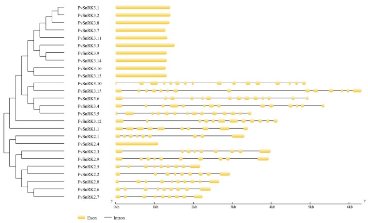
The FvSnRKs exon–intron organizations were analyzed in GSDS. As shown in Figure 1, the FvSnRK1.1 coding sequence was composed of 10 exons. Most of the FvSnRK2s had nine exons or eight introns, except FvSnRK2.4, FvSnRK2.8 and FvSnRK2.9. The order and approximate size of exons among the FvSnRK2s were relatively conserved, but the size of introns was variable, which gave rise to a diversity of gene structures. Clearly, FvSnRK3s can be divided into three clades according to the exon–intron of gene structures. The first clade included ten genes without introns. The second clade had five members. Of these, four members contained 14 exons, while the remaining one contained 16 exons. The third clade contained only one gene, FvSnRK3.12, with 12 exons.
Figure 1.
Gene structure analysis of FvSnRKs. Exon–intron structure was analyzed in the Gene Structure Display Server (GSDS) program. The yellow boxes indicate exons, and the gray horizontal lines indicate introns. The scale bar represents 6 kb.
3.3. Phylogenetic Analysis and Conserved Motifs of FvSnRKs
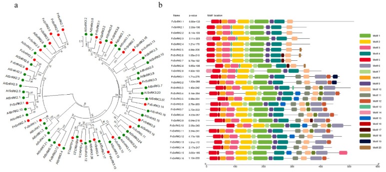
To gain insight into the evolutionary relationship among SnRKs between the strawberry and Arabidopsis, a phylogenetic tree was constructed based on the multiple alignment of the amino acid sequences. All SnRKs were specifically classified into three main clades of SnRK1, SnRK2 and SnRK3 subfamilies (Figure 2a). The results showed FvSnRK1.1 was clustered together with AtSnRK1.1, AtSnRK1.2 and AtSnRK1.3. FvSnRK2s were divided into three subgroups. Two subgroups respectively contained one member FvSnRK2.1 and FvSnRK2.4, and the third one contained the rest seven members. FvSnRK3s were compartmentalized into three subgroups that respectively included 1, 5 and 10 members. Twenty conserved motifs of 26 FvSnRKs were identified using the online MEME program (Supplementary File S5). Clearly, all of SnRKs in the strawberry shared some motifs, but SnRK1, SnRK2 and SnRK3-class had their own idiographic characteristic (Figure 2b).
Figure 2.
Phylogenetic relationships among SnRKs between strawberry and Arabidopsis (a) and conserved motifs of FvSnRKs (b). The evolutionary tree was constructed with the neighbor-joining method based on the Poisson model using 1000 bootstrap replicates. The green and red dots respectively indicate Arabidopsis and strawberry SnRK proteins. Conserved motifs of FvSnRKs in strawberry were analyzed using the MEME web server. Different color boxes represent different types of putative motifs.
3.4. Cis-element Analysis in the Promoter Region of FvSnRK1.1 Gene
The 2000 bp fragment of upstream region from the start codon in FvSnRK1.1 gene was isolated to predict the cis-acting regulatory element. As shown in Table 2, the FvSnRK1.1 promoter included several light responsive elements, such as AE-box, Box 4, G-box, GATA-motif, GT1-motif, L-box, TCT-motif and MRE. It also contained stress-related (anaerobic, drought, low temperature, etc.) cis-elements (ARE, LTR, MBS, TC-rich repeats), hormone-related (abscisic acid, MeJA, gibberellin, salicylic acid) cis-elements (ABRE, CGTCA-motif, P-box, TCA-element, TGACG-motif). Moreover, it was involved in endosperm expression, zein metabolism regulation. In addition, this region existed many core promoter TATA-box and enhancer CAAT-box.
Table 2.
The cis-acting regulatory element in the promoter of FvSnRK1.1.
| Function Class | Cis-Elements | Amount | Function |
|---|---|---|---|
| Stress | ARE | 3 | essential for anaerobic induction |
| LTR | 1 | low-temperature responsiveness | |
| MBS | 1 | MYB binding site involved in drought-inducibility | |
| TC-rich repeats | 3 | defense and stress responsiveness | |
| Hormone | ABRE | 1 | abscisic acid responsiveness |
| CGTCA-motif | 3 | MeJA-responsiveness | |
| P-box | 1 | gibberellin-responsive element | |
| TCA-element | 1 | salicylic acid responsiveness | |
| TGACG-motif | 3 | MeJA-responsiveness | |
| Others | AE-box | 3 | part of a module for light response |
| Box 4 | 2 | light responsiveness | |
| G-box | 3 | light responsiveness | |
| GATA-motif | 2 | part of a light responsive element | |
| GT1-motif | 3 | light responsive element | |
| L-box | 1 | part of a light responsive element | |
| TCT-motif | 2 | part of a light responsive element | |
| MRE | 3 | MYB binding site involved in light responsiveness | |
| GCN4_motif | 1 | endosperm expression | |
| O2-site | 1 | zein metabolism regulation |
3.5. Expression Pattern of FvSnRK1.1 in Different Tissues and During the Fruit Development
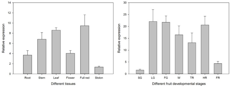
As shown in Figure 3, FvSnRK1.1 constitutively expressed among different tissues. It had much higher expression level in fruit, leaf and stem, followed by flower and root, and had lowest transcript abundance in stolon. To investigate whether the expressions of the FvSnRK1.1 in strawberries were associated with fruit development, its expression pattern in different samples of small green (SG), large green (LG), fade green (FG), white (W), turning red (TR), half red (HR) and full red (FR) was analyzed by qRT-PCR (quantitative real-time PCR). The expression level of FvSnRK1.1 had a remarkable increase from SG to LG. After this, it kept high transcript abundance except in the FR stage. These results indicated that expression of FvSnRK1.1 was related to early strawberry fruit development and might play an important role in regulating fruit ripening.
Figure 3.
Expression level of FvSnRK1.1 in different tissues and during the fruit development. SG, small green; LG, large green; FG, fade green; W, white; TR, turning red; HR, half red; FR, full red.
3.6. Expression Pattern of FvSnRK1.1 in Response to Different Treatment
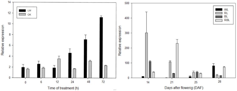
SnRK1s was confirmed to participate widely in response to various environmental elicitors. The expression pattern of the FvSnRK1.1 gene under low temperature and light quality treatments were thus examined. The results showed that FvSnRK1.1 was highly induced after the 12-hour low temperature exposure. Additionally, the transcript level of FvSnRK1.1 had different sensitivities to white, red, blue and red/blue light. It was significantly upregulated by RL, BL and RBL from 14 to 25 DAF. Furthermore, the FvSnRK1.1 expression pattern differed under different light quality during the fruit development. It had a gradually decreasing trend under red and blue light treatment, while an opposite tendency existed under white light (Figure 4).
Figure 4.
Expression level of FvSnRK1.1 under low temperature and light quality treatment. LW, low temperature; WL, white light; RL, red light; BL, blue light; RBL, mixed light (red: blue = 1:1).
4. Discussion
SnRKs as protein kinases represent an interface between metabolic and stress signaling pathways in plants. SnRK1 is closely related to the metabolic regulators of mammals (5’-AMP-activated protein kinase, AMPK) and yeast (sucrose non-fermenting-1, SNF1), with which it shares about 47% amino acid sequence identity and similar substrate specificity. As the plant has evolved, the subfamilies SnRK2 and SnRK3 have emerged, which are larger and relatively more diverse compared with SnRK1 [8]. Bioinformatic analysis of SnRK family has isolated a total of 39 AtSnRKs including three AtSnRK1s, 10 AtSnRK2s and 26 AtSnRK3s in Arabidopsis [21,22,23,24], 44 BdSnRKs including three BdSnRK1s, 10 BdSnRK2s and 31 BdSnRK3s in Brachypodium distachyon [25], 34 EgrSnRK including two EgrSnRK1s, eight EgrSnRK2s and 24 EgrSnRK3s in Eucalyptus grandis [26]. Here, we identified 26 FvSnRKs composed of one FvSnRK1, nine FvSnRK2s, 16 FvSnRK3s in the wild strawberry (Fragaria vesca). Overall, the SnRK3 subfamily has the largest number of members, while the SnRK1 subfamily has the fewest members. In our study, the consequences of sequence feature, subcellular localization, gene structure and phylogeny analysis also indicated the function diversity of SnRKs.
Subcellular localization showed SnRK3s were localized to endoplasmic reticulum and membranes besides cytoplasm and nuclei, indicating it functions in more cellular compartments. The diversification of the exon–intron structure played a pivotal role in the evolution and function of many gene families. The number of exons or introns varied in different subfamilies and genes. The FvSnRK1.1 had 10 exons, which was the same as that reported for EgrSnRK1s and BdSnRK1s [25,26]. Most of FvSnRK2s had nine exons or eight introns, except FvSnRK2.4, FvSnRK2.8 and FvSnRK2.9, which are consistent with that of most Arabidopsis, rice, maize, sorghum, tea and grape SnRK2 genes [27,28,29,30], suggesting that most SnRK2s in plants have a conserved structure with nine exons or eight introns. Like the SnRK3s subfamily in other plant species, SnRK3 genes in strawberries were also divided into intron-free and intron-harboring members, and the number of introns in intron-harboring group ranged from 11 to 15. These findings have showed that the number of SnRKs exon–intron exhibits high conservation during the evolution in each subfamily. Moreover, the phylogenetic analysis showed SnRK1, SnRK2 and SnRK3 respectively clustered together in spite of the different species of strawberry and Arabidopsis, indicating that the genes were established prior to the divergence of the corresponding taxonomic lineages. Meanwhile, conserved motif analyses showed that SnRK sequences that belonged to the same subgroup contained the specific motifs.
The SnRK1 kinases influence plant metabolism, stress tolerance, and a large array of growth and developmental processes by direct phosphorylation of regulatory proteins and metabolic enzymes, and by extensive transcriptional orchestration [31,32,33]. To better understand the function and regulation of SnRK1 in strawberries, the FvSnRK1.1 promoter sequence was analyzed using PlantCARE tool. The results showed that it contained a series of cis-elements involved in stress-responsiveness, hormone-responsiveness and light responsiveness, indicating that FvSnRK1.1 has a diverse role in the strawberry. Clearly, FvSnRK1.1 indeed responded to low temperatures according to their up-regulating expression pattern, which corroborated the result in FvSnRK1.1 promoter analysis. It was reported that SnRK1 could interact with a trihelix family gene ShCIGT to mediate cold tolerance in a tomato [34]. Meanwhile, the FvSnRK1.1 transcript level changed under different light quality treatment, indicating it was sensitive to light signal. The Arabidopsis akinβ1(a subunit of SnRK1) mutant altered multiple genes expression level in response to light and dark [33]. In addition, FvSnRK1.1 transcript accumulated in tissue-specific manner. It has been documented that overexpression of SnRK1 could increase the fruit starch and soluble sugar and promote fruit ripening [35,36,37]. For instance, a tomato overexpressing PpSnRK1α caused higher sugar content and matured approximately 10 days earlier than the WT. It was demonstrated that PpSnRK1α interacted with the MADS-box transcription factor SIRIN, increasing the expression of RIN, regulating the expression of downstream ripening-related genes and promoting the fruit ripening [37]. Here, the high expression level of FvSnRK1.1 during fruit development suggested that FvSnRK1.1 was probably involved in strawberry fruit maturation, but the detailed mechanism requires in-depth study.
In conclusion, we isolated and characterized 26 SnRKs including one SnRK1, nine SnRK2s and 16 SnRK3s from the strawberry genome and analyzed their gene structure, conserved protein motif and evolutionary relationship, which will supply abundant information for functional investigation of SnRK genes. Cis-elements in promoter and expression patterns in response to external signals analysis suggested that FvSnRK1.1 was involved in cold responsiveness and light responsiveness. The expression profiles of FvSnRK1.1 in distinct tissues, stages of fruit development and ripening indicated that the FvSnRK1.1 transcript accumulation was tissue-specific and possibly related to strawberry fruit maturation. These results may advance the understanding of the role of FvSnRK1.1 in the regulation of strawberry development and responses to abiotic stress.
Supplementary Materials
The following are available online at https://www.mdpi.com/2073-4425/11/4/427/s1, Supplementary File S1: Protein sequences of Arabidopsis SnRK genes; Supplementary File S2: Protein sequences of strawberry SnRK genes; Supplementary File S3: Genomic sequences of strawberry SnRK genes; Supplementary File S4: Coding sequences of strawberry SnRK genes; Supplementary File S5: Conserved motifs of strawberry SnRK proteins.
Author Contributions
Conceptualization, Y.Z. and H.T.; Methodology, Y.Z., Q.C. and B.S.; Software, Y.Y., Y.L. and X.G.; Validation, Y.Z., Y.Y. and L.J.; Data Curation, Y.Z. and Y.Y; Writing—Original Draft Preparation, Y.Z.; Writing–Review & Editing, Y.Z., Y.L. and Y.W. Supervision, X.W. and H.T. Funding acquisition, Q.C. and H.T. All authors have read and agreed to the published version of the manuscript.
Funding
The work was supported by the National Natural Science Foundation of China (31872083 and 31972387).
Conflicts of Interest
The authors declare that they have no conflict of interest.
References
- 1.Demidchik V., Maathuis F., Voitsekhovskaja O. Unravelling the plant signalling machinery: An update on the cellular and genetic basis of plant signal transduction. Funct. Plant Boil. 2018;45:1–8. doi: 10.1071/FP17085. [DOI] [PubMed] [Google Scholar]
- 2.Fujita M., Fujita Y., Noutoshi Y., Takahashi F., Narusaka Y., Yamaguchi-Shinozaki K., Shinozaki K. Crosstalk between abiotic and biotic stress responses: A current view from the points of convergence in the stress signaling networks. Curr. Opin. Plant Boil. 2006;9:436–442. doi: 10.1016/j.pbi.2006.05.014. [DOI] [PubMed] [Google Scholar]
- 3.Hashiguchi A., Komatsu S. Impact of post-translational modifications of crop proteins under abiotic stress. Proteomes. 2016;4:42. doi: 10.3390/proteomes4040042. [DOI] [PMC free article] [PubMed] [Google Scholar]
- 4.Tena G., Boudsocq M., Sheen J. Protein kinase signaling networks in plant innate immunity. Curr. Opin. Plant Boil. 2011;14:519–529. doi: 10.1016/j.pbi.2011.05.006. [DOI] [PMC free article] [PubMed] [Google Scholar]
- 5.Coello P., Hey S.J., Halford N.G. The sucrose non-fermenting-1-related (SnRK) family of protein kinases: Potential for manipulation to improve stress tolerance and increase yield. J. Exp. Bot. 2011;62:883–893. doi: 10.1093/jxb/erq331. [DOI] [PubMed] [Google Scholar]
- 6.Broeckx T., Hulsmans S., Rolland F. The plant energy sensor: Evolutionary conservation and divergence of SnRK1 structure, regulation, and function. J. Exp. Bot. 2016;67:6215–6252. doi: 10.1093/jxb/erw416. [DOI] [PubMed] [Google Scholar]
- 7.Hulsmans S., Rodriguez M., De Coninck B., Rolland F. The SnRK1 energy sensor in plant biotic interactions. Trends Plant Sci. 2016;21:648–661. doi: 10.1016/j.tplants.2016.04.008. [DOI] [PubMed] [Google Scholar]
- 8.Halford N.G., Hey S.J. Snf1-related protein kinases (SnRKs) act within an intricate network that links metabolic and stress signalling in plants. Biochem. J. 2009;419:247–259. doi: 10.1042/BJ20082408. [DOI] [PubMed] [Google Scholar]
- 9.Dong X., Cui N., Wang L., Zhao X., Qu B., Li T., Zhang G. The SnRK protein kinase family and the function of SnRK1 protein kinase. Int. J. Agric. Biol. 2012;14:196–200. [Google Scholar]
- 10.Saha J., Chatterjee C., Sengupta A., Gupta K., Gupta B. Genome-wide analysis and evolutionary study of sucrose non-fermenting 1-related protein kinase 2 (SnRK2) gene family members in Arabidopsis and Oryza. Comput. Boil. Chem. 2014;49:59–70. doi: 10.1016/j.compbiolchem.2013.09.005. [DOI] [PubMed] [Google Scholar]
- 11.Anderberg R.J., Walker-Simmons M.K. Isolation of a wheat cDNA clone for an abscisic acid-inducible transcript with homology to protein kinases. Proc. Natl. Acad. Sci. USA. 1992;89:10183–10187. doi: 10.1073/pnas.89.21.10183. [DOI] [PMC free article] [PubMed] [Google Scholar]
- 12.Holappa L.D., Walker-Simmons M.K. The wheat abscisic acid-responsive protein kinase mRNA, PKABA1, is up-regulated by dehydration, cold temperature, and osmotic stress. Plant Physiol. 1995;108:1203–1210. doi: 10.1104/pp.108.3.1203. [DOI] [PMC free article] [PubMed] [Google Scholar]
- 13.Mao X., Li Y., Rehman S.U., Miao L., Zhang Y., Chen X., Yu C., Wang J., Li C., Jing R. The sucrose non-fermenting 1-related protein kinase 2 (SnRK2) genes are multifaceted players in plant growth, development and response to environmental stimuli. Plant Cell Physiol. 2020;61:225–242. doi: 10.1093/pcp/pcz230. [DOI] [PubMed] [Google Scholar]
- 14.Guo Y., Halfter U., Ishitani M., Zhu J.K. Molecular characterization of functional domains in the protein kinase SOS2 that is required for plant salt tolerance. Plant Cell. 2001;13:1383–1400. doi: 10.1105/TPC.010021. [DOI] [PMC free article] [PubMed] [Google Scholar]
- 15.Cheong Y.H., Pandey G.K., Grant J.J., Batistic O., Li L., Kim B.G., Lee S.C., Kudla J., Luan S. Two calcineurin B-like calcium sensors, interacting with protein kinase CIPK23, regulate leaf transpiration and root potassium uptake in Arabidopsis. Plant J. 2007;52:223–239. doi: 10.1111/j.1365-313X.2007.03236.x. [DOI] [PubMed] [Google Scholar]
- 16.Tang R.J., Wang C., Li K., Luan S. The CBL–CIPK calcium signaling network: Unified paradigm from 20 years of discoveries. Trends Plant Sci. 2020 doi: 10.1016/j.tplants.2020.01.009. [DOI] [PubMed] [Google Scholar]
- 17.Nietzsche M., Landgraf R., Tohge T., Börnke F. A protein–protein interaction network linking the energy-sensor kinase SnRK1 to multiple signaling pathways in Arabidopsis thaliana. Curr. Plant Boil. 2016;5:36–44. doi: 10.1016/j.cpb.2015.10.004. [DOI] [Google Scholar]
- 18.Crozet P., Margalha L., Confraria A., Rodrigues A., Martinho C., Adamo M., Elias C.A., Baena-González E. Mechanisms of regulation of SNF1/AMPK/SNRK1 protein kinases. Front. Plant Sci. 2014;5:190. doi: 10.3389/fpls.2014.00190. [DOI] [PMC free article] [PubMed] [Google Scholar]
- 19.Chen C., Chen H., He Y., Xia R. Tbtools, a toolkit for biologists integrating various biological data handling tools with a user-friendly interface. BioRxiv. 2018:289660. doi: 10.1101/289660. [DOI] [Google Scholar]
- 20.Chen Q., Yu H., Wang X., Xie X., Yue X., Tang H. An alternative cetyltrimethylammonium bromide-based protocol for RNA isolation from blackberry (Rubus L.) Genet. Mol. Res. 2012;11:1773–1782. doi: 10.4238/2012.June.29.10. [DOI] [PubMed] [Google Scholar]
- 21.Hrabak E.M., Chan C.W., Gribskov M., Harper J.F., Choi J.H., Halford N.G., Kudla J., Luan S., Nimmo H., Sussman M.R., et al. The Arabidopsis CDPK-SnRK superfamily of protein kinases. Plant Physiol. 2003;132:666–680. doi: 10.1104/pp.102.011999. [DOI] [PMC free article] [PubMed] [Google Scholar]
- 22.Kolukisaoglu Ü., Weinl S., Blazevic D., Batistic O., Kudla J. Calcium sensors and their interacting protein kinases: Genomics of the Arabidopsis and rice CBL-CIPK signaling networks. Plant Physiol. 2004;134:43–58. doi: 10.1104/pp.103.033068. [DOI] [PMC free article] [PubMed] [Google Scholar]
- 23.Weinl S., Kudla J. The CBL-CIPK Ca2+-decoding signaling network: Function and perspectives. New Phytol. 2009;184:517–528. doi: 10.1111/j.1469-8137.2009.02938.x. [DOI] [PubMed] [Google Scholar]
- 24.Lyzenga W.J., Liu H., Schofield A., Muise-Hennessey A., Stone S.L. Arabidopsis CIPK26 interacts with KEG, components of the ABA signalling network and is degraded by the ubiquitin-proteasome system. J. Exp. Bot. 2013;64:2779–2791. doi: 10.1093/jxb/ert123. [DOI] [PMC free article] [PubMed] [Google Scholar]
- 25.Wang L., Hu W., Sun J., Liang X., Yang X., Wei S., Wang X., Zhou Y., Xiao Q., Yang G., et al. Genome-wide analysis of SnRK gene family in Brachypodium distachyon and functional characterization of BdSnRK2.9. Plant Sci. 2015;237:33–45. doi: 10.1016/j.plantsci.2015.05.008. [DOI] [PubMed] [Google Scholar]
- 26.Wang Y., Yan H., Qiu Z., Hu B., Zeng B., Zhong C., Fan C. Comprehensive analysis of SnRK gene family and their responses to salt stress in Eucalyptus grandis. Int. J. Mol. Sci. 2019;20:2786. doi: 10.3390/ijms20112786. [DOI] [PMC free article] [PubMed] [Google Scholar]
- 27.Huai J., Wang M., He J., Zheng J., Dong Z., Lv H., Zhao J., Wang G. Cloning and characterization of the SnRK2 gene family from Zea mays. Plant Cell Rep. 2008;27:1861–1868. doi: 10.1007/s00299-008-0608-8. [DOI] [PubMed] [Google Scholar]
- 28.Li L.B., Zhang Y.R., Liu K.C., Ni Z.F., Fang Z.J., Sun Q.X., Gao J.W. Identification and bioinformatics analysis of SnRK2 and CIPK family genes in sorghum. Agric. Sci. China. 2010;9:19–30. doi: 10.1016/S1671-2927(09)60063-8. [DOI] [Google Scholar]
- 29.Zhang Y.H., Wan S.Q., Wang W.D., Chen J.F., Huang L.L., Duan M.S., Yu Y.B. Genome-wide identification and characterization of the CsSnRK2 family in Camellia sinensis. Plant Physiol. Biochem. 2018;132:287–296. doi: 10.1016/j.plaphy.2018.09.021. [DOI] [PubMed] [Google Scholar]
- 30.Liu J.Y., Chen N.N., Cheng Z.M., Xiong J.S. Genome-wide identification, annotation and expression profile analysis of SnRK2 gene family in grapevine. Aust. J. Grape Wine Res. 2016;22:478–488. doi: 10.1111/ajgw.12223. [DOI] [Google Scholar]
- 31.Crepin N., Rolland F. SnRK1 activation, signaling, and networking for energy homeostasis. Curr. Opin. Plant Boil. 2019;51:29–36. doi: 10.1016/j.pbi.2019.03.006. [DOI] [PubMed] [Google Scholar]
- 32.Feng X., Feng P., Yu H., Yu X., Sun Q., Liu S., Minh T.N., Chen J., Wang D., Zhang Q., et al. GsSnRK1 interplays with transcription factor GsERF7 from wild soybean to regulate soybean stress resistance. Plant Cell Environ. 2020:1–20. doi: 10.1111/pce.13726. [DOI] [PubMed] [Google Scholar]
- 33.Wang Y., Wang L., Micallef B.J., Tetlow I.J., Mullen R.T., Feil R., Lunn J.E., Emes M.J. AKINβ1, a subunit of SnRK1, regulates organic acid metabolism and acts as a global modulator of genes involved in carbon, lipid, and nitrogen metabolism. J. Exp. Bot. 2020;71:1010–1028. doi: 10.1093/jxb/erz460. [DOI] [PubMed] [Google Scholar]
- 34.Yu C., Song L., Song J., Ouyang B., Guo L., Shang L., Wang T., Li H.X., Zhang J., Ye Z. ShCIGT, a Trihelix family gene, mediates cold and drought tolerance by interacting with SnRK1 in tomato. Plant Sci. 2018;270:140–149. doi: 10.1016/j.plantsci.2018.02.012. [DOI] [PubMed] [Google Scholar]
- 35.Li G., Peng F., Zhang L., Shi X., Wang Z. Cloning and characterization of a SnRK1-encoding gene from Malus hupehensis Rehd and heterologous expression in tomato. Mol. Boil. Rep. 2010;37:947–954. doi: 10.1007/s11033-009-9734-9. [DOI] [PubMed] [Google Scholar]
- 36.Wang X., Peng F., Li M., Yang L., Li G. Expression of a heterologous SnRK1 in tomato increases carbon assimilation, nitrogen uptake and modifies fruit development. J. Plant Physiol. 2012;169:1173–1182. doi: 10.1016/j.jplph.2012.04.013. [DOI] [PubMed] [Google Scholar]
- 37.Yu W., Peng F., Xiao Y., Wang G., Luo J. Overexpression of PpSnRK1α in Tomato Promotes Fruit Ripening by Enhancing RIPENING INHIBITOR Regulation Pathway. Front. Plant Sci. 2018;9:1856. doi: 10.3389/fpls.2018.01856. [DOI] [PMC free article] [PubMed] [Google Scholar]
Associated Data
This section collects any data citations, data availability statements, or supplementary materials included in this article.


