Abstract
Background
The chest CT findings of patients with 2019 Novel Coronavirus (2019-nCoV) pneumonia have not previously been described in detail.
Purpose
To investigate the clinical, laboratory, and imaging findings of emerging 2019-nCoV pneumonia in humans.
Materials and Methods
Fifty-one patients (25 men and 26 women; age range 16–76 years) with laboratory-confirmed 2019-nCoV infection by using real-time reverse transcription polymerase chain reaction underwent thin-section CT. The imaging findings, clinical data, and laboratory data were evaluated.
Results
Fifty of 51 patients (98%) had a history of contact with individuals from the endemic center in Wuhan, China. Fever (49 of 51, 96%) and cough (24 of 51, 47%) were the most common symptoms. Most patients had a normal white blood cell count (37 of 51, 73%), neutrophil count (44 of 51, 86%), and either normal (17 of 51, 35%) or reduced (33 of 51, 65%) lymphocyte count. CT images showed pure ground-glass opacity (GGO) in 39 of 51 (77%) patients and GGO with reticular and/or interlobular septal thickening in 38 of 51 (75%) patients. GGO with consolidation was present in 30 of 51 (59%) patients, and pure consolidation was present in 28 of 51 (55%) patients. Forty-four of 51 (86%) patients had bilateral lung involvement, while 41 of 51 (80%) involved the posterior part of the lungs and 44 of 51 (86%) were peripheral. There were more consolidated lung lesions in patients 5 days or more from disease onset to CT scan versus 4 days or fewer (431 of 712 lesions vs 129 of 612 lesions; P < .001). Patients older than 50 years had more consolidated lung lesions than did those aged 50 years or younger (212 of 470 vs 198 of 854; P < .001). Follow-up CT in 13 patients showed improvement in seven (54%) patients and progression in four (31%) patients.
Conclusion
Patients with fever and/or cough and with conspicuous ground-glass opacity lesions in the peripheral and posterior lungs on CT images, combined with normal or decreased white blood cells and a history of epidemic exposure, are highly suspected of having 2019 Novel Coronavirus (2019-nCoV) pneumonia.
© RSNA, 2020
Summary
Fever and/or cough, chest CT with bilateral ground-glass opacities in the posterior and peripheral lungs, and a history of exposure to individuals from Wuhan, China, are characteristic of 2019 Novel Coronavirus, or 2019-nCoV, pneumonia.
Key Results
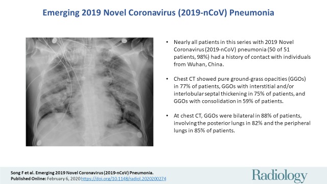
■ Nearly all patients in this series with 2019 Novel Coronavirus, or 2019-nCoV, pneumonia (50 of 51 patients, 98%) had a history of contact with individuals from Wuhan, China.
■ Chest CT showed pure ground-glass opacities (GGOs) in 77% of patients, GGOs with interstitial and/or interlobular septal thickening in 75% of patients, and GGOs with consolidation in 59% of patients.
■ At chest CT, GGOs were bilateral in 88% of patients, involving the posterior lungs in 82% and the peripheral lungs in 85% of patients.
Introduction
On December 30, 2019, a report indicating a cluster of patients with pneumonia of unknown etiology in Wuhan City, Hubei Province, China, was published on ProMED-mail (1). It was possibly related to contact with a local fish and wild animal market (Huanan Seafood Wholesale Market), where there was also sale of live animals. Most of the first reported patients visited the market about 1 month before onset. Deep sequencing analysis from lower respiratory tract samples indicated a novel coronavirus, which was named 2019 Novel Coronavirus (2019-nCoV) by the World Health Organization (2). In mid-January 2020, China began passenger transportation during or around the Chinese New Year, which is equivalent to the Christmas holiday celebration in the West. A large number of people living in Wuhan left the region by air carrier. Since January 17, the confirmed or suspected cases have dramatically increased. By 11:41 PM on February 5, 2020, China has reported 24 363 confirmed cases and 23 260 suspected cases (including health care workers) from all provinces, cities, autonomous regions, and special administrative regions (Hong Kong and Macao). Thailand, Japan, South Korea, and the United States of America (3–6) also reported exported cases. At the time of this writing, there were 492 patients who died in China and 897 cases cured. The 2019-nCoV poses significant threats to international health.
Coronaviruses are enveloped nonsegmented positive-sense RNA viruses belonging to the family Coronaviridae and the order Nidovirales and broadly distributed in humans and other mammals (7). The 2002–2003 pandemic of severe acute respiratory syndrome coronavirus, or SARS-CoV, and the ongoing emergence of the Middle East respiratory syndrome coronavirus, or MERS-CoV, demonstrate that coronaviruses are a significant public health threat (8). The 2019-nCoV is a betacoronavirus of group 2B with at least 70% similarity in genetic sequence to SARS-CoV (9). Different from both MERS-CoV and SARS-CoV, 2019-nCoV is the seventh member of the family of coronaviruses that infect humans (10). It may originate from Chinese horseshoe bats, which are the natural reservoirs of SARS-CoV (8) and can transmit from human to human.
The Shanghai Public Health Clinical Center is the designated hospital for diagnosis and management of infectious diseases and threats against public health in our country, and it is also a World Health Organization–designated training organization for new emerging infectious diseases. At the time of this writing, 56 patients with confirmed 2019-nCoV infection have been admitted to the center.
The purpose of this study was to review the noncontrast chest CT findings in patients with laboratory-confirmed 2019-nCoV infection by using real-time reverse transcription–polymerase chain reaction, or RT-PCR, performed at the Center for Disease Control, Shanghai, China.
Materials and Methods
Patients
The ethical committee of Shanghai Public Health Clinical Center approved this retrospective study. Our inclusion criteria were patients admitted to our center with laboratory-confirmed 2019-nCoV infection by using RT-PCR, patients who underwent thin-section CT, and patients with CT images that demonstrated pneumonia. Excluded were two patients with only bedside chest radiograph (without CT), and three patients with normal findings at chest CT. A total of 51 patients were included. We reviewed the clinical and laboratory data and CT images of the 51 patients with 2019-nCoV pneumonia from January 20, 2020, to January 27, 2020. All patients were confirmed as having positive results by using real-time RT-PCR nucleic acid assay for 2019-nCoV by the Center for Disease Control, Shanghai, China. Other causes of pneumonia from common bacterial and viral pathogens were excluded.
Image Acquisition
All the patients underwent thin-section CT. The median duration from illness onset to CT scan was 4 days, ranging from 1 to 14 days. All CT examinations were performed with a 64-section scanner (Scenaria 64 CT; Hitachi Medical, Kashiwa, Chiba Prefecture, Japan) without the use of contrast material. The CT protocol was as follows: tube voltage, 120 kV; automatic tube current (180 mA–400 mA); iterative reconstruction technique; detector, 64 mm; rotation time, 0.35 second; section thickness, 5 mm; collimation, 0.625 mm; pitch, 1.5; matrix, 512 × 512; and breath hold at full inspiration. Reconstruction kernel used was lung smooth with a thickness of 1 mm and an interval of 0.8 mm. The following windows were used: a mediastinal window with a window width of 350 HU and a window level of 40 HU, and a lung window with a width of 1200 HU and a level of −600 HU.
Three chest radiologists (F.Song, N.S., and Y.S., with approximately 6–32 years of experience in thoracic imaging, especially in the setting of viral pneumonias such as H1N1 and H7N9 pneumonia) reviewed the images independently, with a final finding reached by consensus when there was a discrepancy.
CT images were assessed for the presence and distribution of parenchymal abnormalities including pure ground-glass opacity (GGO), which were defined as a hazy increase in lung attenuation with no obscuration of the underlying vessels; GGO with interlobular septal thickening or reticulation, or intralobular networks in GGO; GGO with consolidation, which was defined as an area of opacification obscuring the underlying vessels in GGO; consolidation; air bronchogram(s); reticulation; lymphadenopathy, which was defined as a lymph node greater than 1 cm in short-axis diameter; and pleural effusion. On the axial CT images, we drew a horizontal line across the axillary midline to divide anterior and posterior parts of the lungs. The outer one-third of the lung was defined as peripheral, and the rest was defined as central.
Chest CT lesions in each patient were identified by the readers. A lesion occupying only one lung segment was counted as one lesion. When a large lesion or fused lesion involved more than one lung segment, the lesion number was recorded as the number of the involved lung segments. For example, a large lesion involving three lung segments was counted as three lesions. Each side of the chest containing pleural fluid was counted as one lesion. A pericardial effusion was counted as one lesion.
Statistical Analysis
All data were statistically analyzed by using Stata software package (version 10.0; StataCorp, College Station, Tex). Data were expressed as means ± standard deviation, or medians for the continuous variables. The comparison of discrete variables between groups was performed by using the Pearson χ2 test. Correlation analysis between two groups was performed by using rank correlation. All analyses were considered significant at P values of less than .05.
Results
Clinical and Laboratory Findings
The demographic and clinical characteristic results of 51 patients are shown in Table 1. The 51 patients included 25 (49%) men and 26 (51%) women, with ages ranging from 16 to 76 years (mean age, 49 years ± 16 [standard deviation]). Fifty patients (98%) had some contact with individuals from Wuhan, China (38 patients traveled to or lived in Wuhan, or were in contact with individuals with confirmed 2019-nCoV pneumonia [eight, 16%], individuals suspected of having 2019-nCoV pneumonia [three, 6%], and a single healthy individual from that city [one, 2.0%]). Only one patient had no definite history of Wuhan contact, but had recently traveled to another city. The most common symptoms were fever (49 of 51, 96%) and cough (24 of 51, 47%). Other symptoms included myalgia or fatigue (16 of 51, 31%), mild headache and dizziness (eight of 51, 16%), and diarrhea (five of 51, 10%). Eleven of 51 (22%) patients had comorbidities including diabetes, hypertension, chronic liver disease, chronic obstructive pulmonary disease, and cardiac disease. Three of the 51 (7%) patients with confirmed 2019-nCoV pneumonia were current cigarette smokers.
Table 1:
Demographic and Clinical Characteristics of 51 Patients with 2019 Novel Coronavirus (2019-nCoV) Pneumonia
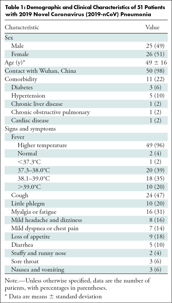
Most of the patients had a normal white blood cell count (37 of 51, 73%) and neutrophil count (44 of 51, 86%). The lymphocyte count was low (33 of 51, 65%) or normal (17 of 51, 35%). C-reactive protein level was elevated in 41 of 51 patients (80%). Thirty-one of 51 (61%) patients had a low CD4+ cell count, with a range of 72–408 cells/µL.
CT Imaging Findings
Data of initial chest thin-section CT imaging findings in 51 patients with 2019-nCoV pneumonia are presented in Tables 2 and 3. There were 44 of 51 (86%) patients with a total of 1284 of 1324 (97%) lesions involving both lungs and 32 of 51 (63%) patients with 1194 of 1324 (90%) lesions involving four to five lobes. There were 46 of 51 (90%) patients with 703 of 1324 (53%) lesions distributed in the lower lobes, 41 of 51 (80%) patients with 1179 of 1324 (89%) lesions distributed in the posterior part of the lung, and 44 of 51 (86%) patients with 1198 of 1324 (91%) lesions distributed in the lung periphery (Fig 1).
Table 2:
Distribution of the Lesions in 51 Patients with 2019 Novel Coronavirus (2019-nCoV) Pneumonia
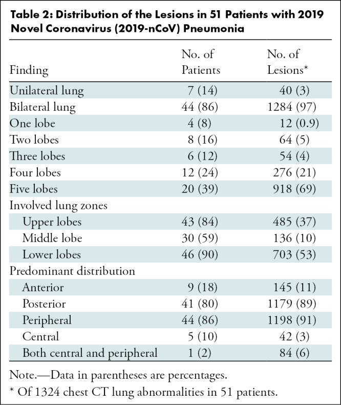
Table 3:
CT Imaging Findings in 51 Patients with 2019 Novel Coronavirus (2019-nCoV) Pneumonia
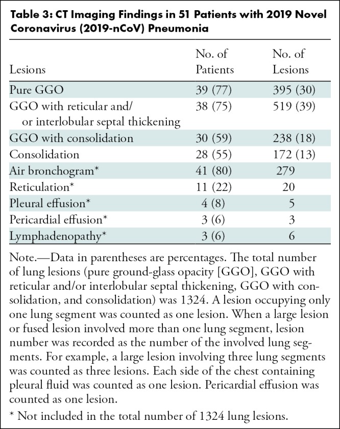
Figure 1a:
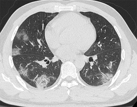
(a) Baseline CT images at admission of a 35-year-old man show multiple patchy areas of pure ground-glass opacity (GGO); GGO with reticular and/or interlobular septal thickening; and patchy, focal, often rounded peribronchovascular and subpleural opacities associated with reticulation and architectural distortion. Lesions are mostly distributed in peripheral and posterior part of lungs. (b, c) Follow-up CT images on day 5 after admission show prominent progression with increased size and density of lesions, greater consolidation, and with interval new bilateral pleural effusions and likely bibasilar compression atelectasis (c).
Figure 1b:
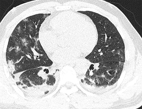
(a) Baseline CT images at admission of a 35-year-old man show multiple patchy areas of pure ground-glass opacity (GGO); GGO with reticular and/or interlobular septal thickening; and patchy, focal, often rounded peribronchovascular and subpleural opacities associated with reticulation and architectural distortion. Lesions are mostly distributed in peripheral and posterior part of lungs. (b, c) Follow-up CT images on day 5 after admission show prominent progression with increased size and density of lesions, greater consolidation, and with interval new bilateral pleural effusions and likely bibasilar compression atelectasis (c).
Figure 1c:
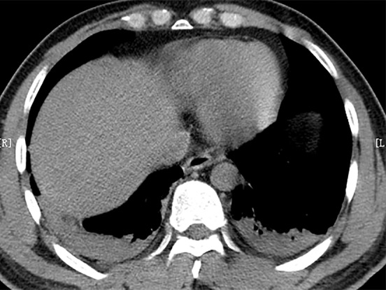
(a) Baseline CT images at admission of a 35-year-old man show multiple patchy areas of pure ground-glass opacity (GGO); GGO with reticular and/or interlobular septal thickening; and patchy, focal, often rounded peribronchovascular and subpleural opacities associated with reticulation and architectural distortion. Lesions are mostly distributed in peripheral and posterior part of lungs. (b, c) Follow-up CT images on day 5 after admission show prominent progression with increased size and density of lesions, greater consolidation, and with interval new bilateral pleural effusions and likely bibasilar compression atelectasis (c).
Pure GGO, GGO with reticular and/or interlobular septal thickening, and GGO with consolidation were the main findings (1152 of 1324, 87%). There were 395 of 1324 (30%) pure GGOs in 39 of 51 (77%) patients, 519 of 1324 (39%) GGO lesions with reticular and/or interlobular septal thickening in 38 (75%) patients, 238 of 1324 (18%) GGO lesions with consolidation in 30 of 51 (59%) patients, 172 of 1324 (13%) consolidation lesions in 28 of 51 (55%) patients (Figs 1–3), and 279 bronchograms in 41 of 51 (80%) patients. Other findings included reticulation, small pleural effusion, small pericardial effusion, and lymphadenopathy. The pleural effusion was bilateral in one patient and unilateral in three patients. One patient had only right pleural with no parenchyma lesions at first chest CT. After 2-day follow-up, the pleural effusion had absorption, but small pure GGO in the lung parenchyma was seen.
Figure 3a:
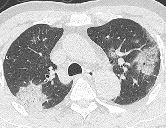
Serial imaging after admission of a 71-year-old man. (a) Baseline CT images on January 21, 2020, show consolidation of right upper lobe and ground-glass opacity (GGO) with consolidation and reticular and/or interlobular septal thickening of left upper lobe of left upper lobe; and patchy, focal, often rounded peribronchovascular and subpleural opacities associated with reticulation and architectural distortion. (b) Two days later, CT images show increased size of lesions in both lungs and decreased density in GGO lesions. (c) However, GGO on both lungs were larger on day 4 after admission. (d) Bedside portable chest radiograph on day 6 following admission shows diffusely increased opacities in both lungs, with relative bibasilar sparing.
Figure 2a:
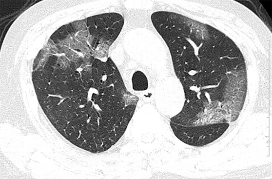
(a, b) Baseline CT images at admission of a 75-year-old man show multiple patchy areas of pure ground-glass opacity (GGO) and GGO with reticular and/or interlobular septal thickening. (c, d) Follow-up CT images on day 3 after admission show overlap of organizing pneumonia with diffuse alveolar damage in that it is more diffuse (not rounded) and associated with underlying reticulation, prominent progression with increased size and density of the lesions, and with more consolidations. Air bronchogram is also shown in d (arrows). Interlobular septal thickening does not seem to be major component. There is reticulation in many of cases of both suspected organizing pneumonia and diffuse alveolar damage.
Figure 2b:
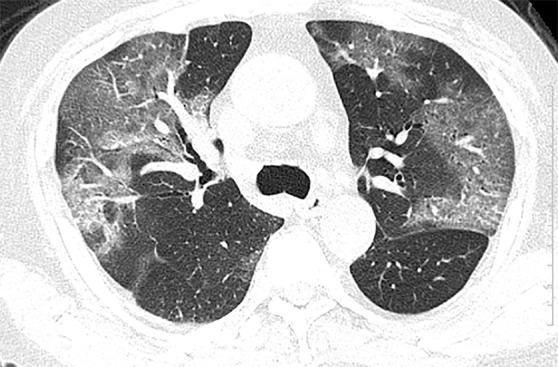
(a, b) Baseline CT images at admission of a 75-year-old man show multiple patchy areas of pure ground-glass opacity (GGO) and GGO with reticular and/or interlobular septal thickening. (c, d) Follow-up CT images on day 3 after admission show overlap of organizing pneumonia with diffuse alveolar damage in that it is more diffuse (not rounded) and associated with underlying reticulation, prominent progression with increased size and density of the lesions, and with more consolidations. Air bronchogram is also shown in d (arrows). Interlobular septal thickening does not seem to be major component. There is reticulation in many of cases of both suspected organizing pneumonia and diffuse alveolar damage.
Figure 2c:
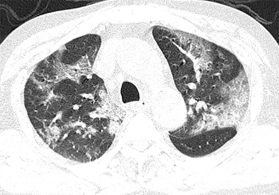
(a, b) Baseline CT images at admission of a 75-year-old man show multiple patchy areas of pure ground-glass opacity (GGO) and GGO with reticular and/or interlobular septal thickening. (c, d) Follow-up CT images on day 3 after admission show overlap of organizing pneumonia with diffuse alveolar damage in that it is more diffuse (not rounded) and associated with underlying reticulation, prominent progression with increased size and density of the lesions, and with more consolidations. Air bronchogram is also shown in d (arrows). Interlobular septal thickening does not seem to be major component. There is reticulation in many of cases of both suspected organizing pneumonia and diffuse alveolar damage.
Figure 2d:
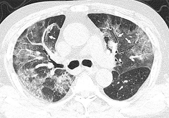
(a, b) Baseline CT images at admission of a 75-year-old man show multiple patchy areas of pure ground-glass opacity (GGO) and GGO with reticular and/or interlobular septal thickening. (c, d) Follow-up CT images on day 3 after admission show overlap of organizing pneumonia with diffuse alveolar damage in that it is more diffuse (not rounded) and associated with underlying reticulation, prominent progression with increased size and density of the lesions, and with more consolidations. Air bronchogram is also shown in d (arrows). Interlobular septal thickening does not seem to be major component. There is reticulation in many of cases of both suspected organizing pneumonia and diffuse alveolar damage.
Figure 3b:
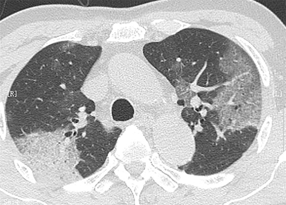
Serial imaging after admission of a 71-year-old man. (a) Baseline CT images on January 21, 2020, show consolidation of right upper lobe and ground-glass opacity (GGO) with consolidation and reticular and/or interlobular septal thickening of left upper lobe of left upper lobe; and patchy, focal, often rounded peribronchovascular and subpleural opacities associated with reticulation and architectural distortion. (b) Two days later, CT images show increased size of lesions in both lungs and decreased density in GGO lesions. (c) However, GGO on both lungs were larger on day 4 after admission. (d) Bedside portable chest radiograph on day 6 following admission shows diffusely increased opacities in both lungs, with relative bibasilar sparing.
Figure 3c:
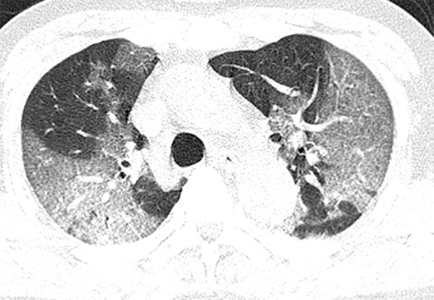
Serial imaging after admission of a 71-year-old man. (a) Baseline CT images on January 21, 2020, show consolidation of right upper lobe and ground-glass opacity (GGO) with consolidation and reticular and/or interlobular septal thickening of left upper lobe of left upper lobe; and patchy, focal, often rounded peribronchovascular and subpleural opacities associated with reticulation and architectural distortion. (b) Two days later, CT images show increased size of lesions in both lungs and decreased density in GGO lesions. (c) However, GGO on both lungs were larger on day 4 after admission. (d) Bedside portable chest radiograph on day 6 following admission shows diffusely increased opacities in both lungs, with relative bibasilar sparing.
Figure 3d:
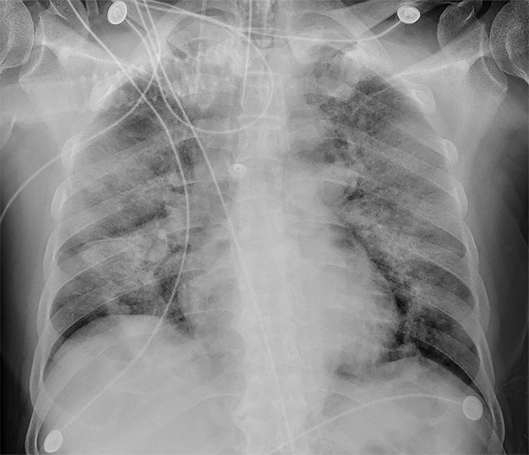
Serial imaging after admission of a 71-year-old man. (a) Baseline CT images on January 21, 2020, show consolidation of right upper lobe and ground-glass opacity (GGO) with consolidation and reticular and/or interlobular septal thickening of left upper lobe of left upper lobe; and patchy, focal, often rounded peribronchovascular and subpleural opacities associated with reticulation and architectural distortion. (b) Two days later, CT images show increased size of lesions in both lungs and decreased density in GGO lesions. (c) However, GGO on both lungs were larger on day 4 after admission. (d) Bedside portable chest radiograph on day 6 following admission shows diffusely increased opacities in both lungs, with relative bibasilar sparing.
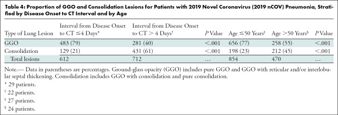
We stratified our patients into two groups: group 1 with an interval of less than or equal to 4 days between symptom onset and the chest CT scan and group 2 with an interval greater than 4 days between symptom onset and the chest CT scan. We found more consolidation (including GGO with consolidation and only consolidation) in group 2 (431 of 712 lesions, 61%) than in group 1 (129 of 612 lesions, 21%) (P < .001). There were significantly more GGOs including pure GGO and GGO with reticular and/or interlobular septal thickening in group 1 (483 of 612 79%) than in group 2 (281 of 712, 40%) (Table 4). The absolute number of lung findings increased with the time from symptom onset (r = 0.33; P = .02). Lesions with consolidations including GGOs with consolidation and pure consolidation showed mildly positive correlation with the time between symptom onset and the CT (r = 0.32; P = .02).
Table 4:
Proportion of GGO and Consolidation Lesions for Patients with 2019 Novel Coronavirus (2019 nCOV) Pneumonia, Stratified by Disease Onset to CT Interval and by Age
We stratified our patients into two age groups (Table 4): age 50 years and younger and age older than 50 years. There were more GGOs (including pure GGOs and GGOs with reticular and/or interlobular septal thickening) in the younger group (656 of 854, 77%) than in the older group (258 of 470, 55%) (P < .001). There was significantly more consolidation with an organizing pneumonia pattern, including GGOs with consolidation and pure consolidation in the older group (212 of 470, 45%) than in the younger group (198 of 854, 23%) (P < .001). The younger patients tended to have more GGOs, while older patients tended to have more areas of lung involvement and more consolidation.
We have followed up chest CT examinations in 13 of 51 of the patients in this series. There was interval progression in four of 13 (31%) patients (Figs 1–3), with an increased size and/or increased consolidation along with more areas of consolidation. At follow-up, pulmonary involvement was improved in seven of 13 (54%) patients, with smaller lesion size and/or a developing organizing pneumonia pattern in what was pure consolidation (Fig 4).
Figure 4a:
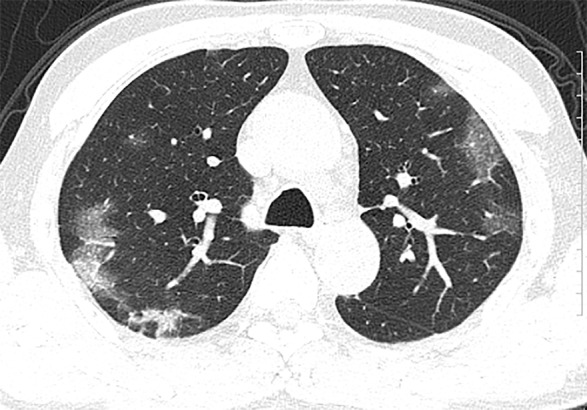
(a, b) Baseline CT images at admission of a 56-year-old man show multiple patchy areas of organizing pneumonia with some areas of interstitial and/or interlobular septal thickening and “strip shaped” consolidation (patchy, focal, often rounded, peribronchovascular and subpleural opacities associated with reticulation and architectural distortion). These abnormalities are mostly distributed in peripheral and posterior parts of lungs. (c, d) Follow-up CT images on day 5 after admission show interval improvement and absorption with fewer lesions and decreased lesion density.
Figure 4b:
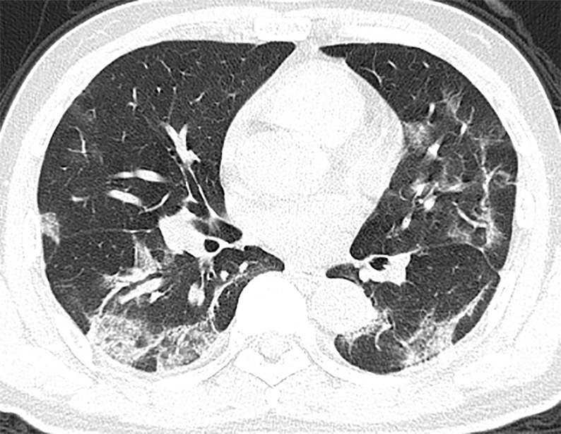
(a, b) Baseline CT images at admission of a 56-year-old man show multiple patchy areas of organizing pneumonia with some areas of interstitial and/or interlobular septal thickening and “strip shaped” consolidation (patchy, focal, often rounded, peribronchovascular and subpleural opacities associated with reticulation and architectural distortion). These abnormalities are mostly distributed in peripheral and posterior parts of lungs. (c, d) Follow-up CT images on day 5 after admission show interval improvement and absorption with fewer lesions and decreased lesion density.
Figure 4c:
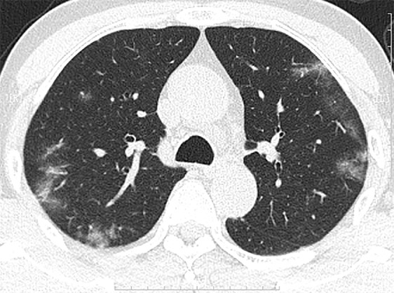
(a, b) Baseline CT images at admission of a 56-year-old man show multiple patchy areas of organizing pneumonia with some areas of interstitial and/or interlobular septal thickening and “strip shaped” consolidation (patchy, focal, often rounded, peribronchovascular and subpleural opacities associated with reticulation and architectural distortion). These abnormalities are mostly distributed in peripheral and posterior parts of lungs. (c, d) Follow-up CT images on day 5 after admission show interval improvement and absorption with fewer lesions and decreased lesion density.
Figure 4d:
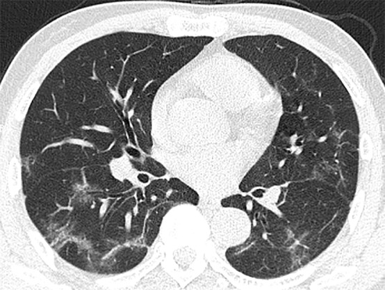
(a, b) Baseline CT images at admission of a 56-year-old man show multiple patchy areas of organizing pneumonia with some areas of interstitial and/or interlobular septal thickening and “strip shaped” consolidation (patchy, focal, often rounded, peribronchovascular and subpleural opacities associated with reticulation and architectural distortion). These abnormalities are mostly distributed in peripheral and posterior parts of lungs. (c, d) Follow-up CT images on day 5 after admission show interval improvement and absorption with fewer lesions and decreased lesion density.
Discussion
All patients except one had a history of contact with individuals from Wuhan, which was very important for diagnosis. The most common symptoms were fever (49, 96%), followed by cough (24, 47%) and myalgia or fatigue (16, 31%), consistent with former report (2,11,12). The laboratory results of normal blood cells could also be helpful to make the diagnosis. In our study, the average age was 49 years, ranging from 16–76 years, and only 11 patients (22%) had coexisting medical conditions such as diabetes, hypertension, chronic liver disease, chronic obstructive pulmonary disease, and cardiac disease. This is different from influenza A (H7N9) virus pneumonia, which tends to affect older adults (aged 66 years, with a range of 48–81 years) with coexisting chronic diseases (13,14).
Most pulmonary lesions involved bilateral lungs with multiple lung lobes, with predominant distribution in posterior and peripheral part of the lungs. Studies showed that influenza pneumonia tend to affect lower lung (15). Wang et al (14) also showed that H7N9 pneumonia had a predominant distribution in the right lower lung. Both H1N1 pneumonia and SARS distributed more peripherally (16,17), while there was no lobar predilection in H5N1 influenza (18). The posterior and peripheral lung predominant distribution in our study is characteristic. Such distribution is conspicuous at first glance of the images.
The observation regarding the high prevalence of bilateral organizing pneumonia in these patients is potentially of importance. This suggests that corticosteroids might be an option to suppress this immune reaction within the lung parenchyma to 2019-nCoV pneumonia.
Our results showed that the most common imaging findings were pure GGO, GGO with reticular and/or interlobular septal thickening, and GGO with consolidation. Complete consolidation was relatively less common (in 55% of patients in 13% of lesions), which might be explained by the early stage of the disease. In H7N9 pneumonia, most cases showed consolidation (14). Each single CT pattern seen in our patients is nonspecific and may overlap with those of other microorganism infections, such as H7N9 pneumonia, H1N1 virus infection, SARS, coronavirus infection, and avian influenza A (H5N1) (14,16,17). GGO, consolidation, and interlobular septal thickening are also the most common thin-section CT findings of H1N1 influenza pneumonia (16). The four patterns in our imaging findings—pure GGO, GGO with reticular and/or interlobular septal thickening, GGO with consolidation, and consolidation—were often mixed, and tended to appear simultaneously in the same patient. While the pure GGO is a common single finding in 77% of patients and in 30% of lesions in the 2019-nCoV pneumonia, the main CT features were all patterns of GGO, including pure GGO, GGO with reticular and/or interlobular septal thickening, and GGO with consolidation, which were observed in all patients and accounted for 87% of lesions. These CT findings combined together, with predominant distribution in posterior and peripheral part of the lungs, were uncommon in other viral pneumonia.
Our study showed that consolidation indicated disease progression. There were more consolidation lesions and fewer GGO lesions in the patients with CT interval greater than or equal to 4 days than in patients with CT interval less than or equal to 4 days, which likely indicates that consolidation increases as disease course extends. In addition, there were significantly more consolidation lesions and less GGO lesions in older patients than in younger patients. Therefore, finding of consolidation lesions would serve as an alert in the treatment of patients.
There were several limitations in our study. First, the sample size was very small with follow-up CT. In addition, the follow-up CT had a short time interval. It would be still inconclusive with CT follow-up findings to evaluate treatment efficacy. Second, there was lack of severe infection, to compare findings with severe infection with mild infection. Third, there was lack of pediatric population. Finally, no lung tissue biopsies were available to investigate correlation between radiologic and histopathologic findings.
In conclusion, the most common patterns of 2019 Novel Coronavirus (2019-nCoV) pneumonia on thin-section CT images are pure ground-glass opacity (GGO), GGO with reticular and/or interlobular septal thickening, GGO with consolidation, and consolidation, with prominent distribution in the posterior and peripheral part of the lungs. Consolidation lesions could serve as a marker of disease progression or more severe disease. Although the positive nucleic acid testing is the diagnostic reference standard, patients with fever and/or cough and with GGO-prominent lesions in the peripheral and posterior part of lungs on CT images, combined with normal or decreased white blood cells and a history of epidemic exposure, should be highly suspected of having 2019-nCoV pneumonia.
Disclosures of Conflicts of Interest: F. Song disclosed no relevant relationships. N.S. disclosed no relevant relationships. F. Shan disclosed no relevant relationships. Z.Z. disclosed no relevant relationships. J.S. disclosed no relevant relationships. H.L. disclosed no relevant relationships. Y.L. disclosed no relevant relationships. Y.J. disclosed no relevant relationships. Y.S. disclosed no relevant relationships.
F. Song and N.S. contributed equally to this work.
Abbreviations:
- GGO
- ground-glass opacity
- 2019-nCOV
- 2019 Novel Coronavirus
References
- 1.ProMED-mail. https://promedmail.org/promed-post/?id=6864153. Accessed January 7, 2020.
- 2.Huang C, Wang Y, Li X, et al. Clinical features of patients infected with 2019 novel coronavirus in Wuhan, China. Lancet 2020 Jan 24 [Epub ahead of print] [Published correction appears in Lancet 2020 Jan 30.]. [DOI] [PMC free article] [PubMed] [Google Scholar]
- 3.World Health Organization . Novel coronavirus – Thailand (ex-China). http://www.who.int/csr/don/14-january-2020-novel-coronavirus-thailand/en/. Published January 14, 2020. Accessed January 19, 2020.
- 4.World Health Organization . Novel coronavirus – Japan (ex-China). http://www.who.int/csr/don/17-january-2020-novel-coronavirus-japan-ex-china/en/. Published January 17, 2020. Accessed January 19, 2020.
- 5.World Health Organization . Novel coronavirus – Republic of Korea (ex-China). http://www.who.int/csr/don/21-january-2020-novel-coronavirus-republic-of-korea-ex-china/en/. Published January 21, 2020. Accessed January 23, 2020.
- 6.Centers for Disease Control and Prevention . First travel-related case of 2019 novel coronavirus detected in United States. https://www.cdc.gov/media/releases/2020/p0121-novel-coronavirus-travel-case.html. Published January 21, 2020. Accessed January 23, 2020.
- 7.Richman DD, Whitley RJ, Hayden FG, eds. Clinical virology. 4th ed. Washington, DC: ASM Press, 2016. [Google Scholar]
- 8.Ge XY, Li JL, Yang XL, et al. Isolation and characterization of a bat SARS-like coronavirus that uses the ACE2 receptor. Nature 2013;503(7477):535–538. [DOI] [PMC free article] [PubMed] [Google Scholar]
- 9.Hui DS, I Azhar E, Madani TA, et al. The continuing 2019-nCoV epidemic threat of novel coronaviruses to global health - The latest 2019 novel coronavirus outbreak in Wuhan, China. Int J Infect Dis 2020;91:264–266. [DOI] [PMC free article] [PubMed] [Google Scholar]
- 10.Zhu N, Zhang D, Wang W, et al. A Novel Coronavirus from Patients with Pneumonia in China, 2019. N Engl J Med 2020 Jan 24 [Epub ahead of print]. [DOI] [PMC free article] [PubMed] [Google Scholar]
- 11.Lei J, Li J, Li X, Qi X. CT Imaging of the 2019 Novel Coronavirus (2019-nCoV) Pneumonia. Radiology 2020 Jan 31 [Epub ahead of print]. [DOI] [PMC free article] [PubMed] [Google Scholar]
- 12.Chen N, Zhou M, Dong X, et al. Epidemiological and clinical characteristics of 99 cases of 2019 novel coronavirus pneumonia in Wuhan, China: a descriptive study. Lancet 2020 Jan 30 [Epub ahead of print]. [DOI] [PMC free article] [PubMed] [Google Scholar]
- 13.Li H, Weng H, Lan C, et al. Comparison of patients with avian influenza A (H7N9) and influenza A (H1N1) complicated by acute respiratory distress syndrome. Medicine (Baltimore) 2018;97(12):e0194. [DOI] [PMC free article] [PubMed] [Google Scholar]
- 14.Wang Q, Zhang Z, Shi Y, Jiang Y. Emerging H7N9 influenza A (novel reassortant avian-origin) pneumonia: radiologic findings. Radiology 2013;268(3):882–889. [DOI] [PubMed] [Google Scholar]
- 15.Koo HJ, Lim S, Choe J, Choi SH, Sung H, Do KH. Radiographic and CT Features of Viral Pneumonia. RadioGraphics 2018;38(3):719–739. [DOI] [PubMed] [Google Scholar]
- 16.Yuan Y, Tao XF, Shi YX, Liu SY, Chen JQ. Initial HRCT findings of novel influenza A (H1N1) infection. Influenza Other Respir Viruses 2012;6(6):e114–e119. [DOI] [PMC free article] [PubMed] [Google Scholar]
- 17.Wong KT, Antonio GE, Hui DS, et al. Severe acute respiratory syndrome: thin-section computed tomography features, temporal changes, and clinicoradiologic correlation during the convalescent period. J Comput Assist Tomogr 2004;28(6):790–795. [DOI] [PubMed] [Google Scholar]
- 18.Qureshi NR, Hien TT, Farrar J, Gleeson FV. The radiologic manifestations of H5N1 avian influenza. J Thorac Imaging 2006;21(4):259–264. [DOI] [PubMed] [Google Scholar]


