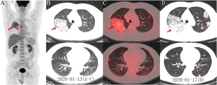
A 55-year-old male smoker in Wuhan developed intermittent fever, fatigue and dry cough for 5 days. Initial chest CT from an outside institution suggested hilar malignancy. 18F-FDG PET/CT was performed for further evaluation. The PET maximum intensity projection image (Figure A) revealed an FDG-avid mass with a maximum standardized uptake value (SUVmax) of 4.9 in the right lung. Increased accumulation of FDG was noted in the right paratracheal and right hilar lymph nodes (arrowheads) as well as bone marrow. On axial images (B, low dose CT; C, PET/CT fusion) there were ground-glass opacities (GGO) with areas of focal consolidation primarily in the right upper lobe (arrows) and a focal opacity in the left upper and right middle lobes (arrows). Influenza, respiratory syncytial virus (RSV) and other common respiratory viral infection were excluded by nasopharyngeal swab tests. Follow-up CT 4 days later revealed progression of lesions in bilateral upper and middle lobes with newly developed focal opacities in the left upper and lower lobes (D, arrows) (1,2). One week later, severe acute respiratory syndrome coronavirus 2 (SARS-CoV-2) infection was confirmed by real-time fluorescence polymerase chain reaction confirming Corona Virus Disease 2019 (COVID-19). After 10 days of treatment, the patient was considered recovered from COVID-19 and was discharged.
Figure:
A, PET maximum intensity projection (MIP) image revealed an FDG-avid mass with a SUVmax of 4.9 in the right lung. Increased accumulation of FDG in the right paratracheal, right hilar lymph nodes (arrowheads) and bone marrow were also noted. On axial images (B, low dose CT; C, PET/CT fusion) there were ground-glass opacities (GGO) with areas of focal consolidation primarily in the right upper lobe (arrows) and a focal opacity in the left upper and right middle lobes (arrows). Follow-up CT 4-day later demonstrated progression of lesions in bilateral upper and middle lobes with newly developed focal opacities in the left upper and lower lobes (D, arrows).
Footnotes
Conflict of Interest: The authors declare that they have no conflict of interest.
This work was supported by the National Natural Science Foundation of China (No. 91959119, 81873903, 81671718) and the Clinical Foundation of Tongji Hospital (No. 2015C013).
References
- 1.Kanne JP, Little BP, Chung JH, Elicker BM, Ketai LH. Essentials for Radiologists on COVID-19: An Update-Radiology Scientific Expert Panel. Radiology. 2020:200527. Doi: 10.1148/radiol.2020200527. PubMed PMID: 32105562. [DOI] [PMC free article] [PubMed] [Google Scholar]
- 2.Ai T, Yang Z, Hou H, Zhan C, Chen C, Lv W, et al. Correlation of Chest CT and RT-PCR Testing in Coronavirus Disease 2019 (COVID-19) in China: A Report of 1014 Cases. Radiology. 2020:200642. Doi: 10.1148/radiol.2020200642. PubMed PMID: 3210151 [DOI] [PMC free article] [PubMed] [Google Scholar]


