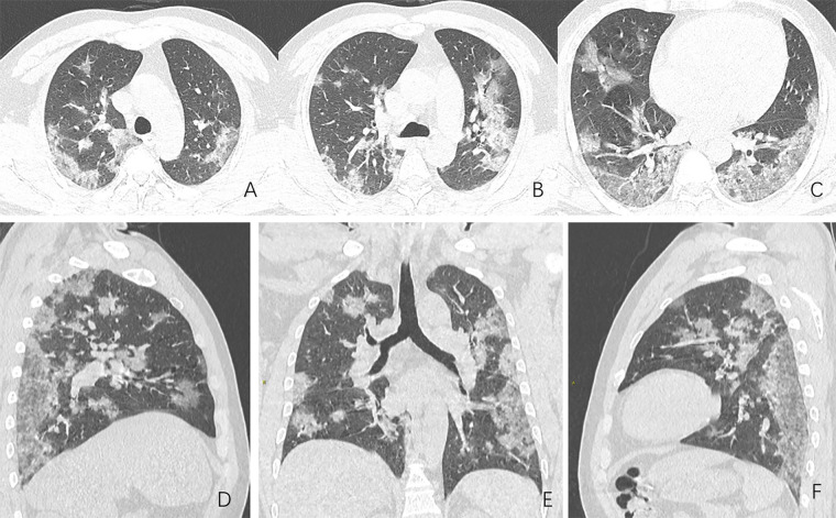
A 44-year-old man who was a transportation staff member in the Huanan seafood market in Wuhan, China, presented with a 13-day history of high fever and cough on December 25, 2019. High-sensitivity C-reactive protein level and erythrocyte sedimentation rate were elevated (>160.0 mg/L and ≥78.6 mm/h, respectively), while blood cell count showed normal white cells (5.29 × 10⁹/L) with decreased lymphocytes (0.22 × 10⁹/L). Chest CT showed patchy bilateral ground-glass opacities with peribronchial and peripheral/subpleural distribution (Fig 1). Serial follow-up chest radiographs showed increased extension of the lung opacities and development of basilar predominant consolidation (Fig 2). He was clinically diagnosed with severe pneumonia and acute respiratory distress syndrome. Usual respiratory pathogens were excluded, and he was eventually diagnosed as a suspected case of COVID-2019 (formerly known as 2019 novel coronavirus) infection. Unfortunately, he died 1 week later after failure of supportive measures.
Figure 1:
Images in a 44-year-old man who presented with fever and suspected COVID-19 pneumonia. A-C, Thin-slice (1-mm) axial CT images showed multiple patchy ground-glass opacity along the peribronchial and subpleural lungs. Some reticular opacities were also found within areas of ground glass (crazy-paving pattern). Lymphadenopathy was absent. D-F, Multiplanar reconstruction showed diffuse distribution of lesions.
Figure 2:
Images in a 44-year-old man who presented with fever and suspected COVID-19 pneumonia. A-C, Serial chest radiographs spanning an interval of 4 days showed rapid progressively increased extension and density of the lung opacities, culminating in confluent basilar predominant bilateral lung consolidation.
This case occurring at the epicenter outbreak of COVID-19 pneumonia illustrates the potential severity of this disease, at the same time that it underscores the role of imaging for monitoring disease progression. Moreover, CT could also have an important diagnostic role, especially when confirmatory tests, such as the real-time RT-PCR are unavailable (1–4).
Footnotes
Disclosures of Conflicts of Interest: L.Q. disclosed no relevant relationships. J.Y. disclosed no relevant relationships. H.S. disclosed no relevant relationships.
References
- 1.Pneumonia Treatment Program for New Coronary Virus Infection (Trial 5th Edition). National Health Commission of the People’s Republic of China Web site. http://www.nhc.gov.cn/. Published February 4, 2020. Updated February 4, 2020. Accessed February 4, 2020.
- 2.Huang C, Wang Y, Li X, et al. Clinical features of patients infected with 2019 novel coronavirus in Wuhan, China. Lancet. 2020;6736(20):1–10. [DOI] [PMC free article] [PubMed] [Google Scholar]
- 3.Chung M, Bernheim A, Mei X, Zhang N, Huang M, Zeng X, Cui J, Xu W, Yang Y, Fayad Z, Jacobi A, Li K, Li S, Shan H. CT Imaging Features of 2019 Novel Coronavirus (2019-nCoV). Radiology 2020 10.1148/radiol.2020200230 (in press). [DOI] [PMC free article] [PubMed] [Google Scholar]
- 4.Kanne JP. Chest CT Findings in 2019 Novel Coronavirus (2019-nCoV) Infections from Wuhan, China: Key Points for the Radiologist. Radiology 2020 10.1148/radiol.2020200241 (in press). [DOI] [PMC free article] [PubMed] [Google Scholar]


