

Abstract
Background and aim of the work: Extensor tendon injuries of the foot in children represent a rare foot injury. We report a case of a 9 year-old male who suffered of a traumatic wound laceration in the distal third of the right leg with a glass the day before in another country, getting a combined injury of tibialis anterior (TA), extensor hallucis longus (EHL) and extensor digitorum longus (EDL). Methods: After an initial clinical and radiological evaluation, antibiotic prophylaxis was immediately started. Surgery was necessary for the repair of the lesions and after rehabilitation the patient recovered a good function with a complete return to a normal life. Results: 5 years follow-up clinical examination revealed a complete and painless range of movement comparable to the other foot. The patient regained active dorsiflexion without functional limitations, deformity or contracture. Conclusions: Early exploration is important to allow full definition of the extent of injury and early surgical repair of tendons is recommended to avoid future disability. (www.actabiomedica.it)
Keywords: extensor tendons, foot, children, injury, rupture
Introduction
The extensor tendons of the foot are vulnerable to laceration because of their subcutaneous. They may be cut when a sharp object lacerates the skin and underlying structures. This kind of injury usually results in a high-stepping, drop-foot gait with weakness of ankle and toes dorsiflexion (1, 2).
Early diagnosis is emphasized, but often it can be confusing and is delayed. Special diagnostic studies, including magnetic resonance imaging (MRI) and sonography can help to estabilish an accurate and prompt diagnosis. Anyway, routine exploration of all dorsal foot and ankle wounds should be performed if there is suspicion of partial or complete tendon laceration. Surgical intervention is often necessary to repair these lesions. These structures heal well and have minimal dysfuction when repaired acutely. While operative management would seem to be indicated in most patients, it is important to consider potential complications from surgery, such as painful scar and joint stiffness from tendon adhesions. Thus, a specific rehabilitation and a close collaboration among different specialists are ultimately required in order to avoid these complications and to return to preinjury status (1, 3-5).
Case report
A 9-years old male came to our emergency department as consequence of inability to move the right foot. Mother told that he injured his right leg with a glass the day before in another country, where the wound was sutured. At the moment of the hospitalization the patient had a cutting wound localized in the anterior distal third of the right leg. Patient was regularly vaccinated against tetanus. In the emergency room an X-ray, an ultrasound and a magnetic resonance imaging (MRI) were done and a total laceration of TA, EHL and EDL was diagnosed. There were no fractures associated. The day after (two days following the traumatic event) surgical repair was performed. Antibiotic therapy with trimethoprim and sulfamethoxazole was administered during hospitalization.
Surgical technique
The patient was placed in the operating room in the supine position, and an ipsilateral thigh tourniquet was applied. The stitches were removed and exploration of the wound was performed (Fig. 1). Tibialis anterior, extensor hallucis longus and extensor digitorum were interrupted. A “Z” extension of the wound was performed and the proximal tendon stumps were found 5 cm proximally. After, the extensor retinaculum was transacted (Fig. 2) and the distal stumps were found (Fig. 3).
Figure 1.
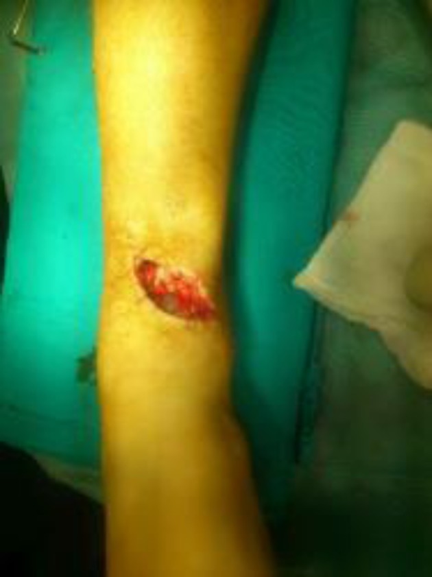
Wound after stitches removal
Figure 2.
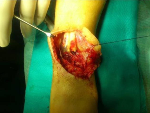
Opening of the extensor retinaculum
Figure 3.
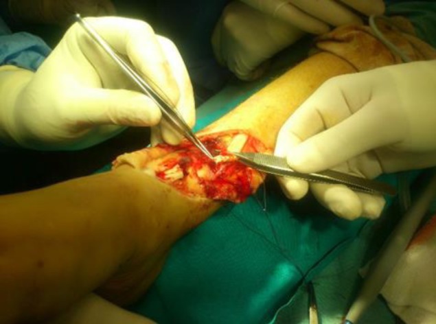
Recognition of tendon stumps
Figure 4.
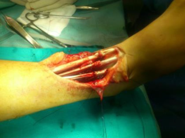
Tenorraphy according to modified Kessler technique
An end-to-end suturing was still possible. TA tenorrhaphy was performed with 3.0 nylon suture wire and overcoat with nylon 5.0, tenorrhaphy of extensor hallucis longus with nylon 3.0 and overcoat with nylon 5.0 and suture of extensor digitorum with 3.0 nylon and overcoat with 5.0. All the surgical tendon repairs were performed according to the modified Kessler technique.
The exploration of anterior neurovascular bundle showed no lesions of the nerve and vascular complete interruption. Vein and artery were micro surgically repaired. The extensor retinacolum was then sutured and the skin was closed in layers. A non–weightbearing removable long leg cast was applied to the lower extremity at the conclusion of the procedure with the knee in flexion of 45° and foot at 90° and slightly in talar position.
Postoperative care and outcomes
The patient was discharged the day after surgery. Patient’s postoperative course involved placement in a removable long-leg cast splint for a period of 4 weeks, during which he was not allowed to weight bear. In this period passive assisted dorsiflexion was permitted.
At the following outpatient visits the wound healed well and the stitches were removed after 15 days. After leg cast removal, the patient started idrokinesis and active rehabilitation in order to recover range of motion (ROM) at the ankle joint and first metatarsal-phalangeal joint. Six weeks postoperatively weightbearing was allowed. After two months and half foot and ankle motility was normal and the patient returned to practice his sport activities. The evaluation 5 years after surgery showed that the scar was good without retraction of the skin, pain was absent and active motion of the foot and toes was complete (Fig. 5). Furthermore, he reported to be satisfied with the surgical outcome and he actually he is playing in a soccer team.
Figure 5.

Follow-up after 5 years; scar, complete extension of the right foot that is equal to the contralateral one
Discussion
Extensor tendon ruptures of the foot represent about 1% of all tendon injuries (6). Lacerations from glass are an important cause of lesions in childhood with high risk of severe soft tissues damages. Frequently, a simple skin wound disguises the extensive nature of the injuries beneath (7). Early diagnosis and surgical repair of the tendons are recommended to avoid future disability (8,9) especially in young active patients with high functional demands. Nonsurgical management could be an appropriate alternative in selected low demanding elderly cases (10). A decrease in ankle joint motion and strength, heel cord contracture, ankle osteoarthritis and pes planus have been described in cases of untreated ruptures (11, 12).
In fact the muscles of the anterior compartment of the leg (tibialis anterior, extensor hallucis longus, extensor digitorum longus and peroneus tertius) are important in order to dorsiflex the ankle and toes and to control the forefoot during the swing phase of gait. Injuries of these anatomical structures and of the anterior neuromuscular bundle may lead to pain, weakness, and drop foot (1).
Primary repair of tendon is possible when their severed ends can be closely approximated, as in our case; however, when primary repair is not possible, a tendon graft will probably be required (13).
In addition to the clinical examination, MRI and also ultrasonography are useful in the diagnosis. Moreover, both can be helpful in evaluating the location of the rupture and the quality of the remaining tendons and the entity of their retraction (14-16).
During the exposure of the tendon, when possible, the superior extensor retinaculum should be left intact, because this structure at the level of the ankle is often thin, and its repair can be difficult. Adhesions of the repaired tendon to the retinaculum are common and difficult to avoid because of the immobilization required in the postoperative protocol. In patients with comorbidities, such as diabetes and chronic vascular insufficiency, the retinaculum may not be repairable, possibly leading to subcutaneous adhesions, bowstringing of the tendon, and wound healing problems (1).
After surgery, the patient has to be immobilized at 0 degrees of plantarflexion; in fact the position of the tendon after repair should be neutral, leaving it without tension. Passive assisted dorsiflexion can be allowed, thus facilitating tendon remodelling and joint motion and avoiding tensioning of the repair zone (1). After a 4-week period of non weight bearing and immobilization, active rehabilitation and progressive assisted walking can be started. Authors believe that a correct postoperative management, programmed based on patient characteristics, is equally important in order to restore a physiological ROM, strength and function.
This case report confirms this assumptions; the young age of the patient suggested to do a less aggressive rehabilitation and to delay the concession of the load. Furthermore, a long chalk was applied in order to discourage him from walking but usually in an adult, a short leg splint is enough (17,18).
Conclusions
Wound exploration in penetrating glass injuries is mandatory. MRI and ultrasound can be useful to confirm the diagnosis and for a preoperative planning. It is preferable to operate this kind of lesions as soon as possible in order to prevent excessive retraction of tendon stumps. In high demanding patients, an anatomical reconstruction and an accurate postoperative management and rehabilitation protocol are essential.
Conflict of interest:
Each author declares that he or she has no commercial associations (e.g. consultancies, stock ownership, equity interest, patent/licensing arrangement etc.) that might pose a conflict of interest in connection with the submitted article
References
- 1.Sammarco VJ, Sammarco GJ. Injuries to the Tibialis Anterior, Peroneal Tendons, and Long Flexors of the Toes. Baxter’s the Foot and Ankle in Sport. 2008;2nd ed:121–46. [Google Scholar]
- 2.Gwynne-Jones D, Garneti N, Wyatt M. Closed tibialis anterior tendon rupture: a case series. Foot & Ankle International. 2009;30:758–62. doi: 10.3113/FAI.2009.0758. [DOI] [PubMed] [Google Scholar]
- 3.Lee MH, Chung CB, Cho JH, Mohana-Borges AV, Pretterklieber ML, Trudell DJ, et al. Tibialis anterior tendon and extensor retinaculum: imaging in cadavers and patients with tendon tear. American Journal of Roentgenology. 2006;187:161–68. doi: 10.2214/AJR.05.0073. [DOI] [PubMed] [Google Scholar]
- 4.Wong JC, Daniel JN, Raikin SM. Repair of acute extensor hallucis longus tendon injuries: a retrospective review. Foot & Ankle Specialist. 2013;7:45–51. doi: 10.1177/1938640013514271. [DOI] [PubMed] [Google Scholar]
- 5.Pedrazzini A, Ceccarelli F, Martelli A, Marenghi L, Petraglia F, Romiti D, et al. Return to run after partial amputation of the ankle: clinical assessment and instrumental evaluation. Acta Biomedica. 2013;84:237–43. [PubMed] [Google Scholar]
- 6.Al-Qattan MM. Surgical treatment and results in 17 cases of open lacerations of the extensor hallucis longus tendon. J Plast Reconstr Aesthet Surg. 2007;60:360–37. doi: 10.1016/j.bjps.2006.05.003. [DOI] [PubMed] [Google Scholar]
- 7.Jackson RH. Laceration from glass in childhood. Br Med J (Clin Res Ed) 1981;283:1310–312. doi: 10.1136/bmj.283.6302.1310. [DOI] [PMC free article] [PubMed] [Google Scholar]
- 8.Griffiths JC. Tendon injuries around the ankle. The journal of bone and joint surgery. 1965;47B:686–89. [PubMed] [Google Scholar]
- 9.Wicks MH, Paterson DC. Tendon Injuries about the foot and ankle in children. Australian and New Zealand Journal of Surgery. 1980;50:158–61. doi: 10.1111/j.1445-2197.1980.tb06656.x. [DOI] [PubMed] [Google Scholar]
- 10.Markarian GG, Kelikian AS, Brage M, Trainor T, Dias L. Anterior tibialis tendon ruptures: an outcome analysis of operative versus nonoperative treatment. Foot & Ankle International. 1998;19:792–02. doi: 10.1177/107110079801901202. [DOI] [PubMed] [Google Scholar]
- 11.Dooley BJ, Kudelka P, Menelaus MB. Subcutaneous rupture of the tendon of tibialis anterior. J Bone Joint Surg Br. 1980;62b:471–72. doi: 10.1302/0301-620X.62B4.7430226. [DOI] [PubMed] [Google Scholar]
- 12.Forst R, Forst J, Heller KD. Ipsilateral peroneus brevis tendon grafting in a complicated case of traumatic rupture of tibialis anterior tendon. Foot Ankle Int. 1995;16:440–44. doi: 10.1177/107110079501600712. [DOI] [PubMed] [Google Scholar]
- 13.Tuncer S, Aksu N, Isiklar Ugur. Delayed rupture of the extensor hallucis longus and extensor digitorum communis tendons after breaching the anterior capsule with a radiofrequency probe during ankle arthroscopy: a case report. The Journal of Foot and Ankle Surgery. 2010;49(9):490.e1–3. doi: 10.1053/j.jfas.2010.05.003. [DOI] [PubMed] [Google Scholar]
- 14.Waizy H, Bouillon B, Stukenborg-Colsman C, et al. Ruptur der Musculus tibialis anterior Sehne. Der Unfallchirurg. 2017;120:1015–19. doi: 10.1007/s00113-017-0417-z. [DOI] [PubMed] [Google Scholar]
- 15.Bianchi S, Zwass A, Abdelwahab IF, Zoccola C. Evaluation of tibialis anterior tendon rupture by ultrasonography. J Clin Ultrasound. 1994;22:564–66. doi: 10.1002/jcu.1870220909. [DOI] [PubMed] [Google Scholar]
- 16.Din R, Therkilsden L. Rupture of tibilais anterior associated with a closed midshaft tibial fracture. J Accid Emerg Med. 1999;16:459. doi: 10.1136/emj.16.6.459. [DOI] [PMC free article] [PubMed] [Google Scholar]
- 17.So E, Black TE, Mehl B. Split peroneus longus free tendon autograft transplantation for the treatment of neglected extensor hallucis longus tendon laceration: a case report. The Journal of Foot and Ankle Surgery. 2018;57:210–14. doi: 10.1053/j.jfas.2017.08.005. [DOI] [PubMed] [Google Scholar]
- 18.Lefebvre B, Beldame J, Bertiaux S, Biga N. Open and subcutaneous recent tibialis anterior tendon ruptures: does postoperative immobilization method influence outcome. Orthopaedics & Traumatology: Surgery & Research. 2011;97:211–16. doi: 10.1016/j.otsr.2010.09.018. [DOI] [PubMed] [Google Scholar]