

Abstract
Background: Modern TKA implants promise to improve functional outcomes, stability, patient satisfaction and operating room efficiency. The purpose of this retrospective study is to evaluate our short-term clinical and radiological results and survival using the ATTUNE Total Knee Replacement System. Methods: The authors reviewed 228 primary cemented TKAs using ATTUNE Total Knee Replacement System which were implanted between 2014 and 2018 concerning short-term clinical and radiographical outcomes and survival. Clinical evaluation was performed using the Knee injury Osteoarthritis Outcome Score (KOOS), the Oxford Score and a Numeric Rating Scale (NRS) for pain. Radiographic analysis was performed using the Modern Knee Society Score Evaluation System. Results: The means of the clinical results as measured with KOOS score were Pain 82,7, Symptoms 79, ADL 78,3, Sport & recreation 51,8 and QOL 78,6. The mean Oxford score was 35 and NRS 2. The mean ROM was 113,4 (SD 9,4 range 90-130). Radiographically mean mechanical axis was 1,97° of Varus and radiolucent lines were detected in 43 knees (22,4%). The survival rate is 98.4% at 2 years and 97.4% at 5 years.Conclusion: At short-term follow-up the ATTUNE Knee Replacement System provide excellent clinical and radiographical outcomes and good results regarding revision rate. Due to high incidence of radiolucent lines, those patients should be closely monitored even though they show no clinical evidence for loosening. (www.actabiomedica.it)
Keywords: TKA, ATTUNE Total Knee Replacement, Radiolucency
Introduction
Total knee arthroplasty (TKA) is believed to be one of the most successful and effective surgical procedures for end-stage osteoarthritis with survivorship more than 94% at 16 years after surgery (1-3). However the incidence of patient dissatisfaction after TKA varies in literature and has been reported to be as high as 20% (4, 5).
There is a constant race between different healthcare companies to advance the technology employed in prostheses to further improve patient outcomes. Newer implants are regularly introduced, or design features of current implants are adjusted in an effort to achieve this (6).
In order to improve functional outcomes, stability, patient satisfaction and operating room efficiency the ATTUNE Total Knee Replacement system was launched as a modified version of a previous prosthesis (Press Fit Condylar Sigma). The theoretical advantages of this prosthesis suggested by the providers are increased conformity between the femoral component and the polyethylene insert with a gradually reducing femoral radius, an innovative s-curve design of the posteriorly stabilized cam for gradual femoral rollback and stability, an extensive range of sizes for a diverse population, optimization of the patellofemoral tracking, an improved polyethylene insert locking mechanism and incorporation of an antioxidant polyethylene insert.
However, despite such advantages, there are some design features that might cause problems.
The thickness of the patellar component is greater and hence the residual bone stock should be shallow with the increased possibility of patella fractures (7). Furthermore, the tibial component stem is located relatively posterior e this might increase the risk of injury to the posterior tibial cortex (8).
Finally recently early aseptic failures at the implant-cement interface were reported in a retrospective study based on data from Manufacter and User Facility Device Experience (MAUDE) database (9).
The purpose of this retrospective study is to evaluate our short-term clinical and radiological results and survival using the ATTUNE Total Knee Replacement System. Our primary objective is to compare our tibial aseptic loosening rate with previous reported studies. Secondly, we show our clinical and radiographic findings with those reported in literature
Material and methods
Patients
All consecutive patients who underwent TKA using ATTUNE between January 2014 and January 2018 were enrolled in this study. For all patients the indication for surgery was based on patient history and physical examination combined with anteroposterior and lateral radiography.
All TKAs were performed by 3 surgeons with certified experience in total joint arthroplasty. All implants were cemented posterior stabilized.
For all patients follow-up, sex, age at operation, revision and revision date, complications, the presence of rheumatoid arthritis, diabetes, smoking status and body mass index (BMI) were registered.
If patients were still alive at follow-up, they were invited to fill out two questionnaires and a pain score as described below.
Operative technique and rehabilitation
All operations were conducted with the patient under spinal or general anaesthesia using the same technique of medial parapatellar approach and capsulotomy with patellar eversion. Femoral and tibial bone resection were made with a modified measured resection technique.
The transepicondylar axis was used as a reference for femoral component rotation. The tibial resection was set to be 0°-3° of posterior slope in sagittal plane. The reference line for tibial rotation was accurately aimed to pass through the medial third of tibial tubercle and the second metatarsal bone. All osteophytes were removed, and any contracted medial or lateral soft tissue was carefully evaluated and selectively released when required.
The bone surface was irrigated with 0.9% saline with a pulsatile high-pressure lavage system (Pulsavac Plus, Zimmer, Warsaw, Indiana, USA) for at least 1 min. After irrigation, preparation of bone cement was initialized according to manufactures specifications. All TKA were implanted with surface cemented components using high-viscosity bone cement (Palacos, Heraeus Medical, Wehrheim, Germany). The bone cement was applied on the tibial bone surface and on the implant surface via cement gun pressurization. The tibial component was then inserted and impacted. The femoral component was inserted and impacted in the same manner. Implantation of tibial and femoral component was performed in a single step. The patella was never replaced.
Patients started with mobilization on the day after surgery dependent on pain. Normal expectancy was unaided walking after 4 weeks of rehabilitation.
Clinical and Radiographic Evaluation
Clinical evaluation at follow-up was performed using the Knee injury Osteoarthritis Outcome Score (KOOS)(10) questionnaire, an Oxford(11) questionnaire and an 11-point Numeric Rating Scale (NRS) for pain ranging from 0 to 10.
The KOOS is a 42-item site specific questionnaire, resulting in five 0-100 scores (higher is better) for Pain, Symptoms, activities of daily life (ADL), Sport & Recreation and quality of life (QOL). The Oxford is a 12-item site specific score, ranging from 0 to 48 (higher is better).
The ROM was measured using a long-armed goniometer.
Radiographic evaluation was performed with anterior-posterior (AP) and lateral x-ray of the knee joint as well as full-length standing AP-radiograph to assess correction of alignment. Detailed analyses of AP and lateral radiographs were conducted on the basis of the Modern Knee Society Radiograph Evaluation System(12) dividing each component in different zones for a standardized documentation of radiolucent lines. Radiolucencies were documented for each radiograph.
Figure 1A.

Radiolucent lines (AP view)
Figure 1B.
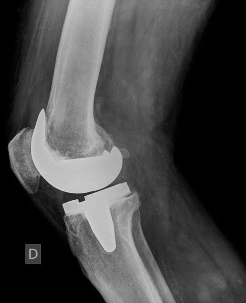
Radiolucent lines (Lateral view)
Statistical analysis
Statistical analysis was performed using Microsoft Excel (2017 version).
Results
During the period considered in the study 228 TKAs were performed with this prosthesis in 218 patients, 89 males and 129 females with a mean age of 70,3 years (SD 6,52; range 43-85).
The mean follow-up was 3,16 years (SD 1,16) ranging from 1 to 5,4 years.
Three patients were deceased at the time of the current study, 22 patients (25 TKAs) were lost to follow-up and 8 withdrew consent. A total of 185 patients and 192 TKAs remained.
The demographic data of the patients are summarized in table 1.
Table 1.
Patient demography
| Sex | Male: n=89 (40.8%) Female: n=129 (59.2%) |
| Side | Right: n=121 (53.1%) Left: n=107 (46.9%) |
| Age | 70.3 y ± 6.52 |
| Body height | 167 cm ± 8 cm |
| Body weight | 86 kg ± 15.2 kg |
| Body Mass Index | 28.6 ± 3.95 |
| Follow up | 3,16 years ± 1,16 years |
| Smoke | Yes: n=17 (7.8%) No: n=201(92.2%) |
Clinical results
The means of the clinical results as measured with KOOS score were Pain 82,7 (SD 14,4 range 44,4-97,2), Symptoms 79 (SD 13,8 range 39,3-100) ADL 78,3 (SD 15,8 range 38,2-98,5) Sport & recreation 51,8 (SD 26,3 range 20-85) and QOL 78,6 (SD 20,9 range 31,2- 97,7). The mean Oxford score was 35 (SD 14,6 range 20-48) and NRS 2 (SD 1,7 range 1-5). The mean ROM was 113,4 (SD 9,4 range 90-130).
Radiographic results
Radiographically, the mean mechanical axis was 1,97° of Varus.
Radiolucent lines were detected in 43 knees (22,4%). Table 2 display the incidence in dependence on the location of the radiolucent lines.
Table 2.
Summary of all radiolucent lines in the anterior-posterior (AP) tibial, lateral tibial and lateral femoral radiograph
| Location, radiolucency | |
| Tibial AP | n=25 (13%) |
| Tibial Lateral | n=17 (8.8%) |
| Femur Lateral | n=23 (12%) |
Complications
Revision surgery for tibial aseptic loosening was performed in 2 cases (1%), respectively 7 and 13 months after surgery, with intra operative finding of failure at tibial implant-cement interface in all cases.
Two patients (1%) developed a periprosthetic infection at 2 and 3 years after surgery respectively and were treated with implant revision in two-stage surgery.
In one patient (0,05%) patient revision surgery was performed for component malposition.
In one case (0,05%) a partial lesion of the patellar tendon during rehabilitation (confirmed with sonography) was observede(13). The patient was treated with 20 days of immobilization in an extended brace. After this period a progressive program of rehabilitation lead to a complete functional recovery.
Other complications included 2 (1%) patients who developed wound infections. Both were superficial and successfully eradicated with antibiotic administration. Following surgery, 3 (1,5%) knees required one manipulation under anesthesia and 1 knee was treated with arthroscopic lysis of adhesions.
Discussion
Aseptic loosening remains a common reason for early revision also with contemporary TKA system(14, 15). Loosening in short-term analyses most likely reflects failure to gain fixation. Retrieval studies have shown that bone resorption stimulated by polyethylene wear particles and stress shielding play an important role in aseptic loosening.(16, 17). Even debonding of the tibial implant-cement interface as a result of cement type and application methods is a reported cause of aseptic loosening (15, 18).
Early aseptic loosening in ATTUNE Total Knee Replacement System is still debated.
Bonutti (9) reported high rate of early tibial aseptic failures at the implant-cement interface in a study based on data from Manufacturer and User Facility Device Experience (MAUDE) database.
In contrast, the Australian registry and the National Joint Registry of the United Kingdom (NJR) reported low rates of aseptic loosening with excellent survivorship rates (19, 20).
Even several recent studies analyzing short-term outcome of ATTUNE implant reported low revision rates (7, 8, 21-23).
In addition, Turgeon used radiostereometric analysis of the components to assess the stability of the ATTUNE prosthesis and showed secure fixation of the tibial baseplate within the first two postoperative years (24).
In this study, the revision rate for tibial aseptic loosening is 1% while overall revision rate is 2.6%. The survival rate is 98.4% at 2 years and 97.4% at 5 years.
These results are congruent with those reported in previous studies and, according to the literature, are acceptable values for primary TKA (14, 25, 26).
However, in our case series, a high number of radiolucent lines was detected at the radiographic analysis. Reasons for the high incidence of radiolucencies remain a matter of speculation.
Staats (21) reported an increased number of radiolucent lines in ATTUNE-patients than PFC Sigma-patients, especially on the tibial component. He hypothesized that it was mostly due to technique-related issues, in particular he assumed that the implant itself may allow too much movement during cement interlocking-phase. He suggested to proceed the cementation of the tibial and femoral component in two separated steps.
Another reason that can explain this high number of radiolucent lines is attributable to the design of this prosthesis, in particular in relation to the cement pockets.
In a recent study peer-reviewed digital imaging method was used to investigate cement adhesion on the ATTUNE tibial tray (27). None of the prosthesis examined in this cohort showed cement attachment at the tibial tray backside and the authors concluded that it may be related to the absence of separate cement pockets.
In the meantime, the company has launched a revised tibial component with additional cement pockets and optimized surface conditions on the tibial base that will have to be studied in the further future.
Literature about the outcome of the ATTUNE Knee Replacement System is scarce due to its recent availability.
Anyhow Ranawat (22) showed excellent clinical results in 90.7% of ATTUNE-patients after 2 years’ follow up with less anterior knee pain and less crepitation than PFC Sigma-patients.
Molloy (23) reported no difference in physical function and most outcomes between ATTUNE and PFC Sigma at short-term follow-up.
Song (7, 8) in his two studies showed more favorable clinical results using the ATTUNE prosthesis than using PFC Sigma prosthesis. However, they reported ad increased risk of posterior tibial cortex injury and residual patellar injury with use of the ATTUNE prosthesis.
Additionally, Takagi (28) showed that the gradually reducing radius design of the ATTUNE prosthesis minimized paradoxical anterior slide in a navigation-based in vivo knee kinematics.
According to our findings we can confirm that this system achieves excellent outcomes at short-term follow-up.
However, the radiographic analysis of the present study doesn’t allow to exclude that the tibial component may have problems even though no evidence for a higher revision rate could be detected in our study.
Conclusion
At short-term follow-up the ATTUNE Knee Replacement System provide excellent clinical and radiographical outcomes and good results regarding revision rate. Due to high incidence of radiolucent lines, those patients should be closely monitored even though they show no clinical evidence for loosening. Further studies with large cohort and long-term follow up are needed to evaluate and improve the application of this modern TKA-system.
Conflict of interest:
Each author declares that he or she has no commercial associations (e.g. consultancies, stock ownership, equity interest, patent/licensing arrangement etc.) that might pose a conflict of interest in connection with the submitted article
References
- 1.Font-Rodriguez DE, Scuderi GR, Insall JN. Survivorship of cemented total knee arthroplasty. Clin Orthop Relat Res [Internet] 1997 Dec [cited 2019 Oct 4];345:79–86. Available from: http://www.ncbi.nlm.nih.gov/pubmed/9418624 . [PubMed] [Google Scholar]
- 2.Abdel MP, Morrey ME, Jensen MR, Morrey BF. Increased long-term survival of posterior cruciate-retaining versus posterior cruciate-stabilizing total knee replacements. J Bone Jt Surg - Ser A. 2011 Nov 16;93(22):2072–8. doi: 10.2106/JBJS.J.01143. [DOI] [PubMed] [Google Scholar]
- 3.Arthur CHC, Wood AM, Keenan ACM, Clayton RAE, Walmsley P, Brenkel I. Ten-year results of the Press Fit Condylar Sigma total knee replacement. Bone Joint J [Internet] 2013 Feb [cited 2019 Oct 4];95-B(2):177–80. doi: 10.1302/0301-620X.95B2.29695. Available from: http://online.boneandjoint.org.uk/doi/10.1302/0301-620X.95B2.29695 . [DOI] [PubMed] [Google Scholar]
- 4.Bourne RB, Chesworth BM, Davis AM, Mahomed NN, Charron KDJ. Patient satisfaction after total knee arthroplasty: who is satisfied and who is not. Clin Orthop Relat Res [Internet] 2010 Jan [cited 2019 Aug 28];468(1):57–63. doi: 10.1007/s11999-009-1119-9. Available from: http://www.ncbi.nlm.nih.gov/pubmed/19844772 . [DOI] [PMC free article] [PubMed] [Google Scholar]
- 5.Scott CEH, Howie CR, MacDonald D, Biant LC. Predicting dissatisfaction following total knee replacement: a prospective study of 1217 patients. J Bone Joint Surg Br [Internet] 2010 Sep [cited 2019 Oct 7];92(9):1253–8. doi: 10.1302/0301-620X.92B9.24394. Available from: http://www.ncbi.nlm.nih.gov/pubmed/20798443 . [DOI] [PubMed] [Google Scholar]
- 6.Causero A, Di Benedetto P, Beltrame A, Gisonni R, Cainero V, Pagano M. Design evolution in total knee replacement: which is the future. Acta Biomed [Internet] 2014 Sep 24[cited 2019 Oct 8];85(Suppl 2):5–19. Available from: http://www.ncbi.nlm.nih.gov/pubmed/25409713 . [PubMed] [Google Scholar]
- 7.Song SJ, Kang SG, Park CH, Bae DK. Comparison of Clinical Results and Risk of Patellar Injury between Attune and PFC Sigma Knee Systems. Knee Surg Relat Res. 2018 Dec 1;30(4):334–40. doi: 10.5792/ksrr.18.020. [DOI] [PMC free article] [PubMed] [Google Scholar]
- 8.Song SJ, Park CH, Liang H, Kang SG, Park JJ, Bae DK. Comparison of Clinical Results and Injury Risk of Posterior Tibial Cortex Between Attune and Press Fit Condylar Sigma Knee Systems. J Arthroplasty. 2018;33(2):391–7. doi: 10.1016/j.arth.2017.09.056. [DOI] [PubMed] [Google Scholar]
- 9.Bonutti PM, Khlopas A, Chughtai M, Cole C, Gwam CU, Harwin SF, et al. Unusually High Rate of Early Failure of Tibial Component in ATTUNE Total Knee Arthroplasty System at Implant-Cement Interface. J Knee Surg. 2017 Jun 1;30(5):435–9. doi: 10.1055/s-0037-1603756. [DOI] [PubMed] [Google Scholar]
- 10.Monticone M, Ferrante S, Salvaderi S, Rocca B, Totti V, Foti C, et al. Development of the Italian version of the knee injury and osteoarthritis outcome score for patients with knee injuries: cross-cultural adaptation, dimensionality, reliability, and validity. Osteoarthr Cartil [Internet] 2012 Apr [cited 2019 Oct 7];20(4):330–5. doi: 10.1016/j.joca.2012.01.001. Available from: http://www.ncbi.nlm.nih.gov/pubmed/22285738 . [DOI] [PubMed] [Google Scholar]
- 11.Dawson J, Fitzpatrick R, Murray D, Carr A. Questionnaire on the perceptions of patients about total knee replacement. J Bone Joint Surg Br [Internet] 1998 Jan [cited 2019 Oct 7];80(1):63–9. doi: 10.1302/0301-620x.80b1.7859. Available from: http://www.ncbi.nlm.nih.gov/pubmed/9460955 . [DOI] [PubMed] [Google Scholar]
- 12.Meneghini RM, Mont MA, Backstein DB, Bourne RB, Dennis DA, Scuderi GR. Development of a Modern Knee Society Radiographic Evaluation System and Methodology for Total Knee Arthroplasty. Journal of Arthroplasty. Churchill Livingstone Inc. 2015;30:2311–4. doi: 10.1016/j.arth.2015.05.049. [DOI] [PubMed] [Google Scholar]
- 13.Ryan JA, Meyers KN, DiBenedetto P, Wright TM, Haas SB. Failure of the Patellar Tendon with the Patella Everted versus Noneverted in a Matched-Pair Cadaver Model. HSS J. 2010;6(2):134–7. doi: 10.1007/s11420-009-9149-0. [DOI] [PMC free article] [PubMed] [Google Scholar]
- 14.Piedade SR, Pinaroli A, Servien E, Neyret P. Revision after early aseptic failures in primary total knee arthroplasty. Knee Surgery, Sport Traumatol Arthrosc. 2009 Mar;17(3):248–53. doi: 10.1007/s00167-008-0667-y. [DOI] [PubMed] [Google Scholar]
- 15.Hazelwood KJ, O’Rourke M, Stamos VP, McMillan RD, Beigler D, Robb WJ. Case series report: Early cement-implant interface fixation failure in total knee replacement. Knee. 2015 Oct 1;22(5):424–8. doi: 10.1016/j.knee.2015.02.016. [DOI] [PubMed] [Google Scholar]
- 16.Rao AR, Engh GA, Collier MB, Lounici S. Tibial interface wear in retrieved total knee components and correlations with modular insert motion. J Bone Jt Surg - Ser A. 2002 Oct 1;84(10):1849–55. doi: 10.2106/00004623-200210000-00017. [DOI] [PubMed] [Google Scholar]
- 17.Zhang QH, Cossey A, Tong J. Stress shielding in periprosthetic bone following a total knee replacement: Effects of implant material, design and alignment. Med Eng Phys. 2016 Dec 1;38(12):1481–8. doi: 10.1016/j.medengphy.2016.09.018. [DOI] [PubMed] [Google Scholar]
- 18.Kopinski JE, Aggarwal A, Nunley RM, Barrack RL, Nam D. Failure at the Tibial Cement-Implant Interface With the Use of High-Viscosity Cement in Total Knee Arthroplasty. J Arthroplasty [Internet] 2016 [cited 2019 Oct 15];31(11):2579–82. doi: 10.1016/j.arth.2016.03.063. Available from: http://www.ncbi.nlm.nih.gov/pubmed/27155996 . [DOI] [PubMed] [Google Scholar]
- 19.No. Hip, Knee & Shoulder Arthroplasty National Joint Replacement Registry. Australian Orthopaedic Assiocation. Annual Report. 2017 [Google Scholar]
- 20.No. National Joint Registry for England, Wales, Northern Ireland and the Isle of Man. 2017;14th Annua [Google Scholar]
- 21.Staats K, Wannmacher T, Weihs V, Koller U, Kubista B, Windhager R. Modern cemented total knee arthroplasty design shows a higher incidence of radiolucent lines compared to its predecessor. Knee Surgery, Sport Traumatol Arthrosc. 2019 Apr 5;27(4):1148–55. doi: 10.1007/s00167-018-5130-0. [DOI] [PMC free article] [PubMed] [Google Scholar]
- 22.Ranawat CS, White PB, West S, Ranawat AS. Clinical and Radiographic Results of Attune and PFC Sigma Knee Designs at 2-Year Follow-Up: A Prospective Matched-Pair Analysis. J Arthroplasty [Internet] 2017 [cited 2019 Oct 14];32(2):431–6. doi: 10.1016/j.arth.2016.07.021. Available from: http://www.ncbi.nlm.nih.gov/pubmed/27600300 . [DOI] [PubMed] [Google Scholar]
- 23.Molloy IB, Keeney BJ, Sparks MB, Paddock NG, Koenig KM, Moschetti WE, et al. Short term patient outcomes after total knee arthroplasty: Does the implant matter. Knee [Internet] 2019 Jun [cited 2019 Oct 14];26(3):687–99. doi: 10.1016/j.knee.2019.01.018. Available from: http://www.ncbi.nlm.nih.gov/pubmed/30910627 . [DOI] [PMC free article] [PubMed] [Google Scholar]
- 24.Turgeon TR, Gascoyne TC, Laende EK, Dunbar MJ, Bohm ER, Richardson CG. The assessment of the stability of the tibial component of a novel knee arthroplasty system using radiostereometric analysis. Bone Jt J. 2018 Dec 1;100B(12):1579–84. doi: 10.1302/0301-620X.100B12.BJJ-2018-0566.R1. [DOI] [PubMed] [Google Scholar]
- 25.Lum ZC, Shieh AK, Dorr LD. Vol. 9. World Journal of Orthopaedics. Baishideng Publishing Group Co: 2018. Why total knees fail-A modern perspective review; pp. 60–4. [DOI] [PMC free article] [PubMed] [Google Scholar]
- 26.Sharkey PF, Lichstein PM, Shen C, Tokarski AT, Parvizi J. Why are total knee arthroplasties failing today--has anything changed after 10 years. J Arthroplasty [Internet] 2014 Sep [cited 2019 Oct 14];29(9):1774–8. doi: 10.1016/j.arth.2013.07.024. Available from: http://www.ncbi.nlm.nih.gov/pubmed/25007726 . [DOI] [PubMed] [Google Scholar]
- 27.Cerquiglini A, Henckel J, Hothi H, Allen P, Lewis J, Eskelinen A, et al. Analysis of the attune tibial tray backside: A comparat ive retrieval study. Bone Jt Res. 2019 Mar 1;8(3):136–45. doi: 10.1302/2046-3758.83.BJJ-2018-0102.R2. [DOI] [PMC free article] [PubMed] [Google Scholar]
- 28.Takagi H, Asai S, Sato A, Maekawa M, Kawashima H, Kanzaki K. Case series report of navigation-based in vivo knee kinematics in total knee arthroplasty with a gradually reducing femoral radius design. Ann Med Surg. 2017 May 1;17:33–7. doi: 10.1016/j.amsu.2017.03.032. [DOI] [PMC free article] [PubMed] [Google Scholar]