

Abstract
Background: Post-traumatic osseous cystic lesions represent a rare complication in children. Usually a post-fracture cyst is a lipid inclusion cyst, which is radiolucent and may be seen adjacent to a healing torus fracture. It is typically asymptomatic and appears just proximal to the fracture line within the area of subperiosteal new bone formation. Case report: We report a case of post-fracture cyst of the distal radius in an 8 year-old girl with spontaneous resolution. A fat-fluid level within the subperiosteal cystic lesion in MRI is a typical feature of post-traumatic cystic lesion in children. Discussion and conclusion: MRI or CT scan is sufficient to confirm the diagnosis of post-traumatic cystic lesions without the need for further management other than reassurance and advise that they may occasionally cause discomfort but resolve with time. (www.actabiomedica.it)
Keywords: distal radius fracture, post-traumatic cyst, torus fracture
Background
Post-traumatic bone cystic lesions have been reported in the orthopaedic, paediatric and radiological literature (1) and represent a rare complication in children. They are radiolucent lesions that appear adjacent to a healing torus fractures (2). The cyst is usually a lipid inclusion developing from an intramedullary fat seepage through the damaged bone cortex and its entrapment within the subperiosteum (3). A typical feature of this kind of lesion is a fat fluid level within the subperiosteal cystic lesion in MRI. These lesions occur with a benign course and usually resolve spontaneously, but failure to recognize this condition can lead to an expensive diagnostic evaluation and create unnecessary apprehension (4). We report the case of an 8 years-old girl in whom such a lesion developed after a torus fracture of the distal radius.
Case Report
An eight-years-old girl presented with a painful right wrist after falling onto her outstretched right arm. The anteroposterior (AP) and lateral (L) X-ray of the right forearm revealed a greenstick fracture of distal radius with minimal dorsal angulation (Fig. 1 A, B). The forearm was immobilized in a below-elbow cast. X-ray examination at one week showed no change in the alignment and the cast was removed at three weeks. The final X-ray showed the complete healing of the fracture (Fig.2 A, B). Patient reported a new trauma on the same arm four months later and new X-rays were taken. While the final radiographs revealed healing process of the fracture, subsequent X-rays four months later showed a well-circumscribed radiolucent lesion without surrounding adjacent sclerosis to the site of the previous fracture (Fig. 3 A, B). The child was comfortable and afebrile with no signs of local inflammation at the cystic site. Whole blood cell count, C-reactive protein (CPR) levels (<1mg/L) and erythrocytes sedimentation rate (VES) were normal. Differential diagnosis needs to be done, included a pre-existing lesion (which was ruled out by the re-examination of the initial X-rays), infection as Brodie abscess formation or a true post-fracture cystic lesion. MRI of the right wrist showed a heterogeneous subperiosteal cystic structure at the dorsal aspect of the right distal radius, measuring 10 mm (Anteroposterior) x 12 mm (Transverse) x 10 mm (Craniocaudal). It contained an upper layer of T1W hyperintensity which was suppressed on fat sat sequence consistent with fat content (Fig. 4 A, B). The lower layer was T1W hypointense and T2W hyperintense and likely represented a chronic subperiosteal haematoma. The MRI features confirmed a fat-fluid level within the subperiosteal cystic lesion in MRI typical of post-traumatic osseous cystic lesion in children. Different from other literature cases, we performed a biopsy to obtain a histological confirmation of the benign nature of the cyst. The histological report of “a fibrous tissue without neoplastic cells and microcalcifications” confirmed the imaging. Clinically the child remained asymptomatic and was allowed to return to her normal sports activities and monitoring her as outpatient at six months. Radiographs at six months and one year from trauma showed that the cyst like lesion had disappeared and distal radius fracture had completely remodelled. (Fig. 5 A, B)
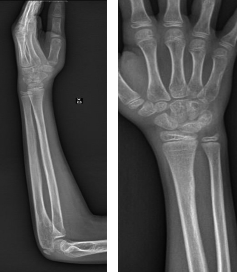
Figure 1.

A, B. Torus distal radius fracture in 8-years old (AP
Figure 3.

A, B. X-ray performed four months later from a new trauma
Figure 4.

A, B. MRI of the cystic lesion
Figure 5.
A, B. X-ray 1 year from the cyst diagnosis
Figure 2.

A, B. Final X-ray three weeks from trauma (bone healing)
Discussion
Despite greenstick or torus fractures are common injuries in children, development of a cystic lesion after these fractures is relatively uncommon. Distal radius fracture seems to be the commonest site of development of these lesions (4, 5). Just over 20 cases of transient post-fracture cyst have been described in the orthopaedic literature. The most common location of the cyst-like lesion was the distal aspect of the radius and only a few of them were located in the distal aspect of the tibia (6, 7). (Tab. 1) These post-fracture cysts are probably more common than the published reports, because most distal radius fractures in children are monitored on clinical signs alone and their exact incidence is unknown (8). They were asymptomatic in all previously reported cases, in fact these lesions can be usually seen on routine follow-up X-rays and even as an incidental finding for evaluation of new re-injury after trivial trauma. They usually appear more than one month after the fracture a characteristically are less than 10 mm in diameter, do not expand, appear rounded or slightly oval shaped and may be multiple (8). There is a debate about the aetiology of transient post-fracture cysts. Pfister-Goedeke et al suggested that they are resorption cysts within an excessive periosteal reaction, related to the subperiosteal haematoma that accompanies greenstick fractures (9). Phillips and Keats attribute post-traumatic cysts to the resorption of intraosseous haemorrhage (6). Malghem et al reported that CT scan of two cases showed a density consistent with a fatty content, supporting the theory of transcortical escape of intramedullary fat. According to this theory the transient cyst could be the result of leakage of intramedullary fat during the fracture event, without disruption of the periosteoum. The entrapped fat could subsequently become visible, while the subperiosteal haematoma becomes calcified. In our study, T1W images, saturation T2W images demonstrated the presence of fat within the rounded cyst-like lesion, which strongly supports the theory of lipid escape from yellow bone marrow into the subperiosteal haematoma (10). This theory of transcortical escape of intramedullary fat is supported by Davids et al, who showed fat within a post-fracture cortical cyst on MRI (4). The rarity of the lesion is explained by the fact that two conditions must be fulfilled in order for development of a post-fracture cyst. The first is that the fracture shouldn’t tear the periosteum but only detach it from bone, an event that usually occurs in children. The second condition is that the cortical defect allows extrusion of the squeezed bone-marrow fat. The time lag of at least 3-4 weeks before the lesion’s first appearance is explained by the fact the surrounding haematoma usually becomes calcified after the follow-up period at which point we tend to examine radiologically a minimally displaced, complete metaphyseal or torus fracture (2).
Table 1.
Review of cases of post-traumatic cyst lesions reported in literature
| Author | Patient number | Age (Years) | Location |
| Caffey (1988) | 1 | 9 | Distal radius (1) |
| Pfister-Goedeke and Braune (1981) | 9 | 2.5-15 | Distal radius (9) |
| Malghem and Maldague (1987) | 2 | 6-10 | Distal radius (1) Distal tibia (1) |
| Phillips and Keats (1986) | 2 | 10-11 | Distal radius (1) Distal tibia (1) |
| Malghem et al. (1990) | 2 | 6-8 | Distal radius (2) |
| Moore et al. (1988) | 1 | 9 | Distal radius (1) |
| Davids et al. (1993) | 1 | 7 | Distal radius (1) |
| Ball et al. (2001) | 2 | 2.5-5.5 | Distal radius (2) |
| Garcia-Alvarez f et al. (1999) | 1 | 10 | Distal radius (1) |
| Wass AR (1996) | 1 | 9 | Distal radius (1) |
| Durr et al. (1997) | 1 | 6 | Distal radius (1) |
| Talawadekar et al. (2009) | 1 | 7 | Distal radius (1) |
| total | 24 | 5.5 |
Distal radius (22) Distal tibia (2) |
The differential diagnosis of cyst-like cortical defects in children included unicameral bone cyst, non-ossifying fibroma, an eosinophilic granuloma, osteomyelitis, cystic bone tumors and transient cyst-like cortical defects (5, 11). Osteomyelitis was excluded in our patient based on clinical absence of signs of inflammation and normal WBC count and PCR. The possibility of a cystic bone tumor was excluded by the supporting history of trauma, X-ray and MRI which showed its localization proximal to the site of the fracture, the lack of sclerotic margins of the lesion and spontaneous disappearance of the lesion.
Conclusion
Post-traumatic cystic lesions of the bone remain an under reported entity and the aetiology remains unclear. These cysts rarely can present clinically with pain and soft tissue swelling at the previous fracture site, up to six months after the fracture. In all the reported literature, including our own, the cyst disappeared spontaneously. In the prototypical setting of post-traumatic cyst in a paediatric patient, MRI or CT scan is sufficient to confirm the diagnosis without the need for further management other than reassurance and advise that they may occasionally cause discomfort but resolve with time.
Conflict of interest:
Each author declares that he or she has no commercial associations (e.g. consultancies, stock ownership, equity interest, patent/licensing arrangement etc.) that might pose a conflict of interest in connection with the submitted article
References
- 1.Talawadekar G D, Muller M, Zahn H. Benign self-limiting cystic lesion after lower end radius fracture in a child. Indian J Orthop. 2009 Jan-Mar;43(1):99–101. doi: 10.4103/0019-5413.45333. [DOI] [PMC free article] [PubMed] [Google Scholar]
- 2.Papadimitriou N G, Christophorides J, Beslikas T A, Doulianaki E G, Papadimitriou A G. Post-traumatic cystic lesion following fracture of the radius. Skeletal Radiol. 2005;34:411–14. doi: 10.1007/s00256-004-0877-4. [DOI] [PubMed] [Google Scholar]
- 3.Beh J, C Y, Hamouda E S M. Paediatric post-traumatic osseous cystic lesion following a distal radial fracture. Radiology Case. 2016 Jul;10(7):23–29. doi: 10.3941/jrcr.v10i7.2838. [DOI] [PMC free article] [PubMed] [Google Scholar]
- 4.Davids JR, Graner KA, Mubarak SJ. Post-fracture lipid inclusion cyst. J Bone Joint Surg (Am) 1993;75:1528–32. doi: 10.2106/00004623-199310000-00014. [DOI] [PubMed] [Google Scholar]
- 5.Garcia-Alvarez F, Bello M, Albareda J, Seral F. Transient cyst-like cortical defect following radius fracture in children. Int Pediatr. 1999;14:179. [Google Scholar]
- 6.Philips Cd, Keats TE. The development of post-traumatic cyst like lesions in bone. Skeletal Radiol. 1986;15:631–4. doi: 10.1007/BF00349859. [DOI] [PubMed] [Google Scholar]
- 7.Malghem J, Maldague B. Occasional correspondence. Posttraumatic cyst-like lesions. Skeletal Radiol. 1987;16:403–406. doi: 10.1007/BF00350968. [DOI] [PubMed] [Google Scholar]
- 8.Wass A R, Sloan J P. Cortical bone cyst following a greenstick radial fracture. J Accid Emerg Med. 1996;13:63–4. doi: 10.1136/emj.13.1.63. [DOI] [PMC free article] [PubMed] [Google Scholar]
- 9.Pfister-Goedeke L, Braune M. Cyst-like cortical defects following fractures in children. Paediatr Radiol. 1981;11:83–6. doi: 10.1007/BF00971785. [DOI] [PubMed] [Google Scholar]
- 10.Malghem J, Maldague B, Claus D, Clapuyt P. Transient cyst-like cortical defects following fractures in children: Medullary fat within the subperiosteal haematoma. J Bone Joint Surg Br. 1990;72:862–5. doi: 10.1302/0301-620X.72B5.2211773. [DOI] [PubMed] [Google Scholar]
- 11.Ball CM, Dawe C. Transient post-traumatic cyst like lesions of bone. J Pediatr Orthop. 2001;21:9–13. doi: 10.1097/00004694-200101000-00004. [DOI] [PubMed] [Google Scholar]