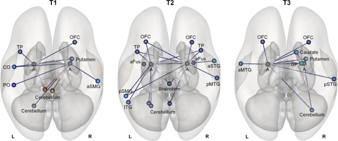
Fig. 3. Whole-brain amygdala connectivity analyses.
Whole-brain analyses exploring treatment-induced changes in amygdala connectivity to other regions of the cortical (n = 91), and subcortical (n = 13) Harvard–Oxford Atlas, and the cerebellar parcellation of the AAL Atlas (n = 26) revealed an overall pattern of reduced amygdala connectivity in the oxytocin group, compared with the placebo group. Connections displaying significant treatment effects (blue connections: oxytocin < placebo; red connections: oxytocin > placebo) are reported separately for each assessment session (T1–T3) at an uncorrected p < 0.05 threshold (two-sided). T1 assessment session immediately after the 4-week treatment (at least 24 h after the final administration), T2 assessment session 4 weeks post treatment, T3 assessment session 1 year post treatment. A amygdala, PO parietal operculum cortex, CO central opercular cortex, aSMG anterior supramarginal gyrus, OFC orbitofrontal cortex, TP temporal pole, pMTG posterior middle temporal gyrus, aFus anterior fusiform cortex, pSMG posterior supramarginal gyrus, aSTG anterior superior temporal gyrus, ITG inferior temporal gyrus, pSTG posterior superior temporal gyrus, aMTG anterior middle temporal gyrus, GP globus pallidus, L left, R right.

