Abstract
CRISPR-associated (Cas) DNA-endonucleases are remarkably effective tools for genome engineering, but have limited target ranges due to their protospacer adjacent motif (PAM) requirements. We demonstrate a critical expansion of the targetable sequence space for a type II-A CRISPR-associated enzyme through identification of the natural 5-NAAN-3 PAM preference of Streptococcus macacae Cas9 (SmacCas9). To achieve efficient editing activity, we graft the PAM-interacting domain of SmacCas9 to its well-established ortholog from Streptococcus pyogenes (SpyCas9), and further engineer an increased efficiency variant (iSpyMac) for robust genome editing activity. We establish that our hybrids can target all adenine dinucleotide PAM sequences and possess robust and accurate editing capabilities in human cells.
Subject terms: Synthetic biology, Computational biology and bioinformatics, CRISPR-Cas9 genome editing
Protospacer adjacent motif (PAM) requirements limit the target range of CRISPR endonucleases. Here, the authors graft the 5-NAAN-3 PAM-interacting domain of SmacCas9 onto SpyCas9 to create adenine dinucleotide targeting chimeras.
Introduction
Biotechnologies based on RNA-guided CRISPR systems have enabled precise and programmable genomic interfacing1. However, CRISPR-associated (Cas) endonucleases are also collectively restrained from localizing to any position along double-stranded DNA (dsDNA) due to their requirement for targets to neighbor a protospacer adajacent motif (PAM)2–4. Current gaps in the PAM sequences that Cas enzymes are known to recognize prevent access to numerous genomic positions for powerful genome editing activities, such as base editing, prime editing, and homology-directed repair5–8. Many adenine-thymine (AT)-rich regions, in particular, have been excluded from compelling CRISPR applications because previously reported endonucleases, such as Cas9 and Cas12a (formerly known as Cpf1), require targets to neighbor guanine-cytosine (GC)-content or more restrictive motifs, respectively9–11.
In this work, we introduce an ortholog of the well-established Cas9 from Streptococcus pyogenes (SpyCas9), derived from Streptococcus macacae NCTC 11558, that can instead recognize a short 5-NAA-3 PAM12. These sequences constitute 18.6% of the human genome, making adjacent adenines the most abundant dinucleotide (Supplementary Fig. 1A–B). The importance of this alternative PAM recognition for a Cas9 enzyme is reinforced by recent work exposing that Type-V DNA-targeting CRISPR nucleases (including Cas12 and Cas14 orthologs), while targeting dsDNA at AT-rich PAM sites with intrinsic high fidelity, will indiscriminately digest single-stranded DNA (ssDNA) once bound to their targets13–16. Such collateral activity may introduce unwanted risks around partially unpaired chromosomal structures, such as transcription bubbles, R-loops, and replication forks.
Here we present engineered nucleases derived from SmacCas9 and characterize their altered specificity and utility by means of transcriptional repression in bacterial culture, in vitro digestion reactions, and gene editing activity in human cells. Our results demonstrate complete 5-NAAN-3 PAM recognition of our engineered variants in all tested contexts.
Results
Discovery of SmacCas9
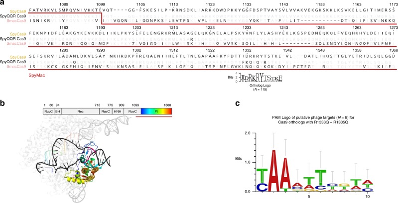
To modify the ancestral 5-NGG-3 PAM specificity of SpyCas9, early and recent reports have employed directed evolution (e.g., "VQR”, "EQR”, "VRER”, and "NRNH” variants) or rational design informed by crystal structure (e.g., "QQR”, "NG”, and "NR” variants)17–22. These reports focused on the PAM-contacting arginine residues R1333 and R1335 that abolish function when exclusively mutated. While those studies identified compensatory mutations resulting in altered PAM specificity, the Cas9 variants that they produced maintained a guanine preference in at least one position of the PAM sequence for reported in vivo editing. Concurrent reports have used evolutionary information to further relax the canonical 5-NNGRRT-3 PAM specificity of Staphylococcus aureus Cas9 (SaCas9) or to discover alternative 5-NNNNCC-3 PAM specificity to the canonical 5-NNNNGHTT-3 PAM of Neisseria meningitidis Cas923,24. The nucleases from both of these new reports, however, still prefer GC-content in at least one position of the PAM sequence. We aimed to lift such GC-content prerequisites via a custom bioinformatics-driven workflow that mines existing PAM diversity in the Streptococcus genus25. Using this strategy, we homed in on SmacCas9 as having the potential to bear altered non-GC PAM specificity upon aligning 115 orthologs of SpyCas9 from UniProt (limited to those with greater than a 70% pairwise BLOSSOM62 score). From the alignment we found SmacCas9 was one of two close homologs, along with a Streptococcus mutans B112SM-A Cas9 (SmutCas9), possessing glutamines at both of the positions aligned to the otherwise highly conserved PAM-contacting arginines (Fig. 1a–b; Supplementary Fig. 2A). Arginine residues are known to strongly prefer guanines in the amino-acid-base interaction landscape, as evidenced by the 5-NGG-3 specificity of SpyCas9. Glutamine residues, on the other hand, preferentially bind to adenines, through interaction with the major groove edge26. We thus hypothesized that SmacCas9 had naturally coevolved the necessary compensatory mutations to gain new adenine-rich PAM recognition. A small sample size of 13 spacers from its corresponding genome’s CRISPR array prevented us from confidently inferring the SmacCas9 PAM in silico. Nevertheless, the possibility for SmacCas9 requiring less GC-content in its PAM was supported by sequence similarities to the "QQR” variant that has 5-NAAG-3 specificity27, in addition to the AT-rich putative consensus PAM for phage-originating spacers in CRISPR arrays associated with highly homologous SmutCas9, which were identified with the aid of our previously-described SPAMALOT pipeline and consistent with previous predictions (Fig. 1c; Supplementary Fig. 2B; Supplementary Fig. 3) 25,28.
Fig. 1. Identification of features from natural PAM divergence through bioinformatics.
a Sequence alignment of SpyCas9, its QQR variant, and SmacCas9. The step in the underlining red line marks the joining of SpyCas9 and SmacCas9 to construct a SpyMac hybrid. The sequence logo (Weblogo online tool) immediately below the alignment depicts the conservation at 11 positions around the PAM-contacting arginines of SpyCas9. b The domain organization of SpyCas9 juxtaposed over a color-coded structure of RNA-guided, target-bound SpyCas9 (PDB ID 5F9R). The two DNA strands are black with the exception of a magenta segment corresponding to the PAM. A blue–green–red color map is used for labeling the Cas9 PI domain and guide spacer sequence to highlight structures that confer sequence specificity and the prevalence of intra-domain contacts within the PI43. c A sequence logo generated online (WebLogo) that was input with putative PAM sequences found in Streptococcus phage and associated with close SmacCas9 homologs.
Engineering and PAM characterization of SpyMac
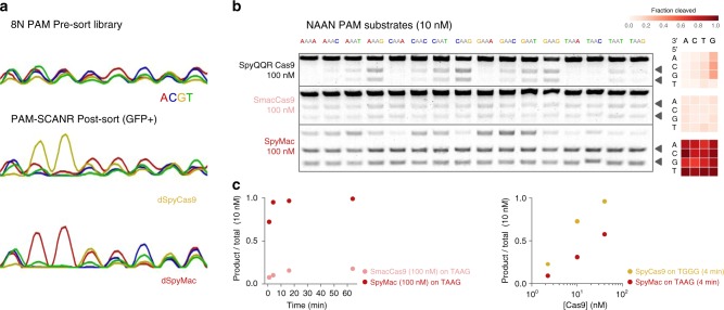
We proceeded to empirically determine the PAM preferences of several Streptococcus orthologs that change one or both of the critical PAM-contacts. Based on demonstrated examples of the PAM-interaction domain (PID) and guide RNA (gRNA) having cross-compatibility between Cas9 orthologs that are closely related and active, we constructed new variants by rationally exchanging the PI region of catalytically "dead” SpyCas9 (dSpyCas9) with those of the selected orthologs (Supplementary Fig. 2A–B)29,30. Assembled variants, including dSpyMacCas9 (herein referred to as dSpyMac), were separately cotransformed into E. coli cells, along with guide RNA derived from S. pyogenes and an 8-mer PAM library of uniform base representation in the PAM-SCANR genetic circuit, established by Leenay et al.31. The circuit upregulates a green fluorescent protein (GFP) reporter in proportion to PAM-binding strength. Therefore, we collected the GFP-positive cell populations by flow cytometry (Supplementary Fig. 4) and Sanger sequenced them around the site of the PAM to determine position-wise base preferences in a corresponding variant’s PAM recognition. dSpyMac, more so than dSpyMutCas9, generated a trace profile that was most consistent with guanine-independent PAM recognition, along with a dominant specificity for adenine dinucleotides (Fig. 2a; Supplementary Fig. 2C).
Fig. 2. Validation of SmacCas9 recognition for adenine dinucleotide PAM sequences.
a Chromatograms representing the PAM-SCANR based enrichment of variant-recognizing PAM sequences from a 5-NNNNNNNN-3 library. b SYBR-stained agarose gels showing in vitro digestion of 10 nM 5-NAAN-3 substrates upon 16 minutes of incubation with 100 nM of purified ribonucleoprotein enzyme assemblies. Arrows distinguish banding of the cleaved products from uncleaved substrate (top band). Matrix plots summarize cleaved fraction calculations, which were carried out in a custom script for processing gel images. Samples were performed in independent biological duplicates (n = 2). c Time course measurements of target DNA substrate cleavage for SmacCas9 and SpyMac. d DNA substrate cleavage plotted as a function of 0.25:1, 1:1, and 4:1 molar ratios of ribonucleoprotein to target for wild-type SpyCas9 and hybrid SpyMac. Source data are provided as a Source Data file.
Next, we purified nuclease-active enzymes to continue probing the DNA target recognition potential and uniqueness of SpyMac. (Supplementary Fig. 5A)27,32. We individually incubated the ribonucleoprotein complex enzymes (composed of Cas9 + crRNA + tracrRNA) with double-stranded target substrates of all 5/3-neighboring base combinations at an adenine dinucleotide PAM (5-NAAN-3; Fig. 2b). A brief 16-min digestion indicated both wild-type SmacCas9 and the hybrid SpyMac cleaved adjacent to 5-NAAN-3 motifs more broadly and evenly than the previously reported QQR variant. SpyMac distinguished itself further with rapid DNA-cutting rates that resemble the fast digest kinetics of SpyCas9 (Fig. 2c–d)33. We ran reactions that used varying crRNA spacer lengths and tracrRNA sequence, as the latter differs slightly between the S. macacae and S. pyogenes genomes (Supplementary Fig. 5B–E). Neither of these two parameters compensated for the slower cleavage rate of SmacCas9, but we did notice marginal improvement in the activity of the wild-type form with its native tracrRNA, which comports with the interface of the guide-Cas9 interaction being mostly outside of the PI domain.
To verify that an adenine dsDNA dinucleotide is sufficient for Cas9 PAM recognition and target cleavage, we assembled target sequences that switch the next four downstream bases to the same nucleotide (e.g., 5-TAAGXXXX-3, for X all fixed to A, C, G, or T; Supplementary Fig. 5F). We confirmed SpyMac remains active across this target set, albeit with some variation in cutting rate. Additionally, we observed moderate yield of cleaved products on examples of 5-NBBAA-3, 5-NABAB-3, 5-NBABA-3 PAM sequences (where B is C, G, or T; Supplementary Fig. 5G), revealing an even broader tolerance for increments to the dinucleotide position or adenine adjacency. We anticipate future measurements of guide-loading, target-dissociation and R-loop expansion/contraction will provide more insights on the serendipitous catalytic benefit over SmacCas9 from grafting its PI domain onto a truncated SpyCas9.
Genome editing with iSpyMac
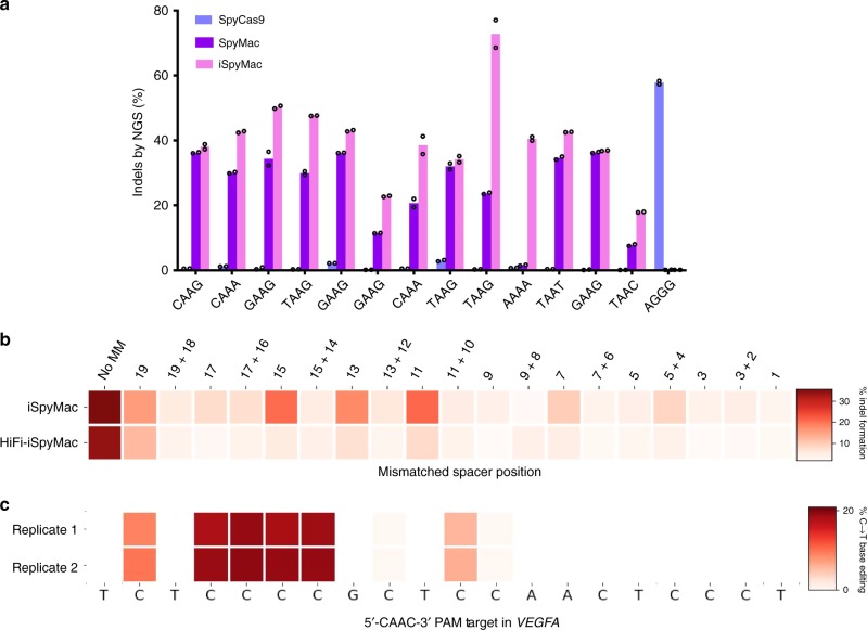
To assess the altered 5-NAAN-3 targeting capabilities of SpyMac, as opposed to SpyCas9, we cotransfected HEK293T cells with plasmids expressing these nucleases alongside one of 12 sgRNAs, targeting PAMs with varying combinations of bases flanking the adenine dinucleotide, within 5 distinct genomic loci. After 5 days post-transfection, we extracted genomic DNA, amplified the target loci, and quantified indel frequencies via next-generation sequencing (NGS). Our results first demonstrate that while SpyCas9 is able to achieve over a 50% modification rate on the 5-NGGN-3 substrate, it has negligible activity on 5-NAAN-3 targets. Alternatively, SpyMac achieves modification on most, but not all, tested 5-NAAN-3 loci. In fact, SpyMac demonstrated negligible editing on the tested 5-AAAA-3 target sequence within the PVALB gene. To address sites with low modification efficiencies, we introduced two mutations (R221K and N394K) into SpyMac that had been previously identified by deep mutational scans of SpyCas9 to increase editing efficiency34. We refer to this variant as an increased editing SpyMac (iSpyMac) due to its elevated modification efficiencies on all tested 5-NAAN-3 targets, even editing on sites that SpyMac is unable to access, such as 5-AAAA-3 PAM sites (Fig. 3a). Additionally, iSpyMac exhibits comparable modification efficiencies to other A-rich CRISPR nucleases, such as Cas12a effectors from Acidaminococcus sp. and Lachnospiraceae bacterium ND2006 (Supplementary Fig. 6).
Fig. 3. Genome editing capabilities of engineered SmacCas9 variants.
a CRISPResso2 indel analysis following NGS of amplified genomic regions to assess on-target editing of iSpyMac in comparison to SpyMac and SpyCas9, on the indicated 5-NAA-3 and 5-NGG-3 PAM sequences. Samples were performed in two independent transfection replicates (n = 2). b Efficiency heatmap of mismatch tolerance assay on genomic targets. Quantified indel frequencies, as assessed by the TIDE algorithm41, are exhibited for each labeled single or double mismatch in the sgRNA sequence for the indicated Cas9 variant and indicated PAM sequence. Samples were performed in two independent transfection replicates (n = 2). c CRISPResso2 genomic base editing analysis following NGS of genomic amplicons to assess conversion of cytosines to thymines by iSpyMac-BE3. Samples were performed in two independent transfection replicates (n = 2). All source data are provided as a Source Data file.
Next, we assessed the tolerance of iSpyMac to mismatched sequences, by employing sgRNAs harboring double or single mismatches to a fixed protospacer within the AAVS gene, possessing a 5-GAAG-3 PAM sequence. iSpyMac demonstrated editing capabilities on targets with single mismatches within the PAM-proximal segment of the sgRNA. To mitigate this supposed off-target propensity, we introduced the R691A mutation, which was previously isolated via bacterial selection for SpyCas9 to maintain high on-target activity while reducing off-target editing35. Our high-fidelity variant, HiFi-iSpyMac, exhibited nearly negligible activity on mismatched sequences, as compared to the original enzyme, with minimal loss of on-target activity (Fig. 3b).
Lastly, we selected a window of four nucleotides in the VEGFA locus in a sequence context such that any other CRISPR endonuclease with reported use for base editing would not allow their base editing with a cytidine deaminase-fused enzyme36. Accordingly, we cotransfected HEK293T cells with a nickase form of iSpyMac derived from the previously reported BE3 architecture for cytosine base editing (iSpyMac-BE3) and the sgRNA plasmid targeting a PAM downstream of the selected nucleotides5. We measured effective base editing levels in harvested cells, which exhibited over 20% cytosine to thymine conversion at these positions via NGS analysis (Fig. 3c).
Discussion
In summary, we have identified a homolog of SpyCas9 in Streptococcus macacae with native 5-NAAN-3 PAM specificity. By leveraging the high similarity in Cas9 sequences between different Streptococcus species and the substantial background in the development and characterization of SpyCas9, we have engineered variants of SmacCas9 that maintain its minimal adenine dinucleotide PAM specificity and achieve efficient and accurate activity for mediating edits on chromosomes in human cells37. This finding sets the path for engineering enzymes like iSpyMac with other desirable properties, control points, effectors, and activities8,38–40. iSpyMac can now open wide access to AT-content PAM sequences in the ever-growing list of genome engineering applications with type II-A CRISPR-Cas systems.
Methods
Selection of Streptococcus Cas9 orthologs of interest
All Cas9 orthologs from the Streptococcus genus were downloaded from the online UniProt database https://www.uniprot.org/. These were the downselected by pairwise alignment to SpyCas9 using a BLOSUM62 cost matrix in Genewiz software, discarding orthologs with less than 70% agreement with the Spy Cas9 sequence. The remaining 115 orthologs were used to generate a sequence logo (Weblogo http://weblogo.threeplusone.com/create.cgi), and were manually selected for divergence at positions aligned to residues critical for the PAM interaction of SpyCas9. The SPAMALOT pipeline was implemented as we previously reported25. Briefly, a set of scripts based around the Bowtie alignment tool (http://bowtie-bio.sourceforge.net) map the spacer sequences from CRISPR cassettes to putative targets in phage genomes. The SPAMALOT software can be downloaded at https://github.com/mitmedialab/SPAMALOT.
PAM-SCANR bacterial fluorescence assay
Sequences encoding the PAM-interaction domains of selected Cas9 orthologs were synthesized as gBlock fragments by Integrated DNA Technologies (IDT) and inserted via a New England Biolabs (NEB) Gibson Assembly reaction into the C-terminus of a low-copy plasmid containing dSpyCas9 (Beisel Lab, NCSU). The hybrid-protein constructs were transformed into electrocompetent E. coli cells with additional PAM-SCANR components as previously established31. Overnight cultures were analyzed and sorted on a Becton Dickinson (BD) FACSAria machine. Sorted GFP-positive cells were grown to sufficient density, and plasmids from the pre-sorted and sorted populations were then isolated. The region flanking the nucleotide library was PCR-amplified and submitted for Sanger sequencing (Genewiz). The chromatograms from received trace files were inspected for post-sorted sequence enrichments relative to the pre-sorted library.
Purification of and DNA cleavage with selected nucleases
The gBlock (IDT) encoding the PAM-interaction domain of S. macacae was inserted into a bacterial protein expression/purification vector containing wild-type S. pyogenes Cas9 fused to the His6-MBP-tobacco etch virus (TEV) protease cleavage site at the N-terminus (pMJ915 was a gift from Jennifer Doudna, Addgene plasmid #69090). The resulting hybrid SpyMac Cas9 protein expression construct was sequence-verified by a next-generation complete plasmid sequencing service (CCBI DNA Core Facility at Massachusetts General Hospital). The hybrid-protein construct was then transformed into BL21 Rosetta 2TM(DE3) (MilliporeSigma), and a single colony was picked for protein expression, inoculated in 1 L 2xYT media, and grown at 37 °C to a cell density of OD600 0.6. The temperature was then lowered to 18 °C and His-MBP-TEV-SpyMac Cas9 expression was induced by supplementing with 0.2 mM IPTG for an additional 18 h of growth before harvest. Cells were then lysed with BugBusterTMProtein Extraction Reagent, supplemented with 1 mg/ml lysozyme solution (MilliporeSigma), 125 Units/gram cell paste of BenzonaseTMNuclease (MilliporeSigma), and complete, EDTA-free protease inhibitors (Roche Diagnostics Corporation). The lysate was clarified by centrifugation, including a final spin with a prechilled SteriflipTM0.45 micron filter (MilliporeSigma). The clarified lysate was incubated with Ni-NTA resin (Qiagen) at 4 °C for 1 h and subsequently applied to an Econo-PacTMchromatography column (Bio-Rad Laboratories). The protein-bound resin was washed extensively with wash buffer (20 mM Tris pH 8.0, 800 mM KCl, 20 mM imidazole, 10% glycerol, 1 mM TCEP) and His-tagged SpyMac protein was eluted in wash buffer (20 mM HEPES, pH 8.0, 500 mM KCl, 250 mM imidazole, 10% glycerol). ProTEVTMPlus protease (Promega, Madison) was added to the pooled fractions and dialyzed overnight into storage buffer (20 mM HEPES, pH 7.5, 500 mM KCl, 20% glycerol) at 4 °C using Slide-A-LyzerTMdialysis cassettes with a molecular weight cutoff of 20 KDa (ThermoFisher Scientific). The sample was then incubated again with Ni-NTA resin for 1 h at 4 °C with gentle rotation and applied to a chromatography column to remove the cleaved His tag. The protein was eluted with wash buffer (20 mM Tris pH 8.0, 800 mM KCl, 20 mM imidazole, 10% glycerol, 1 mM TCEP) and fractions containing cleaved protein were verified once more by SDS-PAGE and Coomassie staining, then pooled, buffer exchanged into storage buffer, and concentrated. The concentrated aliquots were measured based on their light-absorption (Implen Nanophotometer) and flash-frozen at −80 °C for storage or used directly for in vitro cleavage assays. The crRNA and tracrRNA guide components were procured in the form of HPLC-purified RNA oligos (IDT) and resuspended in 1X IDTE pH 7.5 solution (IDT). Duplex crRNA-tracrRNA guides were annealed at 1 uM concentration in duplex buffer (IDT) by a protocol of rapid melting followed by gradual cooling. Target substrates were PCR-amplified from assemblies of the PAM-SCANR plasmid with a fixed PAM sequence. In vitro digestion reactions with 10 nM target and typically a 10-fold excess of enzyme components were prepared on ice and then incubated in a thermal cycler at 37 °C. Reactions were halted after at least 1 min of incubation by subsequent heat denaturation at 65 °C for 5 min and run on a 2% TAE-agarose gel stained with DNA-intercalating SYBR dye (Invitrogen). Gel images were recorded from blue-light exposure and analyzed in a Python script adapted from https://github.com/jharman25/gelquant/. Cleavage fraction measurements were quantified, in ImageJ (imagej.nih.gov) by the relative intensity of substrate and product bands as follows:
Cell culture and DNA modification analysis
HEK293T cells were maintained in DMEM supplemented with 100 units/ml penicillin, 100 mg/ml streptomycin, and 10% fetal bovine serum (FBS). sgRNA plasmids (100 ng) and nuclease plasmids (100 ng) were transfected into cells as duplicates (2 × 10⁴/well in a 96-well plate) with Lipofectamine 3000 (Invitrogen) in Opti-MEM (Gibco). After 5 days post-transfection, genomic DNA was extracted using QuickExtract Solution (Epicentre), and genomic loci were amplified by PCR utilizing the Phusion Hot Start Flex DNA Polymerase (NEB). Amplicons were enzymatically purified and submitted for Sanger sequencing or NGS sequencing. Sanger sequencing ab1 files were analyzed using the TIDE algorithm (tide.deskgen.com)41. in comparison to an unedited control to calculate indel frequencies. NGS FASTQ files were analysed using a batch version of the software CRISPResso2 (https://github.com/pinellolab/CRISPResso2)42. All samples were performed in independent duplicates (n = 2). Standard deviation was used to calculate error bars.
Statistical analysis
Data are shown as the mean of duplicate values, which are indicated by dots for each figure. Data were plotted using Matplotlib and the GraphPad Prism software (graphpad.com/scientific-software/prism/).
Reporting summary
Further information on research design is available in the Nature Research Reporting Summary linked to this article.
Supplementary information
Acknowledgements
This work was supported by the consortia of sponsors of the MIT Media Lab and the MIT Center for Bits and Atoms. NGS work was supported by a grant (GM115911) to E.J.S from the U.S. National Institutes of Health (NIH). We thank E. Boyden for access to cell culture, in addition to N. Gershenfeld and S. Zhang for shared lab equipment. We further thank A. Hennes for technical assistance.
Source data
Author contributions
N.J. identified SmacCas9 and related orthologs as proteins of interest, and L.N. assembled ortholog constructs for PAM characterization. P.C. and N.J. conceived identification strategies for PAM novelty, designed and implemented workflows for PAM discovery, and conducted data analysis for PAM validation. P.C. and N.J. formulated genome editing experiments for mammalian characterization. P.C. carried out genome editing assays, and J.L. conducted NGS experiments and data collection. L.N. optimized protein purification protocols and isolated nucleases for enzymology. S.R.T.K. and E.T. assisted in plasmid construction, transfection, and sample preparation. P.C., N.J., and L.N. wrote the manuscript with input from all authors. N.J. supervised the study, with assistance from E.J.S. and J.M.J.
Data availability
Sequence data that support the findings of this study are available via the NIH Sequence Read Archive via BioProject PRJNA623926. Data underlying Figs. 2-3 and Supplementary Fig. 6 are provided as Source Data.
Competing interests
P.C., N.J., L.N., and J.M.J. are inventors of US Patent WO2019217336A2: "Applications of Streptococcus-Derived Cas9 Nucleases on Minimal Adenine-Rich PAM Targets”.
Footnotes
Peer review information Nature Communications thanks the anonymous reviewer(s) for their contribution to the peer review of this work. Peer reviewer reports are available.
Publisher’s note Springer Nature remains neutral with regard to jurisdictional claims in published maps and institutional affiliations.
Supplementary information
Supplementary information is available for this paper at 10.1038/s41467-020-16117-8.
References
- 1.Komor AC, Badran AH, Liu DR. Crispr-based technologies for the manipulation of eukaryotic genomes. Cell. 2017;168:20–36. doi: 10.1016/j.cell.2016.10.044. [DOI] [PMC free article] [PubMed] [Google Scholar]
- 2.Mojica FJM, Diez-Villasenor C, Garcia-Martinez J, Almendros C. Short motif sequences determine the targets of the prokaryotic crispr defence system. Microbiology. 2009;155:733–740. doi: 10.1099/mic.0.023960-0. [DOI] [PubMed] [Google Scholar]
- 3.Sternberg SH, Redding S, Jinek M, Greene EC, Doudna JA. Dna interrogation by the crispr rna-guided endonuclease cas9. Nature. 2014;507:62–67. doi: 10.1038/nature13011. [DOI] [PMC free article] [PubMed] [Google Scholar]
- 4.Leenay RT, Beisel CL. Deciphering, communicating, and engineering the crispr pam. J. Mol. Biol. 2017;429:177–191. doi: 10.1016/j.jmb.2016.11.024. [DOI] [PMC free article] [PubMed] [Google Scholar]
- 5.Komor AC, Kim YB, Packer MS, Zuris JA, Liu DR. Programmable editing of a target base in genomic dna without double-stranded dna cleavage. Nature. 2016;533:420–424. doi: 10.1038/nature17946. [DOI] [PMC free article] [PubMed] [Google Scholar]
- 6.Gaudelli NM, et al. Programmable base editing of a-t to g-c in genomic dna without dna cleavage. Nature. 2017;551:464–471. doi: 10.1038/nature24644. [DOI] [PMC free article] [PubMed] [Google Scholar]
- 7.Anzalone AV, et al. Search-and-replace genome editing without double-strand breaks or donor DNA. Nature. 2019;576:149–157. doi: 10.1038/s41586-019-1711-4. [DOI] [PMC free article] [PubMed] [Google Scholar]
- 8.Gutschner T, Haemmerle M, Genovese G, Draetta GF, Chin L. Post-translational regulation of cas9 during g1 enhances homology-directed repair. Cell Rep. 2016;14:1555–1566. doi: 10.1016/j.celrep.2016.01.019. [DOI] [PubMed] [Google Scholar]
- 9.Zhang M, et al. Uncovering the essential genes of the human malaria parasite plasmodium falciparum by saturation mutagenesis. Science. 2018;360:eaap7847. doi: 10.1126/science.aap7847. [DOI] [PMC free article] [PubMed] [Google Scholar]
- 10.Jinek M, et al. A programmable dual-rna-guided DNA endonuclease in adaptive bacterial immunity. Science. 2012;337:816–821. doi: 10.1126/science.1225829. [DOI] [PMC free article] [PubMed] [Google Scholar]
- 11.Zetsche B, et al. Cpf1 is a single rna-guided endonuclease of a class 2 crispr-cas system. Cell. 2015;163:759–771. doi: 10.1016/j.cell.2015.09.038. [DOI] [PMC free article] [PubMed] [Google Scholar]
- 12.Richards VP, et al. Phylogenomics and the dynamic genome evolution of the genus streptococcus. Genome Biol. Evol. 2014;6:741–753. doi: 10.1093/gbe/evu048. [DOI] [PMC free article] [PubMed] [Google Scholar]
- 13.Chen JS, et al. Crispr-cas12a target binding unleashes indiscriminate single-stranded dnase activity. Science. 2018;360:436–439. doi: 10.1126/science.aar6245. [DOI] [PMC free article] [PubMed] [Google Scholar]
- 14.Kleinstiver BP, et al. Genome-wide specificities of crispr-cas cpf1 nucleases in human cells. Nat. Biotechnol. 2016;34:869–874. doi: 10.1038/nbt.3620. [DOI] [PMC free article] [PubMed] [Google Scholar]
- 15.Yan WX, et al. Functionally diverse type v crispr-cas systems. Science. 2019;363:88–91. doi: 10.1126/science.aav7271. [DOI] [PMC free article] [PubMed] [Google Scholar]
- 16.Harrington LB, et al. Programmed dna destruction by miniature crispr-cas14 enzymes. Science. 2018;362:839–842. doi: 10.1126/science.aav4294. [DOI] [PMC free article] [PubMed] [Google Scholar]
- 17.Anders C, Bargsten K, Jinek M. Structural plasticity of PAM recognition by engineered variants of the RNA-guided endonuclease cas9. Mol. Cell. 2016;61:895–902. doi: 10.1016/j.molcel.2016.02.020. [DOI] [PMC free article] [PubMed] [Google Scholar]
- 18.Kleinstiver BP, et al. Engineered crispr-cas9 nucleases with altered pam specificities. Nature. 2015;523:481–485. doi: 10.1038/nature14592. [DOI] [PMC free article] [PubMed] [Google Scholar]
- 19.Kleinstiver BP, et al. Broadening the targeting range of staphylococcus aureus CRISPR-cas9 by modifying PAM recognition. Nat. Biotechnol. 2015;33:1293–1298. doi: 10.1038/nbt.3404. [DOI] [PMC free article] [PubMed] [Google Scholar]
- 20.Nishimasu H, et al. Engineered crispr-cas9 nuclease with expanded targeting space. Science. 2018;361:1259–1262. doi: 10.1126/science.aas9129. [DOI] [PMC free article] [PubMed] [Google Scholar]
- 21.Miller SM, et al. Continuous evolution of SpCas9 variants compatible with non-g PAMs. Nat. Biotechnol. 2020;38:471–481. doi: 10.1038/s41587-020-0412-8. [DOI] [PMC free article] [PubMed] [Google Scholar]
- 22.Walton RT, Christie KA, Whittaker MN, Kleinstiver BP. Unconstrained genome targeting with near-PAMless engineered CRISPR-cas9 variants. Science. 2020;368:290–296. doi: 10.1126/science.aba8853. [DOI] [PMC free article] [PubMed] [Google Scholar]
- 23.Edraki A, et al. A compact, high-accuracy cas9 with a dinucleotide pam for in vivo genome editing. Mol. cell. 2019;73:714–726.e4. doi: 10.1016/j.molcel.2018.12.003. [DOI] [PMC free article] [PubMed] [Google Scholar]
- 24.Ma D, et al. Engineer chimeric cas9 to expand pam recognition based on evolutionary information. Nat. Commun. 2019;10:560. doi: 10.1038/s41467-019-08395-8. [DOI] [PMC free article] [PubMed] [Google Scholar]
- 25.Chatterjee, P., Jakimo, N. & Jacobson, J.M.Minimal pam specificity of a highly similar spcas9 ortholog. Sci. Adv.4, eaau0766 (2018). [DOI] [PMC free article] [PubMed]
- 26.Luscombe NM. Amino acid-base interactions: a three-dimensional analysis of protein-DNA interactions at an atomic level. Nucleic Acids Res. 2001;29:2860–2874. doi: 10.1093/nar/29.13.2860. [DOI] [PMC free article] [PubMed] [Google Scholar]
- 27.Anders C, Jinek M. In vitro enzymology of cas9. Methods Enzymol. 2014;546:1–20. doi: 10.1016/B978-0-12-801185-0.00001-5. [DOI] [PMC free article] [PubMed] [Google Scholar]
- 28.van der Ploeg JR. Analysis of crispr in streptococcus mutans suggests frequent occurrence of acquired immunity against infection by m102-like bacteriophages. Microbiology. 2009;155:1966–1976. doi: 10.1099/mic.0.027508-0. [DOI] [PubMed] [Google Scholar]
- 29.Nishimasu H, et al. Crystal structure of cas9 in complex with guide rna and target dna. Cell. 2014;156:935–949. doi: 10.1016/j.cell.2014.02.001. [DOI] [PMC free article] [PubMed] [Google Scholar]
- 30.Briner AE, et al. Guide rna functional modules direct cas9 activity and orthogonality. Mol. cell. 2014;56:333–339. doi: 10.1016/j.molcel.2014.09.019. [DOI] [PubMed] [Google Scholar]
- 31.Leenay RT, et al. Identifying and visualizing functional PAM diversity across CRISPR-cas systems. Mol. Cell. 2016;62:137–147. doi: 10.1016/j.molcel.2016.02.031. [DOI] [PMC free article] [PubMed] [Google Scholar]
- 32.Lin S, Staahl BT, Alla RK, Doudna JA. Enhanced homology-directed human genome engineering by controlled timing of crispr/cas9 delivery. eLife. 2014;3:e04766. doi: 10.7554/eLife.04766. [DOI] [PMC free article] [PubMed] [Google Scholar]
- 33.Gong S, Yu HH, Johnson KA, Taylor DW. DNA unwinding is the primary determinant of CRISPR-cas9 activity. Cell Rep. 2018;22:359–371. doi: 10.1016/j.celrep.2017.12.041. [DOI] [PMC free article] [PubMed] [Google Scholar]
- 34.Spencer, J.M. & Zhang, X. Deep mutational scanning of s. pyogenes cas9 reveals important functional domains. Sci. Rep.7, https://www.nature.com/articles/s41598-017-17081-y (2017). [DOI] [PMC free article] [PubMed]
- 35.Vakulskas CA, et al. A high-fidelity cas9 mutant delivered as a ribonucleoprotein complex enables efficient gene editing in human hematopoietic stem and progenitor cells. Nat. Med. 2018;24:1216–1224. doi: 10.1038/s41591-018-0137-0. [DOI] [PMC free article] [PubMed] [Google Scholar]
- 36.Mir A, Edraki A, Lee J, Sontheimer EJ. Type II-c CRISPR-cas9 biology, mechanism, and application. ACS Chem. Biol. 2017;13:357–365. doi: 10.1021/acschembio.7b00855. [DOI] [PMC free article] [PubMed] [Google Scholar]
- 37.Jiang F, Doudna JA. Crispr-cas9 structures and mechanisms. Annu. Rev. Biophys. 2017;46:505–529. doi: 10.1146/annurev-biophys-062215-010822. [DOI] [PubMed] [Google Scholar]
- 38.Slaymaker IM, et al. Rationally engineered cas9 nucleases with improved specificity. Science. 2016;351:84–88. doi: 10.1126/science.aad5227. [DOI] [PMC free article] [PubMed] [Google Scholar]
- 39.Holtzman L, Gersbach CA. Editing the epigenome: reshaping the genomic landscape. Annu. Rev. Genomics Hum. Genet. 2018;19:43–71. doi: 10.1146/annurev-genom-083117-021632. [DOI] [PubMed] [Google Scholar]
- 40.Hu JH, et al. Evolved cas9 variants with broad pam compatibility and high dna specificity. Nature. 2018;556:57–63. doi: 10.1038/nature26155. [DOI] [PMC free article] [PubMed] [Google Scholar]
- 41.Brinkman, E.K. & van Steensel, B. in Methods in Molecular Biology, 29–44 (Springer New York, 2019).
- 42.Clement K, et al. CRISPResso2 provides accurate and rapid genome editing sequence analysis. Nat. Biotechnol. 2019;37:224–226. doi: 10.1038/s41587-019-0032-3. [DOI] [PMC free article] [PubMed] [Google Scholar]
- 43.Jiang F, et al. Structures of a crispr-cas9 r-loop complex primed for dna cleavage. Science. 2016;351:867–871. doi: 10.1126/science.aad8282. [DOI] [PMC free article] [PubMed] [Google Scholar]
Associated Data
This section collects any data citations, data availability statements, or supplementary materials included in this article.
Supplementary Materials
Data Availability Statement
Sequence data that support the findings of this study are available via the NIH Sequence Read Archive via BioProject PRJNA623926. Data underlying Figs. 2-3 and Supplementary Fig. 6 are provided as Source Data.


