

Abstract
A severe cutaneous drug reaction resembling acute generalized exanthematous pustulosis resulting from ingestion of hydroxychloroquine has been documented. It is distinguishable by its longer incubation period, more varied morphology with initially urticarial and later targetoid and arcuate plaques, recalcitrance to therapy and longer duration. Given the anticipated surge in the use of hydroxychloroquine due to its reported benefits in those with coronavirus disease 2019, specific recognition of this entity is pivotal. We delineate it as generalized pustular figurate erythema.
Keywords: acute generalized exanthematous pustulosis, coronavirus, COVID‐19, DRESS syndrome, drug rash, erythema multiforme, figurate erythema, hydroxychloroquine, SARS‐2, SARS‐CoV‐2, Stevens‐Johnson syndrome, Sweet syndrome, toxic epidermal necrolysis, urticaria
1.
Severe potentially life‐threatening cutaneous drug reactions are a huge concern, most specifically acute generalized exanthematous pustulosis (AGEP), Stevens‐Johnson syndrome (SJS), toxic epidermal necrolysis (TEN), generalized bullous fixed drug eruption, and drug reaction with eosinophilia and systemic symptoms (DRESS) syndrome.1, 2, 3, 4, 5, 6, 7, 8 AGEP was originally misclassified as a form of pustular psoriasis; however, it is not associated with psoriasis. 9 AGEP is a severe cutaneous adverse reaction characterized by the rapid development of sterile nonfollicular pustules on an erythematous base.2, 3 It is usually attributed to drugs, antibiotics being the most common, with an onset typically within 48 hours of ingestion, often with an acute onset of fever and leukocytosis.
There is another rare acute severe generalized disorder, one usually characterized as AGEP, but with an onset of 2 to 3 weeks (range 4‐27 days) rather 1 day after initial drug exposure, typically due to hydroxychloroquine, more severe, more difficult to treat, with a longer duration, and recognized as likely having a different pathogenic mechanism from the usual type of AGEP. 10 This perplexing disorder has been described as atypical AGEP,11, 12 recalcitrant AGEP,13, 14 pustular DRESS syndrome, 15 AGEP/SJS overlap, 16 AGEP/TEN overlap,17, 18 and Sweet's syndrome following hydroxychloroquine.19, 20, 21 We delineate and highlight it as generalized pustular figurate erythema (GPFE). It can be due to a number of medications, but we now emphasize its association with hydroxychloroquine. This antimalarial drug commonly employed for a variety of rheumatic and dermatological disorders is now under evaluation as an antiviral agent against coronavirus disease 2019 (COVID‐19).22, 23 More than 20 cases of GPFE from hydroxychloroquine have been described in the medical literature.
Clinical examination reveals an abrupt onset of a pruritic eruption representing a severe cutaneous drug reaction with fever and neutrophilic leukocytosis.11, 12, 13, 14, 15, 16, 17, 18, 19, 20, 21 GPFE may be first evident as erythematous papules and plaques on the face with facial edema and widespread urticarial or edematous plaques scattered over the entire body, with development of nonfollicular pustules atop what evolve into erythematous and sometimes atypical targetoid erythema multiforme‐like plaques converging into annular and arcuate patterns prominent on the trunk and extremities (Figure 1). Pustular erythema may develop irregularly along active borders. Erythema may fade with scaling, including on the palms and soles. Some cutaneous sloughing and excoriations may also be observed with blisters or erosions occasionally noted. There may be little or no mucosal involvement. Skin biopsy specimens may initially show mainly the changes of urticaria, but evolve into subcorneal and/or intraepidermal neutrophilic pustules sometimes with mild focal acantholysis, exocytosis, spongiosis, and an edematous papillary dermis with a perivascular lymphocytic infiltrate with occasional neutrophils, eosinophils, and mast cells progressing into a dermal neutrophilic infiltrate. No vasculitis is anticipated.
FIGURE 1.
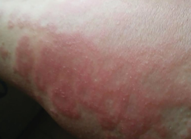
Hydroxychloroquine‐induced GPFE with numerous nonfollicular pustules atop atypical targetoid plaques. GPFE, generalized pustular figurate erythema
Treatments have been varied.11, 12, 13, 14, 15, 16, 17, 18, 19, 20, 21, 24, 25, 26 The first line therapy is topical and systemic steroids, which may be followed by cyclosporine if GPFE is not responsive.13, 14, 21 Other options include potent topical steroids with oral dapsone or etretinate. 25 Additional experience with GPFE and its treatment can be anticipated to surge as hydroxychloroquine becomes widely utilized in the COVID‐19 pandemic.
CONFLICT OF INTEREST
The authors declare no potential conflict of interest.
Schwartz RA, Janniger CK. Generalized pustular figurate erythema: A newly delineated severe cutaneous drug reaction linked with hydroxychloroquine. Dermatologic Therapy. 2020;33:e13380. 10.1111/dth.13380
REFERENCES
- 1. Patel S, John AM, Handler MZ, Schwartz RA. The fixed drug eruptions: an update emphasizing the potentially lethal generalized bullous fixed drug eruption. Am J Clin Dermatol. 2020. 10.1007/s40257-020-00505-3 [Epub ahead of print]. [DOI] [PubMed] [Google Scholar]
- 2. Szatkowski J, Schwartz RA. Acute generalized exanthematous pustulosis: a review and update. J Am Acad Dermatol. 2015;73:843‐848. [DOI] [PubMed] [Google Scholar]
- 3. Miteva L, Kadurina M, Schwartz RA. Childhood acute generalized exanthematous pustulosis induced by oral ketoconazole. Acta Dermatovenerol Croat. 2020;18:267‐270. [PubMed] [Google Scholar]
- 4. Husain Z, Reddy BY, Schwartz RA. Drug rash with eosinophilia and systemic symptoms (DRESS) syndrome: part I: clinical perspectives. J Am Acad Dermatol. 2013;68:693‐705. [DOI] [PubMed] [Google Scholar]
- 5. Husain Z, Reddy BY, Schwartz RA. DRESS syndrome: part II: management and therapeutics. J Am Acad Dermatol. 2013;68:709‐717. [DOI] [PubMed] [Google Scholar]
- 6. Schwartz RA, McDonough PH, Lee BW. Toxic epidermal necrolysis. Part I. introduction, history, classification, clinical features, systemic manifestations, etiology, and immunopathogenesis. J Am Acad Dermatol. 2013;69:173‐184. [DOI] [PubMed] [Google Scholar]
- 7. Schwartz RA, McDonough PH, Lee BW. Toxic epidermal necrolysis. Part II. Prognosis, sequelae, diagnosis, differential diagnosis, prevention, and treatment. J Am Acad Dermatol. 2013;69:187‐202. [DOI] [PubMed] [Google Scholar]
- 8. Dmochowski M, Schwartz RA. Erythema multiforme, Stevens‐Johnson syndrome, and toxic epidermal necrolysis. In: Demis DJ, ed. Clinical Dermatology. Philadelphia, PA: Lippincott Williams & Wilkins; 1999:Unit 7‐3:1‐20. [Google Scholar]
- 9. Hoegler KM, John AM, Handler MZ, Schwartz RA. Generalized pustular psoriasis: a review and update on treatment. J Eur Acad Dermatol Venereol. 2018;32:1645‐1651. [DOI] [PubMed] [Google Scholar]
- 10. Sidoroff A, Dunant A, Viboud C, et al. Risk factors for acute generalized exanthematous pustulosis (AGEP)‐results of a multinational case‐control study (EuroSCAR). Br J Dermatol. 2007;157:989‐996. [DOI] [PubMed] [Google Scholar]
- 11. Duman H, Topal IO, Kocaturk E, Cure K, Mansuroglu I. Acute generalized exanthematous pustulosis induced by hydroxychloroquine: a case with atypical clinical presentation. An Bras Dermatol. 2017;92:404‐406. [DOI] [PMC free article] [PubMed] [Google Scholar]
- 12. Pearson KC, Morrell DS, Runge SR, Jolly P. Prolonged pustular eruption from hydroxychloroquine: an unusual case of acute generalized exanthematous pustulosis. Cutis. 2016;97:212‐216. [PubMed] [Google Scholar]
- 13. Yalçın B, Çakmak S, Yıldırım B. Successful treatment of hydroxychloroquine‐induced recalcitrant acute generalized exanthematous pustulosis with cyclosporine: case report and literature review. Ann Dermatol. 2015;27:431‐434. [DOI] [PMC free article] [PubMed] [Google Scholar]
- 14. İslamoğlu ZGK, Karabağli P. A case of recalcitrant acute generalized exanthematous pustulosis with Sjogren's syndrome: successfully treated with low‐dose cyclosporine. Clin Case Rep. 2019;7(9):1721‐1724. [DOI] [PMC free article] [PubMed] [Google Scholar]
- 15. Girijala RL, Siddiqi I, Kwak Y, Wright D, Patel DB, Goldberg LH. Pustular DRESS syndrome secondary to hydroxychloroquine with EBV reactivation. J Drugs Dermatol. 2019;18:207‐209. [PubMed] [Google Scholar]
- 16. Mercogliano C, Khan M, Lin C, Mohanty E, Zimmerman R. AGEP overlap induced by hydroxychloroquine: a case report and literature review. J Community Hosp Intern Med Perspect. 2018;8:360‐362. [DOI] [PMC free article] [PubMed] [Google Scholar]
- 17. Copaescu AM, Bouffard D, Masse MS. Acute generalized exanthematous pustulosis simulating toxic epidermal necrolysis: case presentation and literature review. Allergy Asthma Clin Immunol. 2020;16:9. 10.1186/s13223-020-0407-5. [DOI] [PMC free article] [PubMed] [Google Scholar]
- 18. Lateef A, Tan KB, Lau TC. Acute generalized exanthematous pustulosis and toxic epidermal necrolysis induced by hydroxychloroquine. Clin Rheumatol. 2009;28:1449‐1452. [DOI] [PubMed] [Google Scholar]
- 19. Manzo C, Pollio N, Natale M. Sweet's syndrome following therapy with hydroxychloroquine in a patient affected with elderly‐onset primary Sjogren's syndrome. Medicines. 2019;6(4):E111. 10.3390/medicines6040111. [DOI] [PMC free article] [PubMed] [Google Scholar]
- 20. Bodard Q, Carre D, Chenal P, Zarnitsky C, Midhat M, Litrowski N. Drug‐induced Sweet's syndrome related to hydroxychloroquine: about 2 cases. Rev Med Interne. 2020;41:289‐292. [DOI] [PubMed] [Google Scholar]
- 21. Falcone LM, Stone RC, Schwartz RA. Drug‐induced neutrophilic dermatoses. In: Wallach D, Vignon‐Pennamen MD, Marzano A, eds. Neutrophilic Dermatoses. Cham, Switzerland: Springer Verlag; 2018:259‐270. [Google Scholar]
- 22. Colson P, Rolain JM, Raoult D. Chloroquine for the 2019 novel coronavirus SARS‐CoV‐2. Int J Antimicrob Agents. 2020;55(3):105923. 10.1016/j.ijantimicag.2020.105923. [DOI] [PMC free article] [PubMed] [Google Scholar]
- 23. Duan YJ, Liu Q, Zhao SQ, et al. The trial of chloroquine in the treatment of Corona Virus Disease 2019 (COVID‐19) and its research progress in forensic toxicology. Fa Yi Xue Za Zhi. 2020;36(2):1‐1. 10.12116/j.issn.1004-5619.2020.02.001. [DOI] [PubMed] [Google Scholar]
- 24. Castner NB, Harris JC, Motaparthi K. Cyclosporine for corticosteroid‐refractory acute generalized exanthematous pustulosis due to hydroxychloroquine. Dermatol Ther. 2018;31(5):e12660. 10.1111/dth.12660. [DOI] [PubMed] [Google Scholar]
- 25. Matsuda‐Hirose H, Sho Y, Yamate T, et al. Acute generalized exanthematous pustulosis induced by hydroxychloroquine successfully treated with etretinate. J Dermatol. 2020;47(2):e53‐e54. [DOI] [PubMed] [Google Scholar]
- 26. Park JJ, Yun SJ, Lee JB, Kim SJ, Won YH, Lee SC. A case of hydroxychloroquine induced acute generalized exanthematous pustulosis confirmed by accidental oral provocation. Ann Dermatol. 2010;22:102‐105. [DOI] [PMC free article] [PubMed] [Google Scholar]