PLOS ONE logoLink to PLOS ONE
. 2020 May 19;15(5):e0233050. doi: 10.1371/journal.pone.0233050

Aortopathies in mouse models of Pompe, Fabry and Mucopolysaccharidosis IIIB lysosomal storage diseases

Maria Paola Belfiore 1,#, Francesca Iacobellis 1,#, Emma Acampora 2, Martina Caiazza 3, Marta Rubino 3, Emanuele Monda 3, Maria Rosaria Magaldi 3, Antonietta Tarallo 2, Marcella Sasso 2, Valeria De Pasquale 4, Roberto Grassi 1, Salvatore Cappabianca 1, Paolo Calabrò 3, Simona Fecarotta 2, Salvatore Esposito 5, Giovanni Esposito 2, Antonio Pisani 2, Luigi Michele Pavone 4, Giancarlo Parenti 2,, Giuseppe Limongelli 3,6,‡,*
Editor: Andrea Dardis7
PMCID: PMC7236983  PMID: 32428018

Abstract

Introduction

Lysosomal storage diseases (LSDs) are rare inherited metabolic diseases characterized by an abnormal accumulation of various toxic materials in the cells as a result of enzyme deficiencies leading to tissue and organ damage. Among clinical manifestations, cardiac diseases are particularly important in Pompe glycogen storage diseases (PD), in glycosphingolipidosis Fabry disease (FD), and mucopolysaccharidoses (MPS). Here, we evaluated the occurrence of aortopathy in knock out (KO) mouse models of three different LSDs, including PD, FD, and MPS IIIB.

Methods

We measured the aortic diameters in 15 KO male mice, 5 for each LSD: 5 GLA-/- mice for FD, 5 NAGLU-/- mice for MPS IIIB, 5 GAA-/- mice for PD, and 15 wild type (WT) mice: 5 for each strain. In order to compare the aortic parameters between KO and WT mice deriving from the same colonies, different diameters were echocardiographically measured: aortic annulus, aortic sinus, sino-tubular junction, ascending aorta, aortic arch and descending aorta. Storage material content and aortic defects of the KO mice were also analyzed by histology, when available.

Results

Compared to their correspondent WT mice: GAA-/- mice showed greater diameters of ascending aorta (1.61mm vs. 1.11mm, p-value = 0.01) and descending aorta (1.17mm vs 1.02mm, p-value 0.04); GLA-/- mice showed greater diameters of aortic annulus (1.35mm vs. 1.22mm, p-value = 0.01), sinus of Valsalva (1.6mm vs. 1.38mm, p-value<0.01), ascending aorta (1.57mm vs. 1.34mm, p-value<0.01), aortic arch (1.36mm vs. 1.22mm, p-value = 0.03) and descending aorta (1.29mm vs. 1.11mm, p-value<0.01); NAGLU-/- mice showed greater diameters of sinus of Valsalva (1.46mm vs. 1.31mm, p-value = 0.05), ascending aorta (1.42mm vs. 1.29mm, p-value<0.01), aortic arch (1.34mm vs. 1.28mm, p-value<0.01) and descending aorta (1.18mm vs. 1.1mm, p-value 0.01).

Conclusions

We evaluated for the first time the aortic diameters in 3 LSD mouse models and identified different aortopathy patterns, in concordance with recent human findings. Our results are relevant in view of using KO mouse models for efficiently testing the efficacy of new therapies on distinct cardiovascular aspects of LSDs.

Introduction

Lysosomal storage diseases (LSDs) are rare inherited metabolic diseases due to defects of lysosomal functions, in most cases, caused by deficiencies of acidic hydrolases involved in the degradation of complex molecules [1,2]. Consequently, in these disorders, storage material accumulates in the lysosomal compartment of the cells, leading to altered cellular processes with variable involvement of multiple tissues and organs [3,4]. Thus, LSDs are multisystem disorders with broad phenotypes that range from early-onset severe forms to late-onset forms characterized by attenuated clinical course, and milder manifestations [1]. Among the clinical symptoms in LSDs, cardiac dysfunction is particularly relevant in lysosomal glycogen storage diseases (Pompe and Danon disease), glycosphingolipidoses (Anderson-Fabry disease), and mucopolysaccharidoses [4]. Hypertrophic and dilated cardiomyopathy, coronary artery disease, and valvular diseases are the most common disease manifestations in affected patients. In this study, we focused on the occurrence of aortopathy in the mouse models of three LSDs: Pompe disease (PD), Fabry disease (FD), and Mucopolysaccharidosis (MPS) IIIB.

PD is a rare autosomal recessive and progressive genetic disorder caused by mutations in the gene encoding for the acid α-glucosidase (GAA), the lysosomal enzyme that degrades glycogen [5]. The clinical spectrum of the disease is broad, ranging from the infantile-onset PD, associated with glycogen accumulation in heart and muscles, leading to premature death, if not treated [68], to the late-onset PD, with predominant skeletal muscle and vascular involvement, including the ascending aorta [9,10].

FD is an X-linked recessive LSD due to mutations in the gene encoding for the α-galactosidase (GLA), leading to the accumulation of globotriaosylceramide in the cells of various tissues with con-sequent multi-organ dysfunction [11,12]. Clinical manifestations of FD include systemic vasculopathy resulting in a markedly increased risk of ischemic stroke, small-fiber peripheral neuropathy, cardiac dysfunction, and chronic kidney disease [1315].

MPSs are inborn errors of metabolism due to the deficiency of lysosomal enzymes involved in the degradation of glycosaminoglycans (GAGs) [16]. Clinical manifestations of MPSs include neurological disorders as well as skeletal, joint, airway and cardiac defects, hearing and vision impairment, and mental retardation. The affected patients usually die in the second or third decade of their life. Depending on the accumulated GAGs, MPSs are classified into seven types (I, II, III, IV, VI, VII, and IX) that are variable in their prevalence, clinical symptoms, and degree of severity. In MPS patient heart, accumulation of GAGs occurs within the cardiac valves, the epicardial coronary arteries, the myocytes, the cardiac interstitium and the walls of the great vessels, thus triggering cardiac valve regurgitation and stenosis, diffuse coronary artery stenosis, myocardial dysfunction and aortic root dilation [1721]. Cardiac disease is highly prevalent (60%-100% of patients) in MPS type I, II, and VI affected patients [22]. However, evidence exists on cardiac involvement in MPS III as well. Indeed, valvulopathies, mainly involving the mitral and aortic valves, cardiomyopathy, arrhythmias, coronary artery disease, aortic root dilatation, and conduction abnormalities have been reported for MPS III affected patients [1718,2324]. The subtype MPS IIIB is due to the deficiency of N-acetyl-α-glucosaminidase (NAGLU), which is required for the degradation of heparan sulfate (HS). We recently demonstrated that heart disease, valvular abnormalities, and cardiac failure is associated with an impaired lysosomal autophagic flux in the mouse model of MPS IIIB [25].

Enzyme replacement therapy (ERT) substantially improves many of the features of the above described LSDs, including some aspects of the cardiac involvement [3,5,26]. However, several disease-modifying treatment options, including chaperone-based therapy, mRNAs, and gene therapies, are currently under investigation [35,2631]. Thus, extensive knowledge of the prevalence and nature of cardiac defects in these LSDs may be relevant. In particular, the presence and characteristics of aortopathy related to LSDs have not been investigated in pre-clinical models of LSDs. This study sought to study aortic size and morphology in knock out (KO) mouse models of 3 different LSDs: PD, FD, and MPS IIIB.

Materials and methods

Animals

Animal studies were performed according to the EU Directive 86/609 regarding the protection of animals used for experimental purposes, and according to Institution Animal Care and use committee (IACUC) guidelines for the Care and Use of animals in research. The study was approved by the Italian Ministry of Health, IACUC n. 523/2015-PR (06/11/2015). Every procedure on the mice was performed to ensure that discomfort, distress, pain, and injury would be minimal.

Thirty mice were examined: 15 were KO male mice for the LSDs: 5 GLA-/-, 5 NAGLU-/-, 5 GAA-/-; the remaining were 15 wild type (WT) mice: 5 for each strain. All of them were examined at 12 months. They were maintained on light/dark cycles of 12/12 h and had free access to food and water.

The NAGLU-/- murine model of MPS IIIB was previously generated by the insertion of the neomycin resistance gene into exon 6 of the NALGU gene on the C57/BL6 background. NAGLU-/- and wildtype (WT) mice derived from the same colony were genotyped by PCR [32].

The GAA-/- murine model of PD was previously created by insertion of the neomycin resistance gene and the herpes virus thymidine kinase gene in the locus encoding GAA. Six independent cell lines containing the disrupted GAA allele were used to make chimeras that were bred to C57/BL6 females to generate heterozygous mice (F1). GAA-/- and wildtype (WT) mice derived from the same colony were genotyped by PCR [33].

The GLA-/- murine model of FD was previously generated by a targeted disruption of the α-GLA gene on the C57/BL6 background. GLA-/- and wildtype (WT) mice derived from the same colony were genotyped by PCR [34].

Ultrasound analysis

M-mode, two-dimensional, Color Doppler echocardiography was performed by a high-resolution ultrasound machine (VisualSonics Vevo 2100 unit, Fujifilm) capable of recording over 750 frames per second and equipped with a high-frequency (40 MHz) transducer. During each imaging session, mice were anesthetized by inhalation with isoflurane in oxygen (3–4% for induction and 1.5% for maintenance).

Mice were allowed to breathe spontaneously. Heart rate and body temperature were appropriately monitored by cardiac electrodes integrated in the heating pad and by a rectal probe and remained constant throughout the examination. In order to prevent imaging artifacts, each mouse was immobilized, and its chest hair was removed, applying a calcium thioglycolate depilatory cream. The transducer was fixed on a special arm. An appropriate amount of heated sonographic gel (Aquasonic 100; Parker Laboratories, Inc, Fairfield, NJ) was applied to the shaved skin.

The transducer was positioned longitudinally to its body, along the right parasternal line, with the index marker pointing to its head and rotated about 35° counterclockwise. The obtained B-mode image represents a section across the long axis of the left ventricle, which allowed visualization of the aortic root and the outflow tract of the left ventricle.

The following echo views were examined: parasternal long axis; parasternal short axis; apical; aortic arch. The last was obtained from a modified right parasternal view, with the transducer positioned longitudinally to the mouse body, along the right parasternal line, almost parallel to the table and with the index marker pointing to the head. This view is optimal to morphologically examine the ascending aorta and the arch, as well as the main arterial branches. Measured diameters were aortic annulus, aortic sinus, sino-tubular junction, ascending aorta, aortic arch, and descending aorta. Annulus dimensions were obtained in the parasternal long-axis view during systole for semilunar valves, and the apical four-chamber view during diastole for atrioventricular valves as previously described [25,35].

Histochemical analysis

Immediately after echocardiographic imaging, mice (n = 5 NAGLU-/- and n = 5 correspondent WT from the same colony, n = 5 GAA-/- and n = 5 correspondent WT from the same colony) were euthanized by cervical dislocation in compliance with the recommendations contained in the American Veterinary Medical Association (AVMA) Guidelines for the Euthanasia of Animals.

Subsequently, hearts were harvested for histological and morphometric analyses. Hearts were isolated, fixed in a bath of 4% aqueous buffered formalin overnight, processed for paraffin embedding [36,37], and coronal sections (10 μm thick), containing right and left ventricles, and aorta, were obtained. Sequential sections from each heart were stained with hematoxylin and eosin (H&E) staining (from Sigma-Aldrich) [38], and periodic acid-Schiff (PAS)-Alcian Blue stain (from Dako) for storage content evaluation. Selected sections were stained with Weiger for elastic fiber evaluation. Image analysis was performed with a charge-coupled device camera coupled with a light microscope.

Statistical analysis

In the post-processing phase, the acquired ultrasound data were exported and analyzed using the dedicated software (Vevo 2100, Visual Sonics). Statistical analyses were performed using SPSS (version 25.0, SPSS Inc., Chicago, IL, USA). Normally distributed continuous data are presented as mean ± standard deviation (SD) and were compared by t-test or ANOVA (if more than two groups). Values of p < 0.05 were considered statistically significant.

Results

The weight of the KO mice for each LSD model was not significantly different compared to their correspondent WT: the medium weight of GLA-/- and of their correspondent WT was 29.2 ±2.2 and 28.7 ±1.2, respectively; the medium weight of NAGLU-/- and of their correspondent WT was 28.9 ±0.7 and 30 ±1.0, respectively; the medium weight of GAA-/- and of their correspondent WT was 28 ±2.0 and 28.2 ±1.0, respectively.

In order to evaluate the aortic parameters in the three different LSD mouse models, the echocardiographic measurement of different portions of the aorta (aortic annulus, aortic sinus, sino-tubular junction ascending aorta, aortic arch and descending aorta) was performed (Tables 13).

Table 1. Comparison between diameters of the different portions of the aorta of GAA knock-out mice (GAA-/-) and their correspondent wild-type (WT) mice.

GAA-/- (Pompe) (n = 5) WT (n = 5) p-value
Aortic annulus 1.51 (±0.22) 1.34 (±1.11) 0.311
Sinus of Valsalva 1.88 (±0.26) 1.6 (±0.08) 0.108
Sino-tubular junction 1.49 (±0.23) 1.26 (±0.07) 0.100
Ascending aorta 1.61 (±0.17) 1.11 (±0.03) 0.010*
Aortic arch 1.51 (±0.24) 1.17 (±0.04) 0.087
Descending aorta 1.17 (±0.09) 1.02 (±0.09) 0.036*

Table 3. Comparison between diameters of the different portions of the aorta of NAGLU knock-out mice (NAGLU-/-) and their correspondent wild-type (WT) mice.

MPS: Mucopolysaccharidosis.

NAGLU-/- (MPS IIIB) (n = 5) WT (n = 5) p-value
Aortic annulus 1.36 (±0.09) 1.26 (±0.1) 0.060
Sinus of Valsalva 1.46 (±0.11) 1.31 (±0.03) 0.049*
Sino-tubular junction 1.16 (±0.08) 1.13 (±0.04) 0.536
Ascending aorta 1.42 (±0.05) 1.29 (±0.06) 0.005*
Aortic arch 1.34 (±0.04) 1.28 (±0.04) 0.002*
Descending aorta 1.18 (±0.04) 1.1 (±0.04) 0.012*

In particular, compared to their correspondent WT mice:

GAA KO mice (GAA-/-) showed greater diameters of ascending aorta (1.61mm vs. 1.11mm, p-value = 0.01) and descending aorta (1.17mm vs. 1.02mm, p-value = 0.04), while no difference statistically significant was reported in the other aortic parameters evaluated (Table 1);

GLA KO mice (GLA-/-) showed greater diameters of aortic annulus (1.35mm vs. 1.22mm, p-value = 0.01), sinus of Valsalva (1.6mm vs. 1.38mm, p-value<0.01), ascending aorta (1.57mm vs. 1.34mm, p-value<0.01), aortic arch (1.36mm vs. 1.22mm, p-value = 0.03) and descending aorta (1.29mm vs. 1.11mm, p-value<0.01), while no difference statistically significant was reported considering the sino-tubular junction (Table 2);

Table 2. Comparison between diameters of the different portions of the aorta of GLA knock-out mice (GLA-/-) and their correspondent wild-type (WT) mice.

GLA-/- (Fabry) (n = 5) WT (n = 5) p-value
Aortic anulus 1.35 (±0–44) 1.22 (±0.01) 0.018*
Sinus of Valsalva 1.6 (±0.05) 1.38 (±0.03) <0.001*
Sino-tubular junction 1.26 (±0.05) 1.22 (±0.05) 0.703
Ascending aorta 1.57 (±0.04) 1.34 (±0.06) <0.001*
Aortic arch 1.36 (±0.03) 1.22 (±0.07) 0.030*
Descending aorta 1.29 (±0.07) 1.11 (±0.04) <0.001

NAGLU KO mice (NAGLU-/-) showed greater diameters of sinus of Valsalva (1.46mm vs. 1.31mm, p-value = 0.05), ascending aorta (1.42mm vs. 1.29mm, p-value<0.01), aortic arch (1.34mm vs. 1.28mm, p-value<0.01) and descending aorta (1.18mm vs. 1.1mm, p-value = 0.01). At the same time, no difference statistically significant was reported considering the aortic annulus and the sino-tubular junction (Table 3).

Moreover, a comparison of the echocardiographic measurements between the 3 LSD KO mouse models was also performed and reported in Table 4.

Table 4. Comparison between diameters of the different portion of aorta in knock-out mouse models of the three different lysosomal storage disease.

MPS: Mucopolysaccharidosis.

GAA -/- (Pompe) (n = 5) GLA -/- (Fabry) (n = 5) NAGLU-/- (MPS IIIB) (n = 5) p-value
Aortic annulus 1.51 (±0.22) 1.35 (±0.44) 1.36 (±0.09) 0.176
Sinus of Valsalva 1.88 (±0.26) 1.6 (±0.05) 1.46 (±0.11) 0.006*
Sino-tubular junction 1.49 (±0.23) 1.26 (±0.05) 1.16 (±0.08) 0.007*
Ascending aorta 1.61 (±0.17) 1.57 (±0.04) 1.42 (±0.05) 0.037*
Aortic arch 1.51 (±0.24) 1.36 (±0.03) 1.34 (±0.04) 0.149
Descending aorta 1.17 (±0.09) 1.29 (±0.07) 1.18 (±0.04) 0.028*

GAA KO mice (GAA-/-) showed greater diameters compared to GLA KO mice (GLA-/-) and NAGLU KO mice (NAGLU-/-) considering aortic sinus (1.88mm, 1.6mm e 1.46mm, respectively, p-value<0.01), the sino-tubular junction (1.49mm, 1.26mm e 1.16mm, respectively, p-value<0.01) and ascending aorta (1.61mm, 1.57mm e 1,42mm, respectively, p-value = 0.04).

GLA KO mice (GLA-/-) showed greater diameters compared to GAA KO mice (GAA-/-) and NAGLU KO mice (NAGLU-/-) considering descending aorta (1.29mm, 1.17mm e 1.18mm, respectively, p-value = 0.03); no difference statistically significant was reported considering the aortic annulus and the aortic arch. Moreover, compared to their correspondent WT and other KO models, NAGLU-/- mice exhibited increased thickening of the aortic valve leaflets.

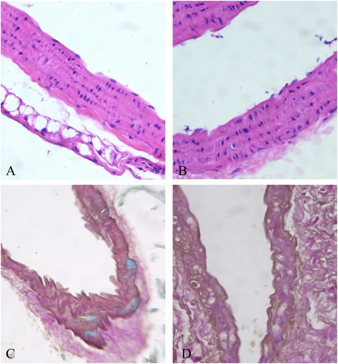
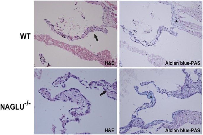
To evaluate aortic structures in the analyzed LSD mouse models (NAGLU-/- and GAA-/-), we performed hematoxylin-eosin (H&E) and Alcian blue staining of heart sections. According to the aortic defects detected by echocardiography at the time point examined, H&E and PAS staining of sections from the hearts of GAA-/- mice evidenced an aortic wall with mild disorganization of lamellar units for the presence of many vacuoles containing fine granular or amorphous material in their reduced inter-lamellar space (Fig 1). The aortic valve was not thickened, nor accumulation was found. H&E and PAS staining of sections from the hearts of NAGLU-/- mice confirmed the aortic valve defects detected by echocardiography at the time point examined. NAGLU-/- mice exhibited significant aortic cuspid thickening with excess/redundant tissue present in the valves, as demonstrated by Alcian blue-PAS staining (Fig 2). By using the H&E staining, NAGLU-/- aortic valve cuspids exhibited a disruption of the normal collagen/proteoglycan boundary interfaces observed in WT animals (Fig 2).

Fig 1. Large empty vacuoles in the external third of the aortic wall in the GAA-/- mouse heart (A and B, H&E 40x), causing lamellar unit disorganization (C, Alcian-Weighert 20x, and D, PAS-Weighert 40x).

Fig 1

Fig 2. Aortic valve morphology alterations in the MPS IIIB mouse model.

Fig 2

Representative images of hematoxylin-eosin (H&E) (left) and Alcian blue-PAS (right) staining of aortic valve sections from WT and NAGLU-/- hearts.

Discussion

In literature, inherited aortopathies are well known to be associated with genetic syndromes (e.g., Marfan syndrome) and collagen disease (e.g., Ehlers Danlos), or with familial diseases (e.g., bicuspid aortic valve aortopathy) [15]. Cardiac involvement in LSD generally involves the heart muscle and cardiac valves [1418]. However, a growing field of interest is the study of the aortic size in LSDs [811]. The concept of “metabolic storage aortopathies” is just emerging with the evidence that mild to moderate aortic enlargement, and few cases of aneurysm or dissection have been reported in LSDs. To date, it is not clear whether aortic dilation is an LSD general feature, or it is typical of specific disorders.

To our knowledge, this is the first report of aortopathy in mouse models of LSDs. Besides, different patterns of dilation have been associated with different models and compared to human findings from literature. In our study, we found a difference between PD vs. WT mice for ascending and descending aorta parameters. This finding is consistent with the data reported in the literature. El-Gharbawy et al. [9] reported dilated arteriopathy involving the ascending thoracic aorta primarily in 5 female patients with late-onset PD, including one patient with bicuspid aortic valve, who developed dissection; another patient with juvenile-onset disease showed both thoracic and basilar artery aneurysms. In the present study, histochemical evaluation in GAA-/- mice evidenced signs of disorganization and storage in the aortic wall. This finding can probably explain the increased aortic stiffness identified in patients with PD [39].

In the FD model, aortic dilation was diffuse (except for sino-tubular junction), and particularly evident at aortic sinus and ascending aorta. Barbey et al. [14], studying a cohort of 106 patients with Fabry disease, showed aortic dilation at the sinus of Valsalva in 32.7% of males and 5.6% of females; aneurysms were present in 9.6% of males and 1.9% of females.

In the MPS IIIB mouse model, except for sino-tubular junction, aortic dilation was diffuse; a non-significant trend was found at the annulus. Moreover, a thickening of the aortic valve was evidenced and confirmed by histochemical analysis. This analysis shows an excess/redundant tissue present in the valves (demonstrated by Alcian blue-PAS staining) and disruption of the regular collagen/proteoglycan boundary interfaces observed in WT animals (H&E staining). In a cohort of MPS I-VII patients (age range: 3.4–25.9 years), about one-third developed aortic dilation, with the highest prevalence in MPS IVA (87.5%). Aortic dilation was prevalent at the annulus (41%; 14/34) and sinus of Valsalva (35%; 12/34) [19]. Poswar et al. [20] studied 69 patients with MPS, showing a prevalence of up to 39%, particularly in MPS IVa and MPS VI. No significant effect of enzyme replacement therapy (ERT) on aortic size was found in 11 patients with available data (2 with MPS I; 4 with MPS II; 2 with MPS IVA, and 3 with MPS VI). The pathogenesis is not clear. However, in MPS VII mice, GAG accumulation seems to activate complement components, which may play a role in signal transduction pathways that upregulate elastases [40]. In MPS canine models, interleukin 6-like cytokine oncostatin M resulted to be increased in the aorta of MPS I and MPS VII dogs, and tumor necrosis factor-α and toll-like receptor 4 were increased in MPS VII dog aortas, suggesting that these cytokines could contribute to the upregulation of the elastases [41]. Similarly, in the NAGLU-/- mouse model also the aortic valve was involved, showing an increased thickening of the aortic valve leaflets compared with WT.

Here, we studied three KO mouse models, which recapitulate LSDs that differ in terms of enzymatic defects, stored substrates (glycogen in PD, glycosphingolipids in FD, and glycosaminoglycans in MPS IIIB), and clinical manifestations. Despite these differences, in all animal models, we found signs of aortic involvement, suggesting that this type of manifestations are common findings in LSDs, and should be carefully looked at in affected patients. Interestingly, ascending aorta dilatation was significantly more relevant in PD mice compared to the other two LSD models. On the other hand, FD mice showed a significant dilation at the descending aorta compared with the other two LSD models. This is a new finding since no data are reported in the literature regarding FD affected patients.

Study limitations

Our study has some limitations, including the small sample size and the single time for the aortic evaluation. Unfortunately, we were not able to have histochemical findings of FD mice (due to technical problems). Moreover, this pilot study was essentially designed to describe the aortic phenotype in different mouse models of lysosomal storage disease. Thus, based on these results, the effect of old and new treatments (ERT, losartan, genetic therapies) on our models will be the objective of future investigations.

Conclusions

In conclusion, we evaluated for the first-time aortic diameters in 3 LSD mouse models and identified different aortopathy patterns, in concordance with recent human findings. Our results are relevant in view of using KO mouse models for efficiently testing the efficacy of new therapies on distinct cardiovascular aspects of LSDs.

Supporting information

S1 Checklist. The ARRIVE guidelines checklist.

(PDF)

Acknowledgments

Author Giuseppe Limongelli is a member of ERN GUARD-HEART (European Reference Network for Rare and Complex Diseases of the Heart; http://guardheart.ern-net.eu).

Data Availability

All relevant data are within the paper and its Supporting Information files.

Funding Statement

The author(s) received no specific funding for this work.

References

  • 1.Sun A. Lysosomal storage disease overview. Ann Transl Med. 2018;6(24): 476 10.21037/atm.2018.11.39 [DOI] [PMC free article] [PubMed] [Google Scholar]
  • 2.Platt FM, d'Azzo A, Davidson BL, Neufeld EF, Tifft CJ. Lysosomal storage diseases. Nat Rev Dis Primers. 2018;4(1): 27 10.1038/s41572-018-0025-4 [DOI] [PubMed] [Google Scholar]
  • 3.Parenti G, Andria G, Ballabio A. Lysosomal storage diseases: from pathophysiology to therapy. Annu Rev Med. 2015;66: 471–86. 10.1146/annurev-med-122313-085916 [DOI] [PubMed] [Google Scholar]
  • 4.Nair V, Belanger EC, Veinot JP. Lysosomal storage disorders affecting the heart: a review. Cardiovasc Pathol. 2019;39: 12–24. 10.1016/j.carpath.2018.11.002 [DOI] [PubMed] [Google Scholar]
  • 5.Kohler L, Puertollano R, Raben N. Pompe Disease: From Basic Science to Therapy. Neurotherapeutics. 2018;15(4): 928–942. 10.1007/s13311-018-0655-y [DOI] [PMC free article] [PubMed] [Google Scholar]
  • 6.Limongelli G, Fratta F. S1.4 Cardiovascular involvement in Pompe disease. Acta Myol. 2011;30(3): 202–203. [Google Scholar]
  • 7.Schoser B. Pompe disease: what are we missing? Ann Transl Med. 2019;7(13): 292 10.21037/atm.2019.05.29 [DOI] [PMC free article] [PubMed] [Google Scholar]
  • 8.Bay LB, Denzler I, Durand C, Eiroa H, Frabasil J, Fainboim A, et al. Infantile-onset Pompe disease: Diagnosis and management. Arch Argent Pediatr. 2019;117(4): 271–278. 10.5546/aap.2019.eng.271 [DOI] [PubMed] [Google Scholar]
  • 9.El-Gharbawy AH, Bhat G, Murillo JE, Thurberg BL, Kampmann C, Mengel KE, et al. Expanding the clinical spectrum of late-onset Pompe disease: dilated arteriopathy involving the thoracic aorta, a novel vascular phenotype uncovered. Mol Genet Metab. 2011;103(4): 362–6. 10.1016/j.ymgme.2011.04.009 [DOI] [PubMed] [Google Scholar]
  • 10.Goeber V, Banz Y, Kaeberich A, Carrel T. Huge aneurysm of the ascending aorta in a patient with adult-type Pompe's disease: histological findings mimicking fibrillinopathy. Eur J Cardiothorac Surg. 2013;43(1): 193–5. 10.1093/ejcts/ezs489 [DOI] [PubMed] [Google Scholar]
  • 11.Chan B, Adam DN. A Review of Fabry Disease. Skin Therapy Lett. 2018;23(2): 4–6. [PubMed] [Google Scholar]
  • 12.Schiffmann R. Fabry disease. Handb Clin Neurol. 2015;132: 231–48. 10.1016/B978-0-444-62702-5.00017-2 [DOI] [PubMed] [Google Scholar]
  • 13.Masarone D, Duro G, Dellegrottaglie S, Colomba P, Rubino M, Cirillo A, et al. Severe hypertrophic cardiomyopathy in a patient with atypical Anderson-Fabry disease. Future Cardiol. 2017;13(6): 521–527. 10.2217/fca-2017-0011 [DOI] [PubMed] [Google Scholar]
  • 14.Barbey F, Qanadli SD, Juli C, Brakch N, Palacek T, Rizzo E, et al. Aortic remodelling in Fabry disease. Eur Heart J. 2010;31(3): 347–53. 10.1093/eurheartj/ehp426 [DOI] [PubMed] [Google Scholar]
  • 15.Limongelli G, Masarone D, Verrengia M, Gravino R, Salerno G, Castelletti S, et al. Diagnostic Clues for the Diagnosis of Nonsarcomeric Hypertrophic Cardiomyopathy (Phenocopies): Amyloidosis, Fabry Disease, and Mitochondrial Disease. J Cardiovasc Echogr. 2018;28(2): 120–123. 10.4103/jcecho.jcecho_2_18 [DOI] [PMC free article] [PubMed] [Google Scholar]
  • 16.Neufeld EF, Muenzer J. The mucopolysaccharidoses In: The metabolic and molecular bases of inherited disease. 8th edition, Scriver C.R., Beaudet A.L., Sly W.S., and Valle D., eds. (NewYork: McGraw-Hill; ), 2001, pp. 3421–3452. [Google Scholar]
  • 17.Braunlin EA, Harmatz PR, Scarpa M, Furlanetto B, Kampmann C, Loehr JP, et al. Cardiac disease in patients with mucopolysaccharidosis: presentation, diagnosis and management. J Inherit Metab Dis. 2011;34: 1183–1197. 10.1007/s10545-011-9359-8 [DOI] [PMC free article] [PubMed] [Google Scholar]
  • 18.Braunlin E, Wang R. Cardiac issues in adults with the mucopolysaccharidoses: current knowledge and emerging needs. Heart. 2016;102(16): 1257–62. 10.1136/heartjnl-2015-309258 [DOI] [PubMed] [Google Scholar]
  • 19.Bolourchi M, Renella P, Wang R. Aortic Root Dilatation in Mucopolysaccharidosis I–VII. Int J Mol Sci. 2016;17(12). [DOI] [PMC free article] [PubMed] [Google Scholar]
  • 20.Poswar FO, de Souza CFM, Giugliani R, Baldo G. Aortic root dilatation in patients with mucopolysaccharidoses and the impact of enzyme replacement therapy. Heart Vessels, 2019;34(2): 290–295. 10.1007/s00380-018-1242-1 [DOI] [PubMed] [Google Scholar]
  • 21.Borgia F, Pezzullo E, Schiano Lomoriello V, Sorrentino R, Lo Iudice F, Cocozza S, et al. Myocardial deformation in pediatric patients with mucopolysaccharidoses: A two-dimensional speckle tracking echocardiography study. Echocardiography. 2017;34(2): 240–249. 10.1111/echo.13444 [DOI] [PubMed] [Google Scholar]
  • 22.Boffi L, Russo P, Limongelli G. Early diagnosis and management of cardiac manifestations in mucopolysaccharidoses: a practical guide for paediatric and adult cardiologists. Ital J Pediatr. 2018;44(Suppl 2): 122 10.1186/s13052-018-0560-3 [DOI] [PMC free article] [PubMed] [Google Scholar]
  • 23.Nijmeijer SCM, de Bruin-Bon RHACM, Wijburg FA, Kuipers IM. Cardiac disease in mucopolysaccharidosis type III. J Inherit Metab Dis. 2019;42(2): 276–285. 10.1002/jimd.12015 [DOI] [PubMed] [Google Scholar]
  • 24.Wilhelm CM, Truxal KV, McBride KL, Kovalchin JP, Flanigan KM. Natural history of echocardiographic abnormalities in mucopolysaccharidosis III. Mol Genet Metab. 2018;124(2): 131–134. 10.1016/j.ymgme.2018.04.010 [DOI] [PMC free article] [PubMed] [Google Scholar]
  • 25.Schiattarella GG, Cerulo G, De Pasquale V, Cocchiaro P, Paciello O, Avallone L, et al. The murine model of mucopolysaccharidosis IIIB develops cardiopathies over time leading to heart failure. PLoS One. 2015;10(7): e0131662 10.1371/journal.pone.0131662 [DOI] [PMC free article] [PubMed] [Google Scholar]
  • 26.Miller JJ, Kanack AJ, Dahms NM. Progress in the understanding and treatment of Fabry disease. Biochim Biophys Acta Gen Subj. 2020;1864(1): 129437 10.1016/j.bbagen.2019.129437 [DOI] [PMC free article] [PubMed] [Google Scholar]
  • 27.Parenti G, Andria G, Valenzano KJ. Pharmacological chaperone therapy: preclinical development, clinical translation, and prospects for the treatment of lysosomal storage disorders. Mol Ther. 2015;23(7): 1138–1148. 10.1038/mt.2015.62 [DOI] [PMC free article] [PubMed] [Google Scholar]
  • 28.Tarallo A, Carissimo A, Gatto F, Nusco E, Toscano A, Musumeci O, et al. microRNAs as biomarkers in Pompe disease. Genet Med. 2019;21(3): 591–600. 10.1038/s41436-018-0103-8 [DOI] [PubMed] [Google Scholar]
  • 29.De Pasquale V, Pavone LM. Heparan sulfate proteoglycans: The sweet side of development turns sour in mucopolysaccharidoses. Biochim Biophys Acta Mol Basis Dis. 2019;1865(11): 165539 10.1016/j.bbadis.2019.165539 [DOI] [PubMed] [Google Scholar]
  • 30.De Pasquale V, Sarogni P, Pistorio V, Cerulo G, Paladino S, Pavone LM. Targeting Heparan Sulfate Proteoglycans as a Novel Therapeutic Strategy for Mucopolysaccharidoses. Mol Ther Methods Clin Dev. 2018;10: 8–16. 10.1016/j.omtm.2018.05.002 [DOI] [PMC free article] [PubMed] [Google Scholar]
  • 31.De Pasquale V, Pezone A, Sarogni P, Tramontano A, Schiattarella GG, Avvedimento VE, et al. EGFR activation triggers cellular hypertrophy and lysosomal disease in NAGLU-depleted cardiomyoblasts, mimicking the hallmarks of mucopolysaccharidosis IIIB. Cell Death Dis. 2018;9(2): 40 10.1038/s41419-017-0187-0 [DOI] [PMC free article] [PubMed] [Google Scholar]
  • 32.Li HH, Yu WH, Rozengurt N, Zhao HZ, Lyons KM, Anagnostaras S, et al. Mouse model of Sanfilippo syndrome type B produced by targeted disruption of the gene encoding alpha-N-acetylglucosaminidase. Proc Natl Acad Sci U S A, 1999;96, 14505–14510. 10.1073/pnas.96.25.14505 [DOI] [PMC free article] [PubMed] [Google Scholar]
  • 33.Raben N, Nagaraju K, Lee E, Kessler P, Byrnei B, Lee L, et al. Targeted disruption of the acid α-Glucosidase gene in mice causes an illness with critical features of both infantile and adult human glycogen storage disease type II. J Biol Chem. 1998;273(30): 19086–19092. 10.1074/jbc.273.30.19086 [DOI] [PubMed] [Google Scholar]
  • 34.Ohshima T, Murray GJ, Swaim WD, Longenecker G, Quirk JM, Cardarelli CO, et al. alpha-Galactosidase A deficient mice: a model of Fabry disease. Proc Natl Acad Sci U S A. 1997;94(6): 2540–2544. 10.1073/pnas.94.6.2540 [DOI] [PMC free article] [PubMed] [Google Scholar]
  • 35.Belfiore MP, Berritto D, Iacobellis F, Rossi C, Nigro G, Rotundo IL, et al. A lon-gitudinal study on BIO14.6 hamsters with dilated cardiomyopathy: micro-echocardiographic evaluation Cardiovasc Ultrasound 2011;9: 39 10.1186/1476-7120-9-39 [DOI] [PMC free article] [PubMed] [Google Scholar]
  • 36.Pavone LM, Rea S, Trapani F, De Pasquale V, Tafuri S, Papparella S, et al. Role of serotonergic system in the pathogenesis of fibrosis in canine idiopathic inflammatory myopathies. Neuromuscul Disord. 2012;22(6): 549–57. 10.1016/j.nmd.2012.01.009 [DOI] [PubMed] [Google Scholar]
  • 37.Cerulo G, Tafuri S, De Pasquale V, Rea S, Romano S, Costagliola A, et al. Serotonin activates cell survival and apoptotic death responses in cultured epithelial thyroid cells. Biochimie. 2014;105: 211–5. 10.1016/j.biochi.2014.06.020 [DOI] [PubMed] [Google Scholar]
  • 38.Spina A, Rea S, De Pasquale V, Mastellone V, Avallone L, Pavone LM. Fate map of serotonin transporter-expressing cells in developing mouse thyroid. Anat Rec (Hoboken). 2011;294(3): 384–90. [DOI] [PubMed] [Google Scholar]
  • 39.Nemes A, Soliman OI, Geleijnse ML, Anwar AM, van der Beek NA, van Doorn PA, et al. Increased aortic stiffness in glycogenosis type 2 (Pompe's disease). Int J Cardiol. 2007;120(1): 138–41. 10.1016/j.ijcard.2006.07.215 [DOI] [PubMed] [Google Scholar]
  • 40.Baldo G, Wu S, Howe RA, Ramamoothy M, Knutsen RH, Fang J, et al. Pathogenesis of aortic dilatation in mucopolysaccharidosis VII mice may involve complement activation. Mol Genet Metab. 2011;104(4): 608–619. 10.1016/j.ymgme.2011.08.018 [DOI] [PMC free article] [PubMed] [Google Scholar]
  • 41.Metcalf JA, Linders B, Wu S, Bigg P, O’Donnell P, Sleeper MM, et al. Upregulation of elastase activity in aorta in mucopolysaccharidosis I and VII dogs may be due to increased cytokine expression. Mol Genet Metab. 2010;99(4): 396–407. 10.1016/j.ymgme.2009.12.003 [DOI] [PMC free article] [PubMed] [Google Scholar]

Decision Letter 0

Andrea Dardis

17 Jan 2020

PONE-D-19-29826

Lysosomal Storage Disease: Pompe, Fabry and Mucopolysaccharidoses. The emerging concept of lysosomal aortopathies

PLOS ONE

Dear Prof., Dr., Limongelli,

Thank you for submitting your manuscript to PLOS ONE. After careful consideration, we feel that it has merit but does not fully meet PLOS ONE’s publication criteria as it currently stands. Therefore, we invite you to submit a revised version of the manuscript that addresses the points raised during the review process.

 Please respond to all points raised by the reviewers.

Additional experiments must be done to render the manuscript suitable for publication.

In particular, since data on the analyzed parameter are already available in human patients, other parameters relevant for the observed aortopathie and not easily measured in human samples must be assessed.

As suggested by both reviewers storage material/lysosomal pathology should be investigated in order to provide some mechanistic explanation to the observed data.

Data showing the response of all investigated parameters to therapy must also be provided.

We would appreciate receiving your revised manuscript by Mar 02 2020 11:59PM. When you are ready to submit your revision, log on to https://www.editorialmanager.com/pone/ and select the 'Submissions Needing Revision' folder to locate your manuscript file.

If you would like to make changes to your financial disclosure, please include your updated statement in your cover letter.

To enhance the reproducibility of your results, we recommend that if applicable you deposit your laboratory protocols in protocols.io, where a protocol can be assigned its own identifier (DOI) such that it can be cited independently in the future. For instructions see: http://journals.plos.org/plosone/s/submission-guidelines#loc-laboratory-protocols

Please include the following items when submitting your revised manuscript:

  • A rebuttal letter that responds to each point raised by the academic editor and reviewer(s). This letter should be uploaded as separate file and labeled 'Response to Reviewers'.

  • A marked-up copy of your manuscript that highlights changes made to the original version. This file should be uploaded as separate file and labeled 'Revised Manuscript with Track Changes'.

  • An unmarked version of your revised paper without tracked changes. This file should be uploaded as separate file and labeled 'Manuscript'.

Please note while forming your response, if your article is accepted, you may have the opportunity to make the peer review history publicly available. The record will include editor decision letters (with reviews) and your responses to reviewer comments. If eligible, we will contact you to opt in or out.

We look forward to receiving your revised manuscript.

Kind regards,

Andrea Dardis, Ph.D.

Academic Editor

PLOS ONE

Journal Requirements:

When submitting your revision, we need you to address these additional requirements.

1. Please ensure that your manuscript meets PLOS ONE's style requirements, including those for file naming. The PLOS ONE style templates can be found at http://www.journals.plos.org/plosone/s/file?id=wjVg/PLOSOne_formatting_sample_main_body.pdf and http://www.journals.plos.org/plosone/s/file?id=ba62/PLOSOne_formatting_sample_title_authors_affiliations.pdf

2. As part of your revision, please complete and submit a copy of the ARRIVE Guidelines checklist, a document that aims to improve experimental reporting and reproducibility of animal studies for purposes of post-publication data analysis and reproducibility: https://www.nc3rs.org.uk/arrive-guidelines. Please also add method of sacrifice and source of animals in the Methods section of your manuscript. Please include your completed checklist as a Supporting Information file. Note that if your paper is accepted for publication, this checklist will be published as part of your article.

3. We noticed you have some minor occurrence(s) of overlapping text with the following previous publication(s), which needs to be addressed:

https://doi.org/10.1038/s41436-018-0103-8

https://doi.org/10.1186/1476-7120-9-39

In your revision ensure you cite all your sources (including your own works), and quote or rephrase any duplicated text outside the Methods section. Further consideration is dependent on these concerns being addressed.

4. PLOS requires an ORCID iD for the corresponding author in Editorial Manager on papers submitted after December 6th, 2016. Please ensure that you have an ORCID iD and that it is validated in Editorial Manager. To do this, go to ‘Update my Information’ (in the upper left-hand corner of the main menu), and click on the Fetch/Validate link next to the ORCID field. This will take you to the ORCID site and allow you to create a new iD or authenticate a pre-existing iD in Editorial Manager. Please see the following video for instructions on linking an ORCID iD to your Editorial Manager account: https://www.youtube.com/watch?v=_xcclfuvtxQ

[Note: HTML markup is below. Please do not edit.]

Reviewers' comments:

Reviewer's Responses to Questions

Comments to the Author

1. Is the manuscript technically sound, and do the data support the conclusions?

The manuscript must describe a technically sound piece of scientific research with data that supports the conclusions. Experiments must have been conducted rigorously, with appropriate controls, replication, and sample sizes. The conclusions must be drawn appropriately based on the data presented.

Reviewer #1: Yes

Reviewer #2: No

**********

2. Has the statistical analysis been performed appropriately and rigorously?

Reviewer #1: I Don't Know

Reviewer #2: Yes

**********

3. Have the authors made all data underlying the findings in their manuscript fully available?

The PLOS Data policy requires authors to make all data underlying the findings described in their manuscript fully available without restriction, with rare exception (please refer to the Data Availability Statement in the manuscript PDF file). The data should be provided as part of the manuscript or its supporting information, or deposited to a public repository. For example, in addition to summary statistics, the data points behind means, medians and variance measures should be available. If there are restrictions on publicly sharing data—e.g. participant privacy or use of data from a third party—those must be specified.

Reviewer #1: Yes

Reviewer #2: Yes

**********

4. Is the manuscript presented in an intelligible fashion and written in standard English?

PLOS ONE does not copyedit accepted manuscripts, so the language in submitted articles must be clear, correct, and unambiguous. Any typographical or grammatical errors should be corrected at revision, so please note any specific errors here.

Reviewer #1: Yes

Reviewer #2: Yes

**********

5. Review Comments to the Author

Please use the space provided to explain your answers to the questions above. You may also include additional comments for the author, including concerns about dual publication, research ethics, or publication ethics. (Please upload your review as an attachment if it exceeds 20,000 characters)

Reviewer #1: This is an interesting manuscript that highlights an aspect of LSDs that is not often considered. I wonder whether the authors can provide a little more data on parameters that are relevant for the aortopathies that they observe? For example cell biological analysis of storage material/lysosomal pathology? Other minor comments are listed below.

- Literature citations and discussion are incomplete, no mentioning has been made on aortic stiffness in human patients. These are relevant to this manuscript.

- The introduction is too short in my opinion. The question: 'aortopathies…have not been investigated in pre-clinical models…' is not well explained. Why is this study relevant? It is in my opinion, but please make this more clear and provide a more comprehensive reasoning to introduce the topic

- please provide information on the genetic background of the mice and corresponding wild type mice.

- Please provide the data for the weight of the ko mouse models at analysis

- The figures are redundant with the tables and could be removed

- how was multiple testing corrected for?

Reviewer #2: The paper by Belfiore et al describes the measurement of some aortic parameters by using echocardiography in 3 different mouse models of LSD, in particular Pompe Disease, Fabry Disease and Sanfilippo IIIB. Although such evaluation, conducted on each single model and also among the 3 models, showed some statistically significant differences between each model vs. wt mice as well as among the models, and although the Authors state this is the first study on this specific topic in these LSD models, this reviewer finds the study not particularly interesting due to the already available data on the patients affected by the same diseases, who can be much more informative than the mouse models. In my opinion, mouse models are worthy to be tested and used when similar data are nor recoverable from the clinics, also taking into consideration the important differences that might sometimes be registered between the models and the human subjects. In addition, the very limited number of mice examined further reduces the interest of the paper.

In my opinion, the paper is not worthy of publication.

Maybe Authors should have complemented the presented data by pathologically characterizing the cardiac region examined and by providing data obtained from the same mice following treatment, as Authors themselves state in the conclusion of the manuscript.

**********

6. PLOS authors have the option to publish the peer review history of their article (what does this mean?). If published, this will include your full peer review and any attached files.

If you choose “no”, your identity will remain anonymous but your review may still be made public.

Do you want your identity to be public for this peer review? For information about this choice, including consent withdrawal, please see our Privacy Policy.

Reviewer #1: No

Reviewer #2: No

[NOTE: If reviewer comments were submitted as an attachment file, they will be attached to this email and accessible via the submission site. Please log into your account, locate the manuscript record, and check for the action link "View Attachments". If this link does not appear, there are no attachment files to be viewed.]

While revising your submission, please upload your figure files to the Preflight Analysis and Conversion Engine (PACE) digital diagnostic tool, https://pacev2.apexcovantage.com/. PACE helps ensure that figures meet PLOS requirements. To use PACE, you must first register as a user. Registration is free. Then, login and navigate to the UPLOAD tab, where you will find detailed instructions on how to use the tool. If you encounter any issues or have any questions when using PACE, please email us at figures@plos.org. Please note that Supporting Information files do not need this step.

PLoS One. 2020 May 19;15(5):e0233050. doi: 10.1371/journal.pone.0233050.r002

Author response to Decision Letter 0


1 Mar 2020

Response to reviewers

Reviewer #1

Question n. 1 - Major

This is an interesting manuscript that highlights an aspect of LSDs that is not often considered. I wonder whether the authors can provide a little more data on parameters that are relevant for the aortopathies that they observe? For example cell biological analysis of storage material/lysosomal pathology? Other minor comments are listed below.

Answer n. 1 – Major

Thank you for your comment. As suggested, in the revised version of the manuscript we have provided available data on the histochemical analysis of the KO mice (NAGLU-/- and GAA-/-), describing unique characteristics of the semilunar valve and vessel in these aortic models. We think that this evidence significantly strengthens the paper.

We specified in the text:” H&E staining and PAS sections in GAA-/- mice evidenced an aortic wall with mild disorganization of lamellar units for the presence of many vacuoles containing fine granular or amorphous material in their reduced inter-lamellar space. The aortic valve was not thickened, nor accumulation was found (data not showed).

H&E staining and PAS sections in in NAGLU-/- mice confirmed the aortic valve defects detected by echocardiography at the time point examined. NAGLU-/- mice exhibited significant aortic cuspid thickening with excess/redundant tissue present in the valves as demonstrated by Alcian blue-PAS staining. By using the H&E staining, NAGLU-/- aortic valve cuspids exhibited a disruption of the normal collagen/proteoglycan boundary interfaces observed in WT animals”

Question n. 2 - Minor

- Literature citations and discussion are incomplete, no mentioning has been made on aortic stiffness in human patients. These are relevant to this manuscript.

Answer n. 2 - Minor

Thank you for your comment. As you suggested, we expanded the literature citations and discussion.

Question n. 3 - Minor

- The introduction is too short in my opinion. The question: 'aortopathies…have not been investigated in pre-clinical models…' is not well explained. Why is this study relevant? It is in my opinion, but please make this more clear and provide a more comprehensive reasoning to introduce the topic

Answer n. 3 - Minor

Thank you for your comment. As suggested, we expanded the introduction, focusing on cardiac and aortic involvement in patients with LSDs, to clarify the aim and the importance of the present study.

Question n. 4 - Minor

- please provide information on the genetic background of the mice and corresponding wild type mice.

Answer n. 4 - Minor

Thank you for your comment. As suggested, we have provided the genetic background of the mice models studied. We specified in the materials and methods as follows: “MPS IIIB knockout mice (NAGLU-/-) were generated by insertion of neomycin resistance gene into exon 6 of NALGU gene on the C57/BL6 background .GAA-/- murine model was created by insertion of plasmid containing both the neomycin-resistance gene and the herpes virus thymidine kinase gene in the pBluescript vector. Six independent cell lines containing the disrupted GAA allele were used to make chimeras that were bred to C57BL/6 females to generate heterozygous mice (F1). Fabry KO mice were generated by a targeted disruption of the α-GAL A gene on the C57Bl/6N back-ground. α-GAL A and wildtype (WT) mice were genotyped by PCR.”

Question n. 5 - Minor

- Please provide the data for the weight of the ko mouse models at analysis

Answer n. 5 - Minor

Thank you for your comment. We have provided the weight of the mouse models investigated. In particular, we specified in the text:” The weight of KO mouse models was not significantly different compared to WT (GLA-/- 29.2 ±2.2, NAGLU-/- 28.9 ±0.7, GAA-/- 28 ±2.0, WT 29 ±1.0).”

Question n. 6 - Minor

- The figures are redundant with the tables and could be removed

Answer n. 6 - Minor

Thank you for your comment. As suggested, we have removed the figures.

Question n. 7 - Minor

- how was multiple testing corrected for?

Answer n. 7 – Minor

Thank you for your comment. To compare the aortic parameters in different mouse models we used t-test or ANOVA (when appropriate). Since the groups were similar, multivariate analysis was not performed.

Reviewer #2

Question n. 1 - Major

The paper by Belfiore et al describes the measurement of some aortic parameters by using echocardiography in 3 different mouse models of LSD, in particular Pompe Disease, Fabry Disease and Sanfilippo IIIB. Although such evaluation, conducted on each single model and also among the 3 models, showed some statistically significant differences between each model vs. wt mice as well as among the models, and although the Authors state this is the first study on this specific topic in these LSD models, this reviewer finds the study not particularly interesting due to the already available data on the patients affected by the same diseases, who can be much more informative than the mouse models. In my opinion, mouse models are worthy to be tested and used when similar data are nor recoverable from the clinics, also taking into consideration the important differences that might sometimes be registered between the models and the human subjects. In addition, the very limited number of mice examined further reduces the interest of the paper.

In my opinion, the paper is not worthy of publication.

Maybe Authors should have complemented the presented data by pathologically characterizing the cardiac region examined and by providing data obtained from the same mice following treatment, as Authors themselves state in the conclusion of the manuscript.

Answer n. 1 - Major

Thank you for your comment. We think that the paper provides important information in the field of LSDs, since the knowledge about the aortic involvement in these conditions are scant. As suggested, we have provided the histochemical analysis of the available KO mice (NAGLU-/- and GAA-/-) describing unique characteristics of the semilunar valve and vessel in these aortic models. We think that this evidence significantly strengthens the paper.

Unfortunately, we were not able to investigate the effects of therapy on our models. This aim, although really interesting, would take years and efforts to be completed.

We specified in the text:” H&E staining and PAS sections in GAA-/- mice evidenced an aortic wall with mild disorganization of lamellar units for the presence of many vacuoles containing fine granular or amorphous material in their reduced inter-lamellar space. The aortic valve was not thickened, nor accumulation was found (data not showed).

H&E staining and PAS sections in in NAGLU-/- mice confirmed the aortic valve defects detected by echocardiography at the time point examined. NAGLU-/- mice exhibited significant aortic cuspid thickening with excess/redundant tissue present in the valves as demonstrated by Alcian blue-PAS staining. By using the H&E staining, NAGLU-/- aortic valve cuspids exhibited a disruption of the normal collagen/proteoglycan boundary interfaces observed in WT animals”

Attachment

Submitted filename: Response to Reviewers.docx

Decision Letter 1

Andrea Dardis

2 Apr 2020

PONE-D-19-29826R1

Aortopathies in mouse models of Pompe, Fabry and Mucopolysaccharidosis lysosomal diseases.

PLOS ONE

Dear Prof., Dr., Limongelli,

Thank you for submitting your manuscript to PLOS ONE. After careful consideration, we feel that it has merit but does not fully meet PLOS ONE’s publication criteria as it currently stands. Therefore, we invite you to submit a revised version of the manuscript that addresses the points raised during the review process.

===========

The reviewers still have concerns and indeed, not all points raised by them in the first revision were addressed. The main concern of the reviewers, besides the  small number of animals used in the study, is that each disease group must be compared to wild types from each respective backgrounds. This comparison must be done to make the manuscript acceptable.

In addition, all disease animals were all evacuated at 12 month although as pointed by the reviewer they may present different disease course. The reason for evaluating all groups at this time must be provided with a discussion of the disease course in each group

the quality of the figures should be improved

==============================

We would appreciate receiving your revised manuscript by May 17 2020 11:59PM. When you are ready to submit your revision, log on to https://www.editorialmanager.com/pone/ and select the 'Submissions Needing Revision' folder to locate your manuscript file.

If you would like to make changes to your financial disclosure, please include your updated statement in your cover letter.

To enhance the reproducibility of your results, we recommend that if applicable you deposit your laboratory protocols in protocols.io, where a protocol can be assigned its own identifier (DOI) such that it can be cited independently in the future. For instructions see: http://journals.plos.org/plosone/s/submission-guidelines#loc-laboratory-protocols

Please include the following items when submitting your revised manuscript:

  • A rebuttal letter that responds to each point raised by the academic editor and reviewer(s). This letter should be uploaded as separate file and labeled 'Response to Reviewers'.

  • A marked-up copy of your manuscript that highlights changes made to the original version. This file should be uploaded as separate file and labeled 'Revised Manuscript with Track Changes'.

  • An unmarked version of your revised paper without tracked changes. This file should be uploaded as separate file and labeled 'Manuscript'.

Please note while forming your response, if your article is accepted, you may have the opportunity to make the peer review history publicly available. The record will include editor decision letters (with reviews) and your responses to reviewer comments. If eligible, we will contact you to opt in or out.

We look forward to receiving your revised manuscript.

Kind regards,

Andrea Dardis, Ph.D.

Academic Editor

PLOS ONE

[Note: HTML markup is below. Please do not edit.]

Reviewers' comments:

Reviewer's Responses to Questions

Comments to the Author

1. If the authors have adequately addressed your comments raised in a previous round of review and you feel that this manuscript is now acceptable for publication, you may indicate that here to bypass the “Comments to the Author” section, enter your conflict of interest statement in the “Confidential to Editor” section, and submit your "Accept" recommendation.

Reviewer #1: (No Response)

Reviewer #2: (No Response)

Reviewer #3: (No Response)

**********

2. Is the manuscript technically sound, and do the data support the conclusions?

The manuscript must describe a technically sound piece of scientific research with data that supports the conclusions. Experiments must have been conducted rigorously, with appropriate controls, replication, and sample sizes. The conclusions must be drawn appropriately based on the data presented.

Reviewer #1: Partly

Reviewer #2: Partly

Reviewer #3: (No Response)

**********

3. Has the statistical analysis been performed appropriately and rigorously?

Reviewer #1: Yes

Reviewer #2: I Don't Know

Reviewer #3: (No Response)

**********

4. Have the authors made all data underlying the findings in their manuscript fully available?

The PLOS Data policy requires authors to make all data underlying the findings described in their manuscript fully available without restriction, with rare exception (please refer to the Data Availability Statement in the manuscript PDF file). The data should be provided as part of the manuscript or its supporting information, or deposited to a public repository. For example, in addition to summary statistics, the data points behind means, medians and variance measures should be available. If there are restrictions on publicly sharing data—e.g. participant privacy or use of data from a third party—those must be specified.

Reviewer #1: Yes

Reviewer #2: Yes

Reviewer #3: (No Response)

**********

5. Is the manuscript presented in an intelligible fashion and written in standard English?

PLOS ONE does not copyedit accepted manuscripts, so the language in submitted articles must be clear, correct, and unambiguous. Any typographical or grammatical errors should be corrected at revision, so please note any specific errors here.

Reviewer #1: No

Reviewer #2: Yes

Reviewer #3: (No Response)

**********

6. Review Comments to the Author

Please use the space provided to explain your answers to the questions above. You may also include additional comments for the author, including concerns about dual publication, research ethics, or publication ethics. (Please upload your review as an attachment if it exceeds 20,000 characters)

Reviewer #1: The authors have significantly improved the manuscript. The histochemical data are partially informative. In the legend to Fig1, no A or B has been described. The quality of Figures 2 (out of focus) and 3 (no details, unclear phenotype, very small and damaged preparations) is not so good. Do the authors have better pictures? Otherwise I would consider removing these.

The English needs to be revised.

Reviewer #2: 1) Although the manuscript was slightly revised, the Authors did not explain why they evaluated a so small number of animals. In addition, Authors did not specify in each Table or Figure the number of animals analyzed in each analysis and did not indicate which Wild Type mice were analyzed. It is known that WT mice deriving from different colonies may not share similar features. Thus, each model should include for comparison wild type mice deriving from the same colony, whereas from the manuscript it seems that Authors used a general group of wild type mice with no specific reference to each colony.

2) All along the manuscript MPS IIIB is called MPS, which is a misleading definition, since features of MPS IIIB may be not extendable to all MPS. We know for example that as for other cardiologic features, as valvulopathies or others, not all MPSs present the same degree of involvement or even the same presence/absence in the patients along their pathological progression, so to ascertain that the described features, related to aortopathies, were only identified in the MPS IIIB mouse model is mandatory. This should be described also in the discussion in more details.

3) Why did the Authors analyze animals at 12 months of age? Authors should explain why they decided to evaluate all animals at the same age while different models may show pathological signs/symptoms at different ages. Very likely at 12 months of age all models present a very pathological phenotype. Which was the disease progression in the different models?

Reviewer #3: In this work, the Belfiore et al have investigated the presence and characteristics of aortopathy related to three lysosomal storage disorder models Pompe Disease (PD), Fabry disease (FD) and mucopolysaccharidoses (MPS). In a series of experiments the authors have reported differences and in the revised version the authors have supplied some additional experimental evidence to answer one of the reviewers.

The second reviewer who recommended rejection based this decision on the small numer of animals used in the experiments and not providing data following ERT treatment.

Although it is understandable that providing data following treatment might take too long it is not clear to this reviewer whether all the different mouse strains were tested with wild-types from each respective background. If this has not been done then it would be important to include it in a eventually revised version of the manuscript

**********

7. PLOS authors have the option to publish the peer review history of their article (what does this mean?). If published, this will include your full peer review and any attached files.

If you choose “no”, your identity will remain anonymous but your review may still be made public.

Do you want your identity to be public for this peer review? For information about this choice, including consent withdrawal, please see our Privacy Policy.

Reviewer #1: No

Reviewer #2: No

Reviewer #3: No

[NOTE: If reviewer comments were submitted as an attachment file, they will be attached to this email and accessible via the submission site. Please log into your account, locate the manuscript record, and check for the action link "View Attachments". If this link does not appear, there are no attachment files to be viewed.]

While revising your submission, please upload your figure files to the Preflight Analysis and Conversion Engine (PACE) digital diagnostic tool, https://pacev2.apexcovantage.com/. PACE helps ensure that figures meet PLOS requirements. To use PACE, you must first register as a user. Registration is free. Then, login and navigate to the UPLOAD tab, where you will find detailed instructions on how to use the tool. If you encounter any issues or have any questions when using PACE, please email us at figures@plos.org. Please note that Supporting Information files do not need this step.

PLoS One. 2020 May 19;15(5):e0233050. doi: 10.1371/journal.pone.0233050.r004

Author response to Decision Letter 1


11 Apr 2020

Response to Reviewers.

Reviewer #1:

The authors have significantly improved the manuscript. The histochemical data are partially informative. In the legend to Fig1, no A or B has been described. The quality of Figures 2 (out of focus) and 3 (no details, unclear phenotype, very small and damaged preparations) is not so good. Do the authors have better pictures? Otherwise I would consider removing these. The English needs to be revised.

Thank you for your comment. Pictures have been modified and improved in their quality. English have been revised all over the text.

Reviewer #2:

1) Although the manuscript was slightly revised, the Authors did not explain why they evaluated a so small number of animals. In addition, Authors did not specify in each Table or Figure the number of animals analyzed in each analysis and did not indicate which Wild Type mice were analyzed.

Thank you for your comment. This study was essentially designed to describe the aortic phenotype in different mouse models of lysosomal storage disease. To this aim, we believe that sample size was efficacious to verify our hypothesis. Moreover, according to your suggestion, the number of animals analyzed in each analysis was specified in each Table or Figure.

It is known that WT mice deriving from different colonies may not share similar features. Thus, each model should include for comparison wild type mice deriving from the same colony, whereas from the manuscript it seems that Authors used a general group of wild type mice with no specific reference to each colony.

Wild type mice were absolutely derived from the same colony of their correspondent LSD mouse model. We corrected the text all over indicating that phenotypic comparisons have been performed against wild type mice deriving from the same colony of their correspondent KO mice.

2) All along the manuscript MPS IIIB is called MPS, which is a misleading definition, since features of MPS IIIB may be not extendable to all MPS. We know for example that as for other cardiologic features, as valvulopathies or others, not all MPSs present the same degree of involvement or even the same presence/absence in the patients along their pathological progression, so to ascertain that the described features, related to aortopathies, were only identified in the MPS IIIB mouse model is mandatory. This should be described also in the discussion in more details.

In agreement with reviewer suggestion, we modified all along the manuscript the meaning of MPS by specifying that our model is referred to the specific MPS IIIB disease. We specified this also in the discussion section.

3) Why did the Authors analyze animals at 12 months of age? Authors should explain why they decided to evaluate all animals at the same age while different models may show pathological signs/symptoms at different ages. Very likely at 12 months of age all models present a very pathological phenotype. Which was the disease progression in the different models?

Thank you for your important comment. Your assumption is right, since at 12 months of age all models are very likely to show pathological phenotype (including a progressive phenotype as aortopathy). The main aim of this study was to describe the aortic phenotype in different mouse models of lysosomal storage disease. On the other hand, this pilot study was not designed to describe aortic progression in different models and to test different therapies (i.e. ERT, chaperone, genetic therapy, etc). Therefore, we do not have data on different aortic parameters progression, and we stated this in the study limitation.

Reviewer #3:

In this work, the Belfiore et al have investigated the presence and characteristics of aortopathy related to three lysosomal storage disorder models Pompe Disease (PD), Fabry disease (FD) and mucopolysaccharidoses (MPS). In a series of experiments the authors have reported differences and in the revised version the authors have supplied some additional experimental evidence to answer one of the reviewers.

The second reviewer who recommended rejection based this decision on the small number of animals used in the experiments and not providing data following ERT treatment.

Although it is understandable that providing data following treatment might take too long it is not clear to this reviewer whether all the different mouse strains were tested with wild-types from each respective background. If this has not been done then it would be important to include it in a eventually revised version of the manuscript.

Thank you for your comment. Wild type mice were absolutely derived from the same colony of their correspondent LSD mouse model. We corrected the text all over indicating that phenotypic comparisons have been performed against wild type mice deriving from the same colony of their correspondent KO mice.

Attachment

Submitted filename: Response to reviewers (10.04 def).docx

Decision Letter 2

Andrea Dardis

28 Apr 2020

Aortopathies in mouse models of Pompe, Fabry and Mucopolysaccharidosis IIIB lysosomal storage diseases.

PONE-D-19-29826R2

Dear Dr. Limongelli,

We are pleased to inform you that your manuscript has been judged scientifically suitable for publication and will be formally accepted for publication once it complies with all outstanding technical requirements.

Within one week, you will receive an e-mail containing information on the amendments required prior to publication. When all required modifications have been addressed, you will receive a formal acceptance letter and your manuscript will proceed to our production department and be scheduled for publication.

Shortly after the formal acceptance letter is sent, an invoice for payment will follow. To ensure an efficient production and billing process, please log into Editorial Manager at https://www.editorialmanager.com/pone/, click the "Update My Information" link at the top of the page, and update your user information. If you have any billing related questions, please contact our Author Billing department directly at authorbilling@plos.org.

If your institution or institutions have a press office, please notify them about your upcoming paper to enable them to help maximize its impact. If they will be preparing press materials for this manuscript, you must inform our press team as soon as possible and no later than 48 hours after receiving the formal acceptance. Your manuscript will remain under strict press embargo until 2 pm Eastern Time on the date of publication. For more information, please contact onepress@plos.org.

With kind regards,

Andrea Dardis, Ph.D.

Academic Editor

PLOS ONE

Additional Editor Comments (optional):

Reviewers' comments:

Reviewer's Responses to Questions

Comments to the Author

1. If the authors have adequately addressed your comments raised in a previous round of review and you feel that this manuscript is now acceptable for publication, you may indicate that here to bypass the “Comments to the Author” section, enter your conflict of interest statement in the “Confidential to Editor” section, and submit your "Accept" recommendation.

Reviewer #1: All comments have been addressed

Reviewer #3: All comments have been addressed

**********

2. Is the manuscript technically sound, and do the data support the conclusions?

The manuscript must describe a technically sound piece of scientific research with data that supports the conclusions. Experiments must have been conducted rigorously, with appropriate controls, replication, and sample sizes. The conclusions must be drawn appropriately based on the data presented.

Reviewer #1: Yes

Reviewer #3: Yes

**********

3. Has the statistical analysis been performed appropriately and rigorously?

Reviewer #1: Yes

Reviewer #3: Yes

**********

4. Have the authors made all data underlying the findings in their manuscript fully available?

The PLOS Data policy requires authors to make all data underlying the findings described in their manuscript fully available without restriction, with rare exception (please refer to the Data Availability Statement in the manuscript PDF file). The data should be provided as part of the manuscript or its supporting information, or deposited to a public repository. For example, in addition to summary statistics, the data points behind means, medians and variance measures should be available. If there are restrictions on publicly sharing data—e.g. participant privacy or use of data from a third party—those must be specified.

Reviewer #1: Yes

Reviewer #3: Yes

**********

5. Is the manuscript presented in an intelligible fashion and written in standard English?

PLOS ONE does not copyedit accepted manuscripts, so the language in submitted articles must be clear, correct, and unambiguous. Any typographical or grammatical errors should be corrected at revision, so please note any specific errors here.

Reviewer #1: Yes

Reviewer #3: Yes

**********

6. Review Comments to the Author

Please use the space provided to explain your answers to the questions above. You may also include additional comments for the author, including concerns about dual publication, research ethics, or publication ethics. (Please upload your review as an attachment if it exceeds 20,000 characters)

Reviewer #1: The authors have addressed my comments and I recommend publication. This is original work that is of value for the scientific community.

Reviewer #3: Authors have clarified my previous observation regarding the type of controls that have been used. The manuscript has also been improved in the remaining areas highlighted by the other reviewer

**********

7. PLOS authors have the option to publish the peer review history of their article (what does this mean?). If published, this will include your full peer review and any attached files.

If you choose “no”, your identity will remain anonymous but your review may still be made public.

Do you want your identity to be public for this peer review? For information about this choice, including consent withdrawal, please see our Privacy Policy.

Reviewer #1: No

Reviewer #3: No

Acceptance letter

Andrea Dardis

1 May 2020

PONE-D-19-29826R2

Aortopathies in mouse models of Pompe, Fabry and Mucopolysaccharidosis IIIB lysosomal storage diseases.

Dear Dr. Limongelli:

I am pleased to inform you that your manuscript has been deemed suitable for publication in PLOS ONE. Congratulations! Your manuscript is now with our production department.

If your institution or institutions have a press office, please notify them about your upcoming paper at this point, to enable them to help maximize its impact. If they will be preparing press materials for this manuscript, please inform our press team within the next 48 hours. Your manuscript will remain under strict press embargo until 2 pm Eastern Time on the date of publication. For more information please contact onepress@plos.org.

For any other questions or concerns, please email plosone@plos.org.

Thank you for submitting your work to PLOS ONE.

With kind regards,

PLOS ONE Editorial Office Staff

on behalf of

Dr. Andrea Dardis

Academic Editor

PLOS ONE

Associated Data

    This section collects any data citations, data availability statements, or supplementary materials included in this article.

    Supplementary Materials

    S1 Checklist. The ARRIVE guidelines checklist.

    (PDF)

    Attachment

    Submitted filename: Response to Reviewers.docx

    Attachment

    Submitted filename: Response to reviewers (10.04 def).docx

    Data Availability Statement

    All relevant data are within the paper and its Supporting Information files.


    Articles from PLoS ONE are provided here courtesy of PLOS

    RESOURCES