Abstract
Introduction
Ethanol infusion (EI) in the vein of Marshall (VOM) has multifactorial effects that could be synergistic to pulmonary vein isolation (PVI) in ablation of atrial fibrillation (AF). The efficacy of radiofrequency (RF) versus cryoablation when combined with a VOM-EI has never been investigated. The aim of this study is to evaluate outcome differences of AF ablation using RF versus cryoablation when combined with a VOM-EI.
Materials and Methods
Consecutive patients (n=132) underwent catheter ablation of paroxysmal AF with either RF or cryoballoon (CB) for PVI combined with VOM-EI. Bi-directional conduction block at the mitral isthmus was attempted. The end-point was the freedom from any atrial arrhythmias documented after a blanking period of 90 days after the procedure.
Results
Kaplan-Meier estimates of the arrhythmia-free survival after 1 year were 63.8 (RF + VOM), and 82.7 % (CB + VOM), respectively. Comparison between CB + VOM versus RF + VOM reached a significance (p=0.0292). The periprocedural complication rate was comparable in both groups (5.0 % RF, 5.8 % CB; p=0.14) with a significant difference in the incidence of phrenic nerve palsy (0 % RF, 2.0 % CB; p<0.05).
Conclusions
PVI with a CB had an increased freedom from AF recurrence compared to RF combined with VOM-EI. The present results suggest a potential additive effect of a VOM-EI to CB application.
Keywords: Atrial fibrillation, Chemical ablation, Catheter ablation, Cryoballoon, Radiofrequency current
Introduction
Catheter ablation is a well-established technique for treating atrial fibrillation (AF) via pulmonary vein (PV) isolation (PVI), with a variety of energy sources used, most commonly radiofrequency (RF) or cryoablation energy [1], [2]. Radiofrequency (RF) ablation has been widely performed and shown to be a highly effective treatment for AF, however, it can be associated with serious complications [3], [4], [5]. The recently introduced therapeutic technology of using a cryoballoon catheter (CB) for PVI has produced satisfactory results in a number of trials providing comparable efficacy [6], [7]. Recently, infusion of ethanol into the vein of Marshall (VOM-EI) has been proposed as a potentially synergistic adjunctive therapeutic strategy for AF [8], [9]. The aim of this study is to investigate whether a VOM-EI translates into an improved rhythm control in paroxysmal AF, and whether the technique chosen for the PVI (RF or CB) has any influence when combined with a VOM-EI.
Methods
Patients Population
Patients 44 to 82 years of age who had experienced multiple episodes of AF within the previous 6 months were consecutively enrolled in the study. Patients were excluded if their left atrium (LA) was >55 mm in diameter, or if there was evidence of any LA thrombus. Further exclusion criteria included unstable angina, myocardial infarctions within the previous 6 months, percutaneous transluminal coronary angioplasty within the previous 6 months, an ejection fraction of <40 %, heart failure grade III or IV (New York Heart Association criteria), and strokes or transient ischemic attacks within the previous 6 months. The patients provided informed consent and the protocol was approved by the local ethical committee of all institutions.
Patient groups
RF only; n=90, CB only; n=120, RF combined with VOM-EI; n=80, CB combined with VOM-EI; n=52.
A total of 132 patients underwent VOM-EI, 80 of which underwent PVI using RF, and 52 using CB (Group 1). For comparison, a total of 210 patients underwent PVI without VOM-EI (Group 2), ninety of which with RF, and 120 with CB. Patients in Group 1 underwent ablation of the mitral isthmus, aiming to achieve bidirectional block. Patients in Group 2 did not undergo any mitral isthmus ablation.
Ablation strategy in each patient was selected by the discretion of the attending physician at each institution in a random fashion. When the VOM was absent assessed by the venography of the coronary sinus, such patients were assigned into Group 2. All centers involved in this study enrolled the patients into these four groups. Data were collected prospectively.
Pre-procedural management
Cardiac computed tomography (CT) and transesophageal echocardiography (TEE) were performed the day before the ablation to analyze the LA and PV anatomy and to rule out any intracardiac thrombus. For patients receiving novel anticoagulant agents, our practice was to stop the anticoagulation as follows: (i) the last dose of rivaroxaban and edoxaban were given in the morning 1 day prior to the procedure, and (ii) the last dose of dabigatran and apixaban were given in the evening 1 day prior to procedure. Warfarin was not interrupted.
Ethanol ablation procedure
Under general anesthesia, a quadripolar catheter was positioned at the His bundle recording site, and a decapolar catheter was inserted into the coronary sinus (CS) via the right jugular vein. A balloon occlusion venogram of the CS was performed to delineate the CS anatomy. The presence of the VOM was established when a posteriorly directed vein branch was visible in the right anterior oblique projection.
The VOM was cannulated as previously described, using outer (GCS aim SL 59cm, St Jude Medical Inc., Minneapolis, MN) and inner sheaths (GCS direct SL II 50cm, St. Jude Medical Inc., Minneapolis, MN) designed for a left ventricular pacing lead delivery that was inserted in the CS via the right jugular vein [8], [9], [10], [11]. The inner sheath was manipulated so that its tip faced posteriorly and superiorly toward the orifice of the VOM. Angiographic contrast was slowly injected and the VOM was identified as an atrial branch of the CS that arose from the level of the valve of Vieussens. A single transseptal puncture was undertaken after venous and arterial access had been achieved. A duodecapolar catheter was inserted in the left atrium through the long guiding sheath and used to pace from the left atrial appendage (LAA). Heparin was administered to maintain the Activated Clotting Time (ACT) between 300 and 350 seconds throughout the procedure. Three-dimensional mapping of the left atrial geometry and bipolar voltage amplitude were performed using the NavX system (St. Jude Medical Inc., Minneapolis, MN).
An angioplasty wire (Runthrough, TERUMO, Tokyo) was advanced into the VOM. An angioplasty balloon (8mm length, 1.5 mm diameter, Ryujin, TERUMO, Tokyo or 10 mm length, 2.0 mm diameter, Ottimo, Japan Lifer Line, Tokyo) was then advanced over the wire as distally as possible. Depending on the length of the VOM, up to three balloon occlusive injections of 98 % ethanol (1.5 cc over 90 seconds) were delivered. Starting in the most distal VOM, the balloon was slightly retracted sequentially after each EI, so that the last EI was undertaken from the most proximal portion of the VOM. After completion of the VOM-EI, a repeat voltage map was performed to delineate the ethanol-induced low voltage area. A low voltage area was defined as a bipolar voltage amplitude of <0.05mV. The VOM-EI was performed as the first step during the ablation procedure. Ablation effects of this procedure on the mitral isthmus area was also investigated.
RF Ablation Procedure
A Thermocool Smart Touch (Biosense Webster, Johnson & Johnson) was introduced into the LA through a transseptal long guiding sheath, and a circumferential PV ablation was performed by systematic RF applications around the PV ostia, consisting of an encirclement of the ipsilateral pairs of the PV antra. The power was limited to 30W at the anterior, superior, and inferior sites (flow rate, 17-20 mL/min) and 25W at all posterior sites (flow rate, 17mL/min) with the temperature limited to 48℃ for each lesion. The power was not adjusted according to the contact force (CF). The CF data were available to the operator throughout the procedure. The aim was to achieve a CF of at least 10 grams (mean) with a vector perpendicular to the tissue. The upper limit was defined as a 50 grams force. These values were chosen based on animal studies showing that to be effective for lesion formation while avoiding perforations [12], [13]. The PVI was first performed anatomically under guidance using CARTO navigation (Biosense Webster, Diamond Bar), and RF applications were not performed at the left-sided PV area where a low voltage zone was already created by the VOM-EI. A circular mapping catheter (Lasso, Biosense Webster, Diamond Bar) was used only after the completion of the anatomic PVI to confirm a full PV disconnection. This was performed by testing for both entrance and exit block with a waiting time of 30 min after the last RF application. In cases of an incomplete PVI, touch-up RF applications were performed until all points of residual PV connections were eliminated.
Cryoablation Procedure
Cryoballoon ablation was performed as previously described [14]. Briefly, a 14 French deflectable sheath (FlexCath, Medtronic) was introduced into the LA after a single transseptal puncture. Only the 28-mm CB was used. The cryoballoon catheter (Arctic Front Advance, Medtronic) was introduced into the sheath, inflated, and advanced to the ostium of each PV and ablation of the PV antra was performed with a single application of 180 seconds per vein. PV angiography and measurement of the PV potentials were carried out both before and after the PVI with the use of a circular mapping catheter (Achieve, Medtronic). Occlusion of each PV was assessed with venous angiography. Continuous monitoring of the phrenic nerve (PN) function during the freezing of the PVs was performed as previously described [14]. In short, PN was electrically stimulated through the electrode catheter positioned at the right and left subclavian veins at a rate of 40 bpm with an output of 10~20 % above the pacing threshold, and the compound motor action potential (CMAP) of the diaphragm was continuously monitored during cryoablation. If the PV remained connected, additional applications were performed using different angulations. The PVI was finally checked at least 30min after the last CB ablation. In cases of an incomplete PVI, touch-up RF applications were performed using irrigated-tip RF ablation catheters with a contact force system in addition to the CB when a gap conduction between the PV and LA was identified until all points of residual PV connections were eliminated. Patients with a left common PV were excluded from the study.
Linear lesion formation at the mitral isthmus
Mitral isthmus (MI) conduction was assessed during pacing from the base of the LA appendage using the ring catheter (Lasso, Biosense Webster). Distal-to-proximal activation of the CS indicated persistent isthmus conduction, and proximal-to-distal activation of the CS indicated conduction block at the MI. In groups A and B, no MI ablation was performed, because we have already reported the easiness of accomplishing MI ablation in patients after undergoing the VOM-EI as previously reported [15]. In Group RF + VOM-EI and the CB + VOM-EI, if the initial ablation lesion created by the VOM-EI failed to yield conduction block at the MI, ablation was performed using the irrigated-tip RF catheter at the conduction gap between the mitral annulus and ostium of the left-sided PVs. If there was a persistent conduction across the MI after the endocardial ablation, RF energy was then delivered in the distal CS. The ablation catheter was advanced into the distal CS to the level of the endocardial lesion, as visualized on the three-dimensional map. If conduction block was not achieved after the initial attempt in the CS, the process was repeated for a maximum of ten attempts for the prevention of damage to the CS as previously reported [16]. Linear block was confirmed by differential pacing [17]. RF energy was delivered at a maximum power of 25 W near the PVs and along the posterior wall, with 35W at the endocardial MI, and 20 W within the CS. The maximum temperature was set at 40℃. All patients were observed at the catheter laboratory for approximately 1 hour after undergoing transthoracic echocardiography, and then transferred to the ward for further observation.
Follow-up
After the index procedure, all patients underwent a cerebral magnetic resonance imaging examination three days after ablation procedure, and were followed for a total of 396 ± 67 days. Endoscopic examination was performed in patients who had a luminal esophageal temperature higher than 39℃ during RF application and lower than 15℃ during CB ablation. Patients were evaluated at 1, 2, 3, 6, 9, 12, and 15 months after the procedure. Information collected during follow-up included a 12-lead electrocardiogram (ECG) and 24-h Holter monitoring at each visit regardless of the symptoms. All patients had been instructed to maintain personal records with a description of every episode of symptomatic palpitations. The first 3 months post-procedure were considered as a blanking period [18]. Anticoagulant medication after the first 3 months was considered based on the CHA2DS2Vasc and HAS-BLED scores. If antiarrhythmic medications (AAD) were prescribed at discharge, they were discontinued at the 3-month office visit.
Outcomes
The procedure endpoint was a successful PVI confirmed by entry and exit block after a waiting time of 30 minutes. Regarding the long-term follow-up, the endpoint was defined as the absence of any symptomatic atrial arrhythmias lasting longer than 30 seconds after the blanking period of 3-months in combination with the absence of any persistent complications during the year after the ablation procedure. Persistent complications were defined as any new PV stenosis, PN palsy, cerebrovascular accidents, bleeding, or vascular complications that occurred during or within 48-hours after the ablation procedure. This was ascertained by Holter monitoring at each outpatient clinic visit or by 12-lead ECGs in the case of symptomatic palpitations during the clinical interview. Recurrence was defined as any symptomatic or asymptomatic AF/atrial tachycardias (AT) after the 3-month blanking period.
Statistical Analysis
Comparisons were performed between the treatment groups. A Student’s t-test was used for the comparison of the continuous variables. The results with a p< 0.05 were regarded as significant. Variables that differed during the baseline between the two treatment groups and their impact on sinus rhythm maintenance were assessed using a Cox Regression. Kaplan-Meier curves were traced to compare the sinus rhythm maintenance among the two treatment strategies and the log-rank test was used for assessing existing differences. JMP Statistics version 10.0 software (SAS, Cary, NC) was used for the descriptive and inferential statistical analysis.
Results
Patient Characteristics
A total of 342 patients were enrolled between January 2014 and July 2018. The population characteristics are detailed in [Table 1]. The general patient characteristics were similar for all treatment arms. The mean age of the patients was 63.15 ± 11.2 years. The majority of the study patients were male (73 %). The mean follow-up was 396 ± 132 days. A repeat ablation procedure was undertaken in 26 (19.7 %) of the Group 1, and in 37 (17.6 %) of the Group 2. There were no significant differences in the symptomatic AF episodes before the inclusion in the registry among all four patients groups (RF, CB, RF + VOM-EI, and CB + VOM-EI).
Table 1. Patient Clinical Characteristics.
AF=atrial fibrillation; TIA=transient ischemic attack; BMI=body mass index BNP=brain natriuretic peptide; LA=left atrium
| RF | CRYO | RF+VOM | CRYO+VOM | P value | |
|---|---|---|---|---|---|
| Patients, n | 90 | 120 | 80 | 52 | - |
| Age (years) | 62.2±9.6 | 63.1±11.7 | 63.5±10.0 | 62.8±14.2 | 0.73 |
| Male (%) | 61 (69) | 89 (74) | 57 (71) | 39 (75) | 0.64 |
| AF duration, months | 28.8±29.6 | 27.4±27.9 | 25.9±32.8 | 28.3±31.9 | 0.69 |
| heart failure (%) | 2 (2.2) | 5 (4.2) | 4 (5) | 5 (10) | 0.32 |
| Hypertension (%) | 19 (21.1) | 28 (23.3) | 25 (31.2) | 16 (30.8) | 0.87 |
| Diabetes mellitus (%) | 11 (12.2) | 17 (14.1) | 6 (8) | 5 (10) | 0.75 |
| Stroke/TIA (%) | 5 (5.5) | 7 (5.8) | 5 (6) | 2 (4) | 0.70 |
| CHADS2 score | 0.87±0.66 | 0.91±0.78 | 0.76±0.83 | 0.88±0.83 | 0.41 |
| BMI, kg/m2 | 22.7±2.9 | 22.9±3.2 | 23.8±3.5 | 24.5±3.6 | 0.29 |
| BNP, pg/mL | 101±33.3 | 98.8±78.6 | 96.1±182.6 | 72.4±87.7 | 0.39 |
| LA diameter, mm | 41.1±4.3 | 40.8±2.9 | 38.7±7.3 | 39.9±6.2 | 0.33 |
| Ejection fraction, % | 58.6±4.8 | 63.4±5.8 | 66.6±8.2 | 65.0±9.4 | 0.31 |
Acute success
The VOM was absent in 15 out of 132 patients (89 %), which prohibited us from performing the EI into the VOM. We were able to successfully perform the EI-VOM in 117 out of 132 patients (88.6 %). PVI was acutely successful for 100 % PVs in Group 1 (80 with RF and 52 with CB). 5 CB patients (0.96 %) required RF touch-up ablation for successful PVI. PVI was acutely successful in 100 % of PVs in Group 2 (90 with RF and 120 with CB). 11 CB patients (0.92 %) also required RF touch-up ablation for accomplishment of PVI. The mean procedure duration for a successful PVI including all repeat PV assessments were 45 ± 15 minutes in Group 1, 48 ± 15 minutes Group 2, (p<0.01). Subgroup (CB vs RF) procedural metrics are shown in the Table. The mean procedure duration for the VOM-EI was 17.8 ± 5.6 minutes in Group 1. The VOM-EI was successfully performed in all study patients of groups RF + VOM-EI and CB + VOM-EI, even though the anatomical chraracteristics of the VOM varied. The mean total volume of the injected ethanol into the VOM was 4.4 ± 0.9 cc.
Dormant conduction assessment
The presence of dormant conduction (DC) was assessed by the injection of adenosine. DC was found in 34 % of Group RF, 4 % of Group CB, 38 % of Group RF + VOM-EI, and 4 % of Group CB + VOM-EI patients. All those all DCs were successfully eliminated by touch-up irrigated RF ablation procedures as previously reported [19].
Conduction block at the MI
In 10 out of 132 (7.7 %) Group 1 patients bidirectional MI block (MIB) was achieved solely by a VOM-EI. Conduction gaps at the MI were identified in the area where a low voltage zone (scar area) could not be created solely by the VOM-EI, and the viable portion of the MI area predominantly remained at its most annular portion. In 85 out of 132 (64.4 %) Group 1 patients, linear block was obtained after only an endocardial ablation. A CS ablation was performed in 30 out of 132 (22.7 %) Group 1 patients for a complete MIB. There were no significant differences between RF and CB in terms of the success rate of creating the MIB (68/80=83 % in Group RF + VOM-EI, and 47/52=90 % in Group CB + VOM-EI, p= 0.23) [Table 2]. The mean procedure duration for the successful construction of the MIB was 13.5 ±11.2 minutes in Group 1. Ablation of the cavotricuspid isthmus in addition to the PVI was performed in all patients, and resulted in complete success in all patients using irrigated-tip RF catheters.
Table 2. Procedural data.
PV=pulmonary vein, EI=ethanol infusion, VOM=vein of Marshall, TIA=transient ischemic attack, MIB=mitral isthmus conduction block
* p<0.05 vs CRYO, ** p<0.05 vs CRYP+VOM
| RF | CRYO | RF+VOM | CRYO+VOM | P value | |
|---|---|---|---|---|---|
| Isolated PVs, n(%) | 360 (100) | 480 (100) | 320 (100) | 208 (100) | 1.0 |
| Procedure time PV isolation, min EI into VOM, min Mitral isthmus ablation, min | 59±18* | 36±12 | 54±19** 18±8 11±9 | 35±12 17±6 15±12 | 0.09 0.09 |
| Complications Phrenic nerve paralysis, n (%) Cardiac tamponade, n (%) Pericardial effusion, n (%) Stroke/TIA, n (%) Coronary sinus dissection during EI into VOM, n (%) | 0 (0%) 3 (6.7%) 0 (0%) 2 (2.2%) | 0 (0%) 0 (0%) 1 (0.8%) 21(21%) | 0 (0%) 1 (1%) 1 (1%) 0 (0%) 2 (3%) | 1 (2%) 0 (0%) 2 (4%) 19 (19%) 0(0%) | 0.39 1.0 0.56 1.0 0.51 |
| Success rate of MIB (%) | 68/80 (83%) | 47/52 (90%) | 0.23 |
Long-term outcomes
The follow-up period was 402 ± 99 days in Group RF, and 388 ± 87 days in Group CB, 381 ± 152 days in the Group RF + VOM-EI, and 398 ± 139 days in Group CB + VOM-EI (p= n.s.).
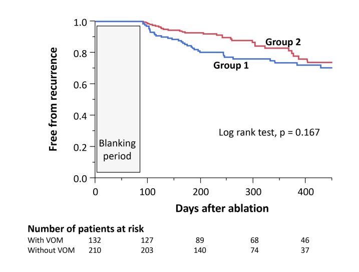
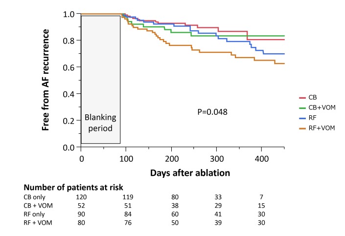
There was a significant association among the types of treatment received and the rate of freedom from AF as shown in [Figure 1A]. Kaplan-Meier curve estimates of the freedom from AF after approximately 450 days were 0.73 for Group 1 and 0.77 for Group 2 (p=0.167). Per subgroups of RF vs cryo 58 out of 90 (64.5 %) in Group RF, 92 out of 120 (79.6 %) in Group CB, 51 out of 80 (63.8 %) in Group RF + VOM-EI, and 43 out of 52 (82.7 %) in Group CB + VOM-EI patients. Among the treatment arms, However, recurrent AF occurred in a significantly larger proportion of patients in Groups RF, CB and C RF + VOM-EI, than in Group CB + VOM-EI, and those differences reached statistical significance [Figure 1B](p= 0.048). The comparison data between each treatment arm were as follows: Group RF vs. CB; p= 0.3091, Group RF vs. RF + VOM-EI; p= 0.2644, Group CB vs. RF + VOM-EI; p= 0.0033, Group CB vs. CB + VOM-EI; p= 0.5205, and Group RF + VOM-EI vs. CB + VOM-EI; p= 0.0292, Group RF + CB vs. Group RF + VOM-EI plus CB + VOM-EI; p= 0.1896. There were significant differences between Group CB and RF + VOM-EI, and between Groups RF + VOM-EI and CB + VOM-EI as shown in [Table 3].
Figure 1A. Kaplan-Meier curve comparing the freedom from AF/AT recurrence between Group 1 and 2.
After approximately 1-year of follow-up, there was no significant association between the treatment methods and survival (0=0.167).
Figure 1B. Kaplan-Meier curve comparing the freedom from AF/AT recurrence across the four study sub-groups.
After approximately 1-year of follow-up, there was significant differences between the treatment methods and survival (0=0.048), with the highest freedom from AF/AT seen in the CB ablation group with a PVI concomitant with a VOM-EI.
CB=cryoballoon, RF=radiofrequency, VOM=vein of Marshall, EI=ethanol infusion
Table 3. Comparison of the freedom from any atrial arrhythmias between each treatment arm.
RF=radiofrequency, CB=cryoballoon, VOM=ethanol infusion into the vein of Marshal
| p value | |||
|---|---|---|---|
| RF only | vs. | RF+VOM | 0.2644 |
| RF only | vs. | CB+VOM | 0.2381 |
| CB only | vs. | RF only | 0.3091 |
| CB only | vs. | RF+VOM | 0.0033 |
| CB only | vs. | CB+VOM | 0.5205 |
| RF+VOM | vs. | CB+VOM | 0.0292 |
| RF only and CB only | vs. | RF+VOM and CB+VOM | 0.1896 |
In the Group RF, CB, and CB + VOM-EI patients, recurrent AF was managed with a repeat catheter ablation using RF energy in 25 patients (Group RF), 12 patients (Group CB), and 9 patients (Group CB + VOM-EI), respectively, after 317 ± 48 days. Reconduction of >1 PV was observed in 21 patients (84 %) in Group RF, 3 patients (25 %) of Group CB, and 4 out of 9 (44.4 %) in Group CB + VOM-EI, and a single reconnection site was observed in 4 patients (16 %) in Group RF, 9 patients (75 %) in Group CB, and 5 out of 9 (55.6 %) in Group CB + VOM-EI, and a complete PVI could be accomplished in all patients as a result [Table 4].
Table 4. Data of the reconnections of the PVs among each treatment arm.
PV=pulmonary vein, LSPV=left superior PV, LIPV=left inferior PV, RSPV=right superior PV, RIPV=right inferior PV
| RF | CRYO | RF+VOM | CRYO+VOM | ||
|---|---|---|---|---|---|
| Re-do ablation (%) | 25/90 (28) | 12/120 (10) | 17/80 (21) | 9/52 (17) | |
| Reconnected PVs | P value | ||||
| LSPV (%) | 5 (20) | 2 (16.7) | 2 (11.8) | 2 (22.2) | 0.877 |
| LIPV (%) | 9 (36) | 4 (33.3) | 16 (94.1) | 4 (44.4) | 0.0009 |
| RSPV (%) | 5 (20) | 2 (16.7) | 1 (5.9) | 1 (11.1) | 0.62 |
| RIPV (%) | 8 (32) | 7 (58.3) | 4 (23.5) | 5 (55.6) | 0.159 |
| Reconnection of PV>1 PV (%) | 21/25 (84) | 3/12 (25) | 1/17 (6) | 4/9 (44) | |
| Single reconnection of PV (%) | 4/25 (16) | 9/12 (75) | 16/17 (94) | 5/9 (56) | |
| Endpoint (%) | 24/90 (26.7%) | 15/120 (12.5%) | 23/80 (28.8%) | 9/52 (17.3%) |
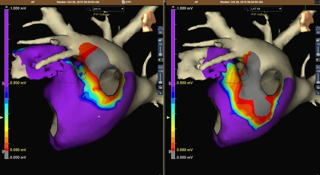
In contrast, in the Group RF + VOM-EI patients, reconnection sites of PVs were observed in 23/65 (35.4 %) in the left inferior PVs (16/23=69.6 %) predominantly at the inferior aspect of the LIPV where low voltage areas had already been created by the VOM-EI and no further touch-up RF applications had not been required for a successful PVI in the first session in Group RF + VOM-EI. In 3 patients in Group RF + VOM-EI (3.8 %), such low voltage areas created by the VOM-EI were found at the posterior antral wall of the left superior PV (LSPV) as shown in [Figure 2]. Of note, no AT whose arrhythmogenic substrate involved the MI area had never been observed in Groups RF + VOM-EI and CB + VOM-EI during the follow-up period.
Figure 2. Low voltage scar creation in the mitral isthmus area.

Left Panel shows the voltage map at baseline, prior to an ethanol injection into the vein of Marshall. Right Panel shows a map after the ethanol infusion, and it demonstrated low voltage areas covering the posterior antral wall of the left superior as well as inferior pulmonary vein along the vein of Marshall.
LAA= left atrial appendage, LSPV=left superior pulmonary vein, LIPV=left inferior pulmonary vein, VOM=vein of Marshall, CS=coronary sinus LSPV=left superior pulmonary vein, LIPV=left inferior pulmonary vein, RSPV=right superior pulmonary vein, RIPV=right inferior pulmonary vein.
Predictors of recurrence
We assessed the prognostic role of the ablation approach. In a univariate Cox regression analysis, the use of the CB during the ablation procedure (HR 0.31; 95 % CI 0.13-0.73; p= 0.005) and Diabetes Mellitus (HR 2.94; 95 % CI 0.84-10.29; p= 0.09) were significant predictors of AF recurrence. In the multivariate Cox regression analysis, the only significant predictor of an AF-free survival was the use of the CB for the ablation (HR 0.29; 95 % CI 0.12-0.69; p= 0.005). The use of a VOM-EI was not a predictor of success.
Safety outcomes
Pericardial tamponade requiring percutaneous drainage occurred in 3/210 (1.4 %) Group 2, and 1/123 (0.8 %) Group 1 patients. Tamponade occurred approximately 60 minutes after acomplishment of the successful PVI procedure. No instances of arterial injury, symptomatic thromboembolism, or esophageal injury were observed. However, sustained phrenic nerve palsy was provoked in one Group 1 patient during right-sided PVI with the CB, and an asymptomatic cerebral infarction was provoked in approximately 20 % of the CB treated patients as reported previously [20], [21]. Dissection of the CS was also provoked by the VOM-EI procedure in two Group 1 patients, and the EI could not be performed in those patients (Table 2).
Discussion
[21]We report a large, prospective, multicenter series of paroxysmal AF patients undergoing 4 different treatment strategies including the largest experience using a VOM-EI over a 12-month follow-up period.
Major findings
The main results of this study were: (1) Overall, the VOM-EI failed to significantly improve the clinical efficacy of the PVI for paroxysmal AF. (2) The success rate of RF combined with a VOM-EI was significantly lower than that of a CB alone (88 and 66 %, p= 0.0033). (3) The success rate of the MIB afforded by the VOM-EI was high and did not differ between Groups RF + VOM-EI and CB + VOM-EI. (4) Using the CB was the only predictor of an AF-free survival among all treatment arms.
We previously demonstrated that a VOM-EI was useful for treating perimitral atrial flutter by reliably achieving bidirectional MI block [15]. We were also able to show that there was regional parasympathetic denervation around the MI area [22]. These clinical aspects of the VOM-EI may be expected to facilitate the clinical efficacy of the ablation of AF.
The CB technology offers the possibility of the PVI with a single energy application as an alternative to a point-by-point RF current ablation [23], [24], [25], and the CB has been increasingly used for treating AF because of the relative technical simplicity and steeper learning curve [26]. However, the clinical results have been hampered by left-sided ATs in approximately 8~10 %, and those ATs were associated with the MI area [27], [28]. Macroreentrant tachycardia involving the mitral annulus-PMF causes 33 % to 60 % of post-PV isolation atrial flutters [29], [30]. Therefore, construction of MIB could be regarded as one of the necessary ablation procedures even in paroxysmal AF.
At 12-months of follow-up, approximately 65 % of the Group RF and 80 % of the Group CB patients were free of AF recurrences in the present study, and that was in line with the recent articles analysing the mid-term outcomes [4], [5], [27], [28]. However, no left-sided ATs have ever been documented in all four groups of patients during the follow-up period, and these results could be associated with the antiarrhythmic effects of preventiong AF recurrences by a VOM-EI procedure [8]. [9]. Lyan et al., reported that 26 out of 238 (10.9 %) AF patients underwent a redo ablation for AT. Nine out of 26 (34.6 %) patients had perimetral reentrant AT after the CB application. An additive EI-VOM might be beneficial in terms of preventing perimetral AT [31].
In Group RF + VOM-EI, reconnected PVs sites were observed in 20/52 (38.5 %) patients predominantly in the left inferior PVs where a VOM-EI had already created low voltage areas and no further touch-up RF applications were performed in the first session, because a successful PVI could be performed by applying RF energy at the antral area where the residual atrial electrograms remained. In general, the anatomical course of the VOM arises from the middle or proximal portion of the CS and runs toward the inferior aspect of the LIPV. Therefore, the antral atrial tissue at the inferior aspect of the LIPV is susceptible to be ablated by the VOM-EI. In those cases, our approach was to spare the RF energy applications in the area where no atrial electrograms were recorded from the tip of the ablation catheter for a successful PVI of the LIPV. In sporadic cases, such low voltage areas were detected at the posterior wall of the LSPV, and that kind of characteristic could be expected in cases that had a relatively long VOM coursing toward the LSPV area.
A high reconnection rate of the LIPV meant that the durability of the lesion around the PVs created by the VOM-EI was not sufficient. This might be the reason why the long-term clinical outcome of Group CB was significantly better than that of Group RF + VOM-EI. Considering that the VOM is an epicardial structure, the VOM-EI is expected to ablate the epicardium first and may not reach to the endocardial myocardium as effectively or as durably. CB ablation in the LIPV, when combined with a VOM-EI, did not have a high LIPV reconnection rate. It is possible that if RF energy applications had been performed at those “silent” areas, the rate of the freedom from AF in Group CB could have been comparable to that of Group RF. In addition, Guler et al., demonstrated that the CB provided additional substrate modification owing to the balloon and PV mismatch and could provide a lesion formation at the posterior wall of the left atrium, which is the most affected part of left atrium [32] That might inflience on the results of the present study.
A previous report demonstrated the necessity of a combined epicardial (inside CS) and endocardial RF catheter ablation of the VOM area was required to eliminate the VOM potentials [33]. In the present study, the success rate of constructing an MIB was 68 out of 80 (83 %) in Group RF + VOM-EI and 47 out of 52 (90 %) in Group CB + VOM-EI, respectively (p= 0.23). This was overall a higher success rate than usually reported in the literature [34] with a lower rate of RF application inside the CS as compared to the other study [33], and may highlight a unique utility of this procedure.
In this study, we failed to demonstrate any significant further improvement in the clinical efficacy in terms of preventing AF recurrences by adding a VOM-EI to the PVI by ablation using the CB or RF energy in patients with paroxysmal AF. According to the recent report, the one-year clinical outcome using the CB for the PVI in patients with persistent AF was not satisfactory [35]. Even though the acute success rate of the PVI was 100 %, the 1-year clinical success rate was 69 % with the use of the second-generation CB. Of note, left-sided ATs were documented in 13 % of the study patients. Those ATs could have been prevented by adding a VOM-EI procedure to the PVI. A VOM-EI has multifactorial effects for facilitating the clinical efficacy of the catheter ablation of AF. As we previously reported, the VOM-EI was able to abolish the non-pulmonary vein ectopy, which degenerated into AF by the VOM-EI [36]. In addition, this procedure might be expected to provoke significant parasympathetic denervation effects in the left atrium, which could contribute to the improvement in the success rate of vagally mediated AF [9], [37]. A durable PVI in addition to a VOM-EI might be expected to improve the rate of freedom from AF recurrences, and further investigation will be required.
Limitations
This study constituted a nonrandomized analysis of consecutive patients, including our initial patients treated with the second-generation CB device. However, all operators were well trained in CB ablation and beyond the learning curve, minimizing any time-dependent confounders. (2) The group sizes were small. However, a number of the efficacy parameters differed significantly between the groups. (3) The assignment of the study patients into the four groups might not be appropriate, because grouping of patients depended on whether the VOM was present or not. (4) A comparison of PVI vs. PVI plus VOM-EI might be unfair due to the additive effects of creating a lesion in the mitral isthmus region. (5) The evaluation of the success and complication rates was complex. (6) A procedure trying to construct the MIB was not performed in Group RF+ VOM-EI, and this might have affected the rate of freedom from AF/ AT. However, all recurrent arrhythmias were AF instead of left-sided ATs rotating along the mitral annulus. Therefore, it was unlikely that the absence of the MIB could affect the clinical results in Group RF + VOM-EI. (7) By definition, successful maintenance of sinus rhythm strongly relies on the length of the follow-up, which varies from center to center. Because we reported on the 12-month follow-up data from outpatient clinic visits or telephone interviews, we could not exclude that AF recurrent episodes could have been missed in some patients. (8) The examinations to evaluate the efficacy of ablation procedure such as periodic 12-lead ECG and Holter monitoring recording might not be sufficient to strictly investigate the clinical efficacy of each ablation modality. (9) There was no comparison between a control group in the present study.
Conclusions
Overall, the VOM-EI failed to demonstrate any significant improvement in the ablation long-term results of paroxysmal AF. However, CB ablation combined with a VOM-EI had a significantly improved outcome compared to a PVI with an RF ablation combined with a VOM-EI.
References
- 1.Cappato Riccardo, Calkins Hugh, Chen Shih-Ann, Davies Wyn, Iesaka Yoshito, Kalman Jonathan, Kim You-Ho, Klein George, Packer Douglas, Skanes Allan. Worldwide survey on the methods, efficacy, and safety of catheter ablation for human atrial fibrillation. Circulation. 2005 Mar 08;111 (9):1100–5. doi: 10.1161/01.CIR.0000157153.30978.67. [DOI] [PubMed] [Google Scholar]
- 2.Neumann Thomas, Vogt Jürgen, Schumacher Burghard, Dorszewski Anja, Kuniss Malte, Neuser Hans, Kurzidim Klaus, Berkowitsch Alexander, Koller Marcus, Heintze Johannes, Scholz Ursula, Wetzel Ulrike, Schneider Michael A E, Horstkotte Dieter, Hamm Christian W, Pitschner Heinz-Friedrich. Circumferential pulmonary vein isolation with the cryoballoon technique results from a prospective 3-center study. J. Am. Coll. Cardiol. 2008 Jul 22;52 (4):273–8. doi: 10.1016/j.jacc.2008.04.021. [DOI] [PubMed] [Google Scholar]
- 3.Hakalahti Antti, Biancari Fausto, Nielsen Jens Cosedis, Raatikainen M J Pekka. Radiofrequency ablation vs. antiarrhythmic drug therapy as first line treatment of symptomatic atrial fibrillation: systematic review and meta-analysis. Europace. 2015 Mar;17 (3):370–8. doi: 10.1093/europace/euu376. [DOI] [PubMed] [Google Scholar]
- 4.Cosedis Nielsen Jens, Johannessen Arne, Raatikainen Pekka, Hindricks Gerhard, Walfridsson Håkan, Kongstad Ole, Pehrson Steen, Englund Anders, Hartikainen Juha, Mortensen Leif Spange, Hansen Peter Steen. Radiofrequency ablation as initial therapy in paroxysmal atrial fibrillation. N. Engl. J. Med. 2012 Oct 25;367 (17):1587–95. doi: 10.1056/NEJMoa1113566. [DOI] [PubMed] [Google Scholar]
- 5.Morillo Carlos A, Verma Atul, Connolly Stuart J, Kuck Karl H, Nair Girish M, Champagne Jean, Sterns Laurence D, Beresh Heather, Healey Jeffrey S, Natale Andrea. Radiofrequency ablation vs antiarrhythmic drugs as first-line treatment of paroxysmal atrial fibrillation (RAAFT-2): a randomized trial. JAMA. 2014 Feb 19;311 (7):692–700. doi: 10.1001/jama.2014.467. [DOI] [PubMed] [Google Scholar]
- 6.Jourda François, Providencia Rui, Marijon Eloi, Bouzeman Abdeslam, Hireche Hassiba, Khoueiry Ziad, Cardin Christelle, Combes Nicolas, Combes Stéphane, Boveda Serge, Albenque Jean-Paul. Contact-force guided radiofrequency vs. second-generation balloon cryotherapy for pulmonary vein isolation in patients with paroxysmal atrial fibrillation-a prospective evaluation. Europace. 2015 Feb;17 (2):225–31. doi: 10.1093/europace/euu215. [DOI] [PubMed] [Google Scholar]
- 7.Luik Armin, Radzewitz Andrea, Kieser Meinhard, Walter Marlene, Bramlage Peter, Hörmann Patrick, Schmidt Kerstin, Horn Nicolas, Brinkmeier-Theofanopoulou Maria, Kunzmann Kevin, Riexinger Tobias, Schymik Gerhard, Merkel Matthias, Schmitt Claus. Cryoballoon Versus Open Irrigated Radiofrequency Ablation in Patients With Paroxysmal Atrial Fibrillation: The Prospective, Randomized, Controlled, Noninferiority FreezeAF Study. Circulation. 2015 Oct 06;132 (14):1311–9. doi: 10.1161/CIRCULATIONAHA.115.016871. [DOI] [PMC free article] [PubMed] [Google Scholar]
- 8.Valderrábano Miguel, Liu Xiushi, Sasaridis Christine, Sidhu Jasvinder, Little Stephen, Khoury Dirar S. Ethanol infusion in the vein of Marshall: Adjunctive effects during ablation of atrial fibrillation. Heart Rhythm. 2009 Nov;6 (11):1552–8. doi: 10.1016/j.hrthm.2009.07.036. [DOI] [PMC free article] [PubMed] [Google Scholar]
- 9.Báez-Escudero José L, Keida Takehiko, Dave Amish S, Okishige Kaoru, Valderrábano Miguel. Ethanol infusion in the vein of Marshall leads to parasympathetic denervation of the human left atrium: implications for atrial fibrillation. J. Am. Coll. Cardiol. 2014 May 13;63 (18):1892–901. doi: 10.1016/j.jacc.2014.01.032. [DOI] [PMC free article] [PubMed] [Google Scholar]
- 10.Valderrábano Miguel, Chen Harvey R, Sidhu Jasvinder, Rao Liyun, Ling Yuesheng, Khoury Dirar S. Retrograde ethanol infusion in the vein of Marshall: regional left atrial ablation, vagal denervation and feasibility in humans. Circ Arrhythm Electrophysiol. 2009 Feb;2 (1):50–6. doi: 10.1161/CIRCEP.108.818427. [DOI] [PMC free article] [PubMed] [Google Scholar]
- 11.Keida Takehiko, Fujita Masaki, Okishige Kaoru, Takami Mitsuaki. Elimination of non-pulmonary vein ectopy by ethanol infusion in the vein of Marshall. Heart Rhythm. 2013 Sep;10 (9):1354–6. doi: 10.1016/j.hrthm.2013.07.019. [DOI] [PubMed] [Google Scholar]
- 12.Shah Dipen C, Lambert Hendrik, Nakagawa Hiroshi, Langenkamp Arne, Aeby Nicolas, Leo Giovanni. Area under the real-time contact force curve (force-time integral) predicts radiofrequency lesion size in an in vitro contractile model. J. Cardiovasc. Electrophysiol. 2010 Sep;21 (9):1038–43. doi: 10.1111/j.1540-8167.2010.01750.x. [DOI] [PubMed] [Google Scholar]
- 13.Squara Fabien, Latcu Decebal Gabriel, Massaad Youssef, Mahjoub Marouane, Bun Sok-Sithikun, Saoudi Nadir. Contact force and force-time integral in atrial radiofrequency ablation predict transmurality of lesions. Europace. 2014 May;16 (5):660–7. doi: 10.1093/europace/euu068. [DOI] [PubMed] [Google Scholar]
- 14.Okishige Kaoru, Aoyagi Hideshi, Kawaguchi Naohiko, Katoh Nobutaka, Yamashita Mitsumi, Nakamura Tomofumi, Kurabayashi Manabu, Suzuki Hidetoshi, Asano Mitsutoshi, Gotoh Kentarou, Shimura Tsukasa, Yamauchi Yasuteru, Kanazawa Toshirou, Sasano Tetsuo, Hirao Kenzo. Novel method for earlier detection of phrenic nerve injury during cryoballoon applications for electrical isolation of pulmonary veins in patients with atrial fibrillation. Heart Rhythm. 2016 Sep;13 (9):1810–6. doi: 10.1016/j.hrthm.2016.05.003. [DOI] [PubMed] [Google Scholar]
- 15.Báez-Escudero José L, Morales Percy Francisco, Dave Amish S, Sasaridis Christine M, Kim Young-Hoon, Okishige Kaoru, Valderrábano Miguel. Ethanol infusion in the vein of Marshall facilitates mitral isthmus ablation. Heart Rhythm. 2012 Aug;9 (8):1207–15. doi: 10.1016/j.hrthm.2012.03.008. [DOI] [PMC free article] [PubMed] [Google Scholar]
- 16.Nakamura Tomofumi, Aoyagi Hideshi, Yamauchi Yasuteru, Okishige Kaoru. Coronary Sinus Stenosis as Collateral Damage of Radiofrequency Ablation in Patients With Atrial Fibrillation. JACC Clin Electrophysiol. 2016 Nov;2 (6):759–761. doi: 10.1016/j.jacep.2016.08.002. [DOI] [PubMed] [Google Scholar]
- 17.Jaïs Pierre, Hocini Mélèze, Hsu Li-Fern, Sanders Prashanthan, Scavee Christophe, Weerasooriya Rukshen, Macle Laurent, Raybaud Florence, Garrigue Stéphane, Shah Dipen C, Le Metayer Philippe, Clémenty Jacques, Haïssaguerre Michel. Technique and results of linear ablation at the mitral isthmus. Circulation. 2004 Nov 09;110 (19):2996–3002. doi: 10.1161/01.CIR.0000146917.75041.58. [DOI] [PubMed] [Google Scholar]
- 18.Camm A John, Lip Gregory Y H, De Caterina Raffaele, Savelieva Irene, Atar Dan, Hohnloser Stefan H, Hindricks Gerhard, Kirchhof Paulus. 2012 focused update of the ESC Guidelines for the management of atrial fibrillation: an update of the 2010 ESC Guidelines for the management of atrial fibrillation--developed with the special contribution of the European Heart Rhythm Association. Europace. 2012 Oct;14 (10):1385–413. doi: 10.1093/europace/eus305. [DOI] [PubMed] [Google Scholar]
- 19.Okishige Kaoru, Aoyagi Hideshi, Ihara Kensuke, Iwai Shinsuke, Nakamura Tomofumi, Yamashita Mitsumi, Katoh Nobutaka, Hasegawa Tomoaki, Kawaguchi Naohiko, Keida Takehiko, Sasano Tetsuo, Hirao Kenzo. Reappraisal of the clinical implications of adenosine triphosphate in terms of the prediction of reconnection sites in cases with electrical isolation of the pulmonary veins. J Interv Card Electrophysiol. 2015 Nov;44 (2):171–8. doi: 10.1007/s10840-015-0019-z. [DOI] [PubMed] [Google Scholar]
- 20.Okishige Kaoru, Nakamura Tomofumi, Aoyagi Hideshi, Kawaguchi Naohiko, Yamashita Mitsumi, Kurabayashi Manabu, Suzuki Hidetoshi, Asano Mitsutoshi, Shimura Tsukasa, Yamauchi Yasuteru, Sasano Tetsuo, Hirao Kenzo. Comparative study of hemorrhagic and ischemic complications among anticoagulants in patients undergoing cryoballoon ablation for atrial fibrillation. J Cardiol. 2017 Jan;69 (1):11–15. doi: 10.1016/j.jjcc.2016.04.009. [DOI] [PubMed] [Google Scholar]
- 21.Nakamura Tomofumi, Okishige Kaoru, Kanazawa Toshiro, Yamashita Mitsumi, Kawaguchi Naohiko, Kato Nobutaka, Aoyagi Hideshi, Yamauchi Yasuteru, Sasano Tetsuo, Hirao Kenzo. Incidence of silent cerebral infarctions after catheter ablation of atrial fibrillation utilizing the second-generation cryoballoon. Europace. 2017 Oct 01;19 (10):1681–1688. doi: 10.1093/europace/euw191. [DOI] [PubMed] [Google Scholar]
- 22.Báez-Escudero José L, Keida Takehiko, Dave Amish S, Okishige Kaoru, Valderrábano Miguel. Ethanol infusion in the vein of Marshall leads to parasympathetic denervation of the human left atrium: implications for atrial fibrillation. J. Am. Coll. Cardiol. 2014 May 13;63 (18):1892–901. doi: 10.1016/j.jacc.2014.01.032. [DOI] [PMC free article] [PubMed] [Google Scholar]
- 23.Andrade Jason G, Khairy Paul, Guerra Peter G, Deyell Marc W, Rivard Lena, Macle Laurent, Thibault Bernard, Talajic Mario, Roy Denis, Dubuc Marc. Efficacy and safety of cryoballoon ablation for atrial fibrillation: a systematic review of published studies. Heart Rhythm. 2011 Sep;8 (9):1444–51. doi: 10.1016/j.hrthm.2011.03.050. [DOI] [PubMed] [Google Scholar]
- 24.Kojodjojo Pipin, O'Neill Mark D, Lim Phang Boon, Malcolm-Lawes Louisa, Whinnett Zachary I, Salukhe Tushar V, Linton Nicholas W, Lefroy David, Mason Anthony, Wright Ian, Peters Nicholas S, Kanagaratnam Prapa, Davies D Wyn. Pulmonary venous isolation by antral ablation with a large cryoballoon for treatment of paroxysmal and persistent atrial fibrillation: medium-term outcomes and non-randomised comparison with pulmonary venous isolation by radiofrequency ablation. Heart. 2010 Sep;96 (17):1379–84. doi: 10.1136/hrt.2009.192419. [DOI] [PMC free article] [PubMed] [Google Scholar]
- 25.Schmidt Martin, Dorwarth Uwe, Andresen Dietrich, Brachmann Johannes, Kuck Karlheinz, Kuniss Malte, Willems Stephan, Deneke Thomas, Tebbenjohanns Jürgen, Gerds-Li Jin-Hong, Spitzer Stefan, Senges Jochen, Hochadel Matthias, Hoffmann Ellen. German ablation registry: Cryoballoon vs. radiofrequency ablation in paroxysmal atrial fibrillation--One-year outcome data. Heart Rhythm. 2016 Apr;13 (4):836–44. doi: 10.1016/j.hrthm.2015.12.007. [DOI] [PubMed] [Google Scholar]
- 26.Chun Kyoung-Ryul Julian, Schmidt Boris, Metzner Andreas, Tilz Roland, Zerm Thomas, Köster Ilka, Fürnkranz Alexander, Koektuerk Buelent, Konstantinidou Melanie, Antz Matthias, Ouyang Feifan, Kuck Karl Heinz. The 'single big cryoballoon' technique for acute pulmonary vein isolation in patients with paroxysmal atrial fibrillation: a prospective observational single centre study. Eur. Heart J. 2009 Mar;30 (6):699–709. doi: 10.1093/eurheartj/ehn570. [DOI] [PMC free article] [PubMed] [Google Scholar]
- 27.Irfan Ghazala, de Asmundis Carlo, Mugnai Giacomo, Poelaert Jan, Verborgh Christian, Umbrain Vincent, Beckers Stefan, Hacioglu Ebru, Hunuk Burak, Velagic Vedran, Stroker Erwin, Brugada Pedro, Chierchia Gian-Battista. One-year follow-up after second-generation cryoballoon ablation for atrial fibrillation in a large cohort of patients: a single-centre experience. Europace. 2016 Jul;18 (7):987–93. doi: 10.1093/europace/euv365. [DOI] [PubMed] [Google Scholar]
- 28.Straube Florian, Dorwarth Uwe, Hartl Stefan, Bunz Benedikt, Wankerl Michael, Ebersberger Ullrich, Hoffmann Ellen. Outcome of paroxysmal atrial fibrillation ablation with the cryoballoon using two different application times: the 4- versus 3-min protocol. J Interv Card Electrophysiol. 2016 Mar;45 (2):169–77. doi: 10.1007/s10840-015-0084-3. [DOI] [PubMed] [Google Scholar]
- 29.Deisenhofer Isabel, Estner Heidi, Zrenner Bernhard, Schreieck Juergen, Weyerbrock Sonja, Hessling Gabriele, Scharf Konstanze, Karch Martin R, Schmitt Claus. Left atrial tachycardia after circumferential pulmonary vein ablation for atrial fibrillation: incidence, electrophysiological characteristics, and results of radiofrequency ablation. Europace. 2006 Aug;8 (8):573–82. doi: 10.1093/europace/eul077. [DOI] [PubMed] [Google Scholar]
- 30.Chugh Aman, Oral Hakan, Lemola Kristina, Hall Burr, Cheung Peter, Good Eric, Tamirisa Kamala, Han Jihn, Bogun Frank, Pelosi Frank, Morady Fred. Prevalence, mechanisms, and clinical significance of macroreentrant atrial tachycardia during and following left atrial ablation for atrial fibrillation. Heart Rhythm. 2005 May;2 (5):464–71. doi: 10.1016/j.hrthm.2005.01.027. [DOI] [PubMed] [Google Scholar]
- 31.Lyan Evgeny, Yalin Kivanc, Abdin Amr, Sawan Noureddin, Liosis Spyridon, Lange Stefan A, Eitel Ingo, Heeger Christian-H, Meyer-Saraei Roza, Eitel Charlotte, Tilz Roland Richard. Mechanism, underlying substrate and predictors of atrial tachycardia following atrial fibrillation ablation using the second-generation cryoballoon. J Cardiol. 2019 Jun;73 (6):497–506. doi: 10.1016/j.jjcc.2019.02.006. [DOI] [PubMed] [Google Scholar]
- 32.Guler Tümer Erdem, Aksu Tolga, Yalin Kivanc, Golcuk Sukriye Ebru, Mutluer Ferit Onur, Bozyel Serdar. Combined Cryoballoon and Radiofrequency Ablation Versus Radiofrequency Ablation Alone for Long-Standing Persistent Atrial Fibrillation. Am. J. Med. Sci. 2017 Dec;354 (6):586–596. doi: 10.1016/j.amjms.2017.08.010. [DOI] [PubMed] [Google Scholar]
- 33.Katritsis D, Ioannidis J P, Anagnostopoulos C E, Sarris G E, Giazitzoglou E, Korovesis S, Camm A J. Identification and catheter ablation of extracardiac and intracardiac components of ligament of Marshall tissue for treatment of paroxysmal atrial fibrillation. J. Cardiovasc. Electrophysiol. 2001 Jul;12 (7):750–8. doi: 10.1046/j.1540-8167.2001.00750.x. [DOI] [PubMed] [Google Scholar]
- 34.Jaïs Pierre, Hocini Mélèze, Hsu Li-Fern, Sanders Prashanthan, Scavee Christophe, Weerasooriya Rukshen, Macle Laurent, Raybaud Florence, Garrigue Stéphane, Shah Dipen C, Le Metayer Philippe, Clémenty Jacques, Haïssaguerre Michel. Technique and results of linear ablation at the mitral isthmus. Circulation. 2004 Nov 09;110 (19):2996–3002. doi: 10.1161/01.CIR.0000146917.75041.58. [DOI] [PubMed] [Google Scholar]
- 35.Lemes Christine, Wissner Erik, Lin Tina, Mathew Shibu, Deiss Sebastian, Rillig Andreas, Heeger Christian, Wohlmuth Peter, Reissmann Bruno, Tilz Roland, Ouyang Feifan, Kuck Karl-Heinz, Metzner Andreas. One-year clinical outcome after pulmonary vein isolation in persistent atrial fibrillation using the second-generation 28 mm cryoballoon: a retrospective analysis. Europace. 2016 Feb;18 (2):201–5. doi: 10.1093/europace/euv092. [DOI] [PubMed] [Google Scholar]
- 36.Keida Takehiko, Fujita Masaki, Okishige Kaoru, Takami Mitsuaki. Elimination of non-pulmonary vein ectopy by ethanol infusion in the vein of Marshall. Heart Rhythm. 2013 Sep;10 (9):1354–6. doi: 10.1016/j.hrthm.2013.07.019. [DOI] [PubMed] [Google Scholar]
- 37.Pappone Carlo, Santinelli Vincenzo, Manguso Francesco, Vicedomini Gabriele, Gugliotta Filippo, Augello Giuseppe, Mazzone Patrizio, Tortoriello Valter, Landoni Giovanni, Zangrillo Alberto, Lang Christopher, Tomita Takeshi, Mesas Cézar, Mastella Elio, Alfieri Ottavio. Pulmonary vein denervation enhances long-term benefit after circumferential ablation for paroxysmal atrial fibrillation. Circulation. 2004 Jan 27;109 (3):327–34. doi: 10.1161/01.CIR.0000112641.16340.C7. [DOI] [PubMed] [Google Scholar]


