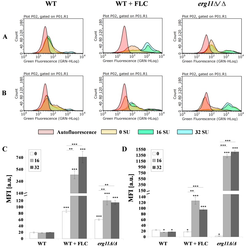
Figure 5.
Exposure of (A,C) chitin and (B,D) β-glucan in C. albicans CAF2-1 (WT) or KS028 (erg11Δ/Δ) strains grown for 24h in YPD without and the addition of 16 or 32 µg/mL surfactin (0 SU, yellow; 16 SU, green and 32 SU, blue). The CAF2-1 strain was simultaneously treated with 1 µg/mL fluconazole (FLC) and SU. In each case, cells were fixed and stained with Fc-hDectin-1 and Alexa Fluor 448-conjugated anti-human IgG Fc antibodies or wheat germ agglutinin conjugated with FITC (WGA-FITC) and quantified by FACS (A,B). All results were compared to unstained CAF2-1 or KS028 cells (autofluorescence, red). Presented data are representative of three independent experiments. Median fluorescence intensities (MFIs) (C,D) were quantified for all three experiments. Autofluorescence of either CAF2-1 or KS028 unstained cells was subtracted. Statistical analyses were performed relative to control experiments using CAF2-1 untreated with SU (above bars) or in the case of WT + FLC and erg11∆/∆ additionally between SU-treated cells (16SU or 32SU) or SU-nontreated (0SU) (above lines) (*, P < 0.05; **, P < 0.01; ***, P < 0.001).

