Abstract
The Qinghai-Tibetan Plateau Area (QTPA) is a plateau with the highest average altitude, located in Northwestern China. There is a risk for interspecies disease transmission, such as spotted fever rickettsioses. However, information on the molecular characteristics of the spotted fever group (SFG) Rickettsia spp. in the area is limited. This study performed screenings, and detected the DNA of human pathogen, SFG Rickettsia spp., with 11.3% (25/222) infection rates in yaks (Bos grunniens). BLASTn analysis revealed that the Rickettsia sequences obtained shared 94.3–100% identity with isolates of Rickettsia spp. from ticks in China. One Rickettsia sequence (MN536161) had 100% nucleotide identity to two R. raoultii isolates from Chinese Homo sapiens, and one isolate from Qinghai Dermacentor silvarum. Meanwhile, another Rickettsia sequence (MN536157) shared 99.1–99.5% identity to one isolate from Dermacentor spp. in China. Furthermore, the phylogenetic analysis of SFG Rickettsia spp. ompA gene revealed that these two sequences obtained from yaks in the present study grouped with the R. slovaca and R. raoultii clades with isolates identified from Dermacentor spp. and Homo sapiens. Our findings showed the first evidence of human pathogen DNA, SFG Rickettsia spp., from animals, in the QTPA.
Keywords: fever rickettsioses, human SFG Rickettsia, Qinghai-Tibetan Plateau, yak
1. Introduction
Rickettsioses are a known infectious diseases around the world, caused by obligate intracellular bacteria, the spotted fever group (SFG), Rickettsia [1,2,3]. Recently, the SFG Rickettsia species has caused diseases with varying clinical presentations in humans, domestic animals, and wildlife, and these diseases are considered an emerging global threat [1,4].
In China, eleven SFG Rickettsia species, Rickettsia sibirica, Rickettsia heilongjiangensis, Rickettsia aeschlimannii, Rickettsia conorii, Rickettsia felis, Rickettsia massiliae, Rickettsia monacensis, Rickettsia rickettsia, Candidatus Rickettsia jingxinensis, Rickettsia raoultii and Rickettsia slovaca, have been identified in tick vectors, domestic animals and wildlife [5,6,7,8,9,10,11,12,13]. Several Rickettsia genotypes have been characterized as etiological factors of human rickettsiosis in the country, such as R. heilongjiangensis [14,15], R. sibirica subsp. sibirica BJ-90 [16], R. raoultii [17,18,19], Rickettsia japonica [20,21], Rickettsia sp. XY99 [22], and Candidatus Rickettsia tarasevichiae [23]. These findings suggest that rickettsioses are emerging zoonoses in China.
The Qinghai-Tibetan Plateau Area (QTPA) is the largest plateau on the planet [24]. There is a special climate, comprised of a lower average annual temperature and rainfall that fluctuates, therefore, a variety of unique livestock are grazed there [25]. Among these livestock, the yak (Bos grunniens), which belongs to the bovine species, can survive in extreme environmental conditions, such as cold, harsh, and oxygen-poor [26]. The Qinghai plateau, located on the northeastern side of the QTPA, has a unique and vigorous natural ecosystem because of its high altitude, cold climate, and oxygen deficiency [27]. There is an abundant yak genetic resource with more than five million individuals in the Qinghai province [27]. Previous studies revealed that Qinghai was considered one of the origins and domestication locales for yaks based on mitochondrial DNA analyses [28].
Among the Rickettsia species, there are several important emerging pathogens in humans and animals. Although one study confirmed the infection of SFG Rickettsia in ticks from Qinghai province, northwestern China [27], information on the infection and molecular characteristics of these pathogens in humans and yaks in the province are still limited. Therefore, in this study, we screened yaks in Qinghai plateau for the existence of human pathogens and found that the DNA of Rickettsia was present in yak blood samples.
2. Results
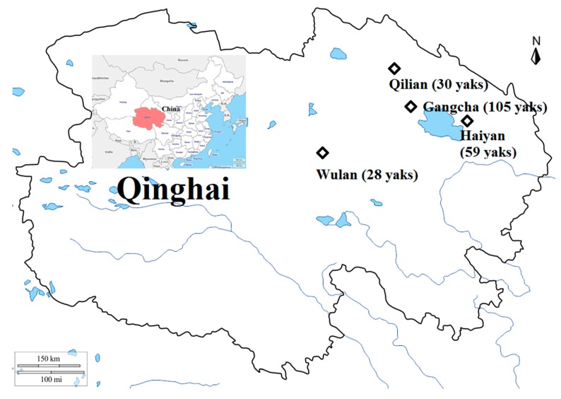
In the present study, a total of 222 blood samples were collected from apparently healthy yaks in four counties on the Qinghai plateau, including Wulan (36°19′-37°20′ N and 97°01′-99°27′E, n = 28), Qilian (37°25′-39°05′N and 98°05′-101°02′E, n = 30), Haiyan (36°44′-37°39′N and 100°23′-101°20′E, n = 59), and Gangcha (36°58′-38°04′N and 99°20′-100°37′E, n = 105) (Figure 1). One pasture was chosen from each sampling site, and in each pasture, 10–20% of the stock was randomly selected for blood sampling from March to May 2018. Geographically, most of the livestock is raised in the northeastern pastoral region of the province. PCR screening based on outer membrane protein A (ompA) gene revealed that the overall infection rate was 11.3% (25/222) for SFG Rickettsia spp. (Table 1). Furthermore, from the four selected areas, the infection rates of SFG Rickettsia spp. in yaks are 10.7% (3/28) in Wulan, 13.3% (4/30) in Qilian, 5.1% (3/59) in Haiyan, and 14.3% (15/105) in Gangcha. The infection rate of SFG Rickettsia spp. in yaks from four areas was not significantly different (p > 0.05).
Figure 1.
Map of Qinghai and China showing sampling areas and the number of animals indicated by the black quadrilateral. The map was generated using GIMP 2.8.10 (https://www. gimp.org).
Table 1.
The infection rate of spotted fever group (SFG) Rickettsia in yaks.
| Rickettsia spp. | Infection Rate (%) in Each Current Area | ||||
|---|---|---|---|---|---|
| Wulan (n = 28) | Qilian (n = 30) | Haiyan (n = 59) | Gangcha (n = 105) | Total (n = 222) | |
| Total positive | 3 (10.7) | 4 (13.3) | 3 (5.1) | 15 (14.3) | 25 (11.3) |
| Negative samples | 25 (89.3) | 26 (86.7) | 56 (94.9) | 90 (85.7) | 197 (88.7) |
Furthermore, a total of 25 SFG Rickettsia spp. sequences based on ompA gene were generated in the present study. Sequence analysis indicated that the SFG Rickettsia spp. infecting the yaks shared 94.3–100% identities to identified R. raoultii or R. slovaca isolates, as shown in Table 2. Meanwhile, SFG Rickettsia spp. sequences based on ompA gene were of three sizes (208, 209, and 212 bp). Additionally, BLASTn analysis of the ompA gene showed that the Rickettsia spp. obtained in this study had 85.4% to 100% identities to either sequence. Interestingly, one Rickettsia sequence (MN536161) obtained in the present study was 100% nucleotide identical to two R. raoultii isolates from Chinese Homo sapiens (JX945525 and KY474580), and three R. raoultii isolates from Chinese Dermacentor spp. (KF003015, KT899076 and MG228272) which include one isolate of R. raoultii from Dermacentor silvarum in Qinghai, China (MG228272). Furthermore, another Rickettsia spp. ompA sequence (MN536157) shared 99.5% identity to one R. slovaca isolate from Dermacentor marginatus (MF002535), and 99.1% identity to another R. slovaca isolate from D. silvarum (JN400402) in China. The five representative sequences obtained were submitted to GenBank and shown in Table 2.
Table 2.
DNA sequences obtained in this study.
| Obtained DNA Sequence | The Cosest Blastn Match | ||||||
|---|---|---|---|---|---|---|---|
| Pathogen | Gene | Accession Number | Length (bp) | Sequencing | Identity (%) | Species | Accession Number (Host, Country) |
| Rickettsia spp. | ompA | MN536157 | 212 | Rickettsia | 99.5 | R. slovaca | MF002535 (tick, China) |
| MN536158 | 209 | Rickettsia | 99.5 | R. raoultii | MF511260 (tick, China) | ||
| MN536159 | 208 | Rickettsia | 94.3 | R. raoultii | MN450415 (tick, China) | ||
| MN536160 | 209 | Rickettsia | 100 | R. raoultii | MG811700 (tick, China) | ||
| MN536161 | 209 | Rickettsia | 100 | R. raoultii | KY474580 (human, China) | ||
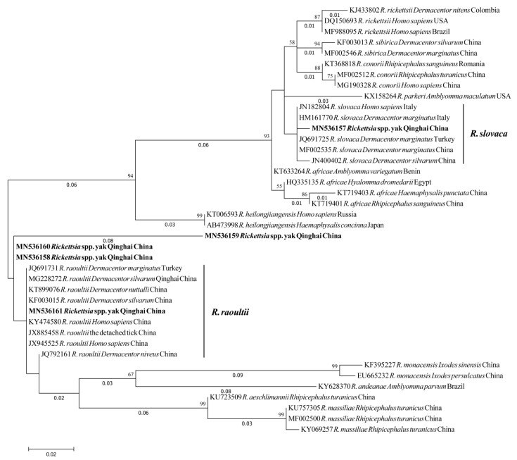
The phylogenetic analysis of SFG Rickettsia spp. ompA gene revealed that two sequences obtained from yaks in the present study (MN536157 and MN536161) grouped with the R. slovaca and R. raoultii clades with isolates identified from Dermacentor spp. and Homo sapiens from China, Turkey, and Italy (Figure 2).
Figure 2.
Molecular Phylogenetic analysis by Maximum Likelihood method based on Rickettsia spp. ompA partial sequences. Sequences from current study are marked in bold. The evolutionary history was inferred by using the Maximum Likelihood method based on the Kimura 2-parameter model. The tree with the highest log likelihood (−864.84) is shown. The percentage of trees in which the associated taxa clustered together is shown next to the branches. Initial tree(s) for the heuristic search were obtained automatically by applying Neighbor-Join and BioNJ algorithms to a matrix of pairwise distances estimated using the Maximum Composite Likelihood approach, and then selecting the topology with superior log likelihood value. A discrete Gamma distribution was used to model evolutionary rate differences among sites (5 categories (+G, parameter = 0.5571)). The tree is drawn to scale, with branch lengths measured in the number of substitutions per site (next to the branches). The analysis involved 40 nucleotide sequences. Codon positions included were 1st+2nd+3rd+Noncoding. All positions containing gaps and missing data were eliminated. There were a total of 186 positions in the final dataset. Evolutionary analyses were conducted in MEGA7.
3. Discussion
This investigation reported the DNA detection of human Rickettsia species in the Qinghai-Tibetan Plateau Area. A previous study reported that SFG rickettsiae, including R. raoultii, R. sibirica subspecies sibirica, Candidatus R. tibetan, and Candidatus R. gannanii Y27 and F107 had a high overall infection rate in ticks from Qinghai, the sampling province of this study [27]. Our results document the first direct evidence of DNA detection of SFG Rickettsia pathogens in animals in this area. Importantly, sequencing analysis showed that two DNA sequences were 99.5% identity to R. slovaca and 100% nucleotide identical to R. raoultii isolates identified in China.
In China, R. raoultii infection cases with increasing numbers have been reported in humans [17,18,19,29]. These rickettsiose patients present not only lethargy, fever, and headache, but also neurological abnormalities [19]. Among these reports, a previous case which identified R. raoultii with 100% sequence identity between the isolate from patient and detached tick is very interesting [17], as the R. raoultii sequence obtained in this study also shared 100% identity to those previously identified from that patient (JX945525) and detached tick (JX885458). Furthermore, previous findings showed that R. raoultii was closely related with Dermacentor spp. [30]. The characterization of R. raoultii in yaks in the current study might indicate successful pathogen transmission from this tick species to yak, since one of the obtained Rickettsia sequences was 100% identical to those previously identified in Dermacentor spp. in Qinghai [27]. On the other hand, this Rickettsia sequence obtained also showed 100% identity with another isolate from human and four isolates from Dermacentor spp. from China. This suggests that humans and animals are both susceptible to this pathogen due to exposure to the same tick vectors.
R. slovaca, a member of human SFG rickettsiae, was first identified in D. marginatus in 1968 in Slovakia [31], and is now considered as the causative factor of tick-borne lymphadenopathy [32]. This human pathogen was reported for the first time in China in 2012 in D. silvarum ticks in Xinjiang province, which is the adjacent province to Qinghai [33]. In addition, R. slovaca was molecularly detected in shepherd blood DNA in Xinjiang Province [19]. However, compared to R. raoultii infections in humans and tick vectors in China, R. slovaca infection rate appears to be low and mild [17,33]. Previously, no reports have identified R. slovaca in ticks in Qinghai. In the present study, despite identifying only one yak positive, the DNA detection closely associated with R. slovaca provides evidence that this pathogen may exist in Qinghai.
In summary, this study found human pathogens, SFG Rickettsia spp., circulating in yaks in Qinghai of the Qinghai-Tibetan Plateau, China. The phylogenetic analysis of ompA gene revealed that sequences obtained from yaks in the present study were closely related to R. slovaca and R. raoultii isolates identified from Dermacentor spp. and Homo sapiens. Therefore, future studies are needed to provide data to the suspected importance of these ticks in transmitting the human pathogens to animals and humans in this plateau area.
4. Materials and Methods
Yak blood samples were collected into tubes, including anticoagulant (EDTA), and genomic DNA. The blood was extracted using and according to the manufacturer’s manual of the QIAamp DNA Blood Mini Kit (QIAGEN, Hilden, Germany). All yak DNA samples were screened with genus-specific primers (forward primer 5’–TGCGCCTTCGAGTTGTACAAGAG–3’ and reverse primer 5’–GACGGGTTGCRTAGGCTGAC–3’) based on Rickettsia spp. ompA gene [34]. The 10 μL PCR reaction mixture was used in the present study, containing 3 μL yak DNA template of blood, 0.1 μL Taq polymerase (0.5 U; New England BioLab, Ipswich, MA, USA), 0.5 μL each of forward and reverse primer (100 μM), 1 μL 10×ThermoPol Reaction Buffer (New England BioLab), 0.2 μL deoxyribonucleotide triphosphate (200 μM; New England BioLab), and double-distilled water up to 10 μL. Positive animal DNA samples from a previous study [8] were used as positive controls, and double-distilled water was used as a negative control.
All positive samples detected in this study were selected and used for sequencing analysis. The PCR products were purified using the QIAquick Gel Extraction Kit (QIAGEN), and then cloned into E. coli DH5α strain using the pGEM-T Easy Vector system (Promega, San Luis Obispo, CA, USA). At least three positive clones were selected to run sequencing by using Dye Terminator Cycle Sequencing Kit (Applied Biosystems, Foster City, CA, USA) by using ABI PRISM 3100 Genetic Analyzer (Applied Biosystems). The obtained sequences were confirmed by BLASTn search in GenBank, and then submitted to GenBank to get accession numbers. Phylogenetic trees were constructed using whole sequence fragment obtained by maximum likelihood statistical method with Kimura 2-parameter model and Gamma Distributed (G), and bootstrap analysis with 500 replications using MEGA7 [35].
The Chi-square test was performed to compare proportions of sample positivity in different sampling areas by Prism 7 software. Observed differences were considered to be statistically significant when p-values were <0.05.
All the procedures of the present study were accorded to the ethical guidelines of Obihiro University of Agriculture and Veterinary Medicine (280149). The protocol of the current study was also reviewed and approved by the Institutional Animal Care and Use Committee of the Qinghai Academy of Animal Sciences and Veterinary Medicine.
Acknowledgments
The authors would like to thank all the people who were involved in making this project a success.
Author Contributions
L.M. and X.X. designed the study. Y.J. and J.L. carried out the experiments. Q.C., X.L., G.W. (Geping Wang), and G.W. (Guanghua Wang) contributed reagents and materials. J.L., Y.J., P.F.A.M., X.Z., M.A.T., M.L., and Y.L., wrote and modified the manuscript. All authors have read and agreed to the published version of the manuscript.
Funding
This research was funded by the Japan Society for the Promotion of Science (JSPS) Core-to-Core Program, the Ministry of Education, Culture, Sports, Science, and Technology of Japan, and the Basic Scientific Independent Research Project of Qinghai Academy of Animal Science and Veterinary Medicine (MKY-2018-05).
Conflicts of Interest
The authors declare that they have no competing interests.
References
- 1.Parola P., Paddock C.D., Socolovschi C., Labruna M.B., Mediannikov O., Kernif T., Abdad M.Y., Stenos J., Bitam I., Fournier P.-E., et al. Update on Tick-Borne Rickettsioses around the World: A Geographic Approach. Clin. Microbiol. Rev. 2013;26:657–702. doi: 10.1128/CMR.00032-13. [DOI] [PMC free article] [PubMed] [Google Scholar]
- 2.Chisu V., Leulmi H., Masala G., Piredda M., Foxi C., Parola P. Detection of Rickettsia hoogstraalii, Rickettsia helvetica, Rickettsia massiliae, Rickettsia slovaca and Rickettsia aeschlimannii in ticks from Sardinia, Italy. Ticks Tick-Borne Dis. 2017;8:347–352. doi: 10.1016/j.ttbdis.2016.12.007. [DOI] [PubMed] [Google Scholar]
- 3.Maina A.N., Jiang J., Omulo S.A., Cutler S.J., Ade F., Ogola E., Feikin D.R., Njenga M.K., Cleaveland S., Mpoke S., et al. High Prevalence of Rickettsia africae Variants in Amblyomma variegatum Ticks from Domestic Mammals in Rural Western Kenya: Implications for Human Health. Vector-Borne Zoonotic Dis. 2014;14:693–702. doi: 10.1089/vbz.2014.1578. [DOI] [PMC free article] [PubMed] [Google Scholar]
- 4.Merhej V., Angelakis E., Socolovschi C., Raoult D. Genotyping, Evolution and Epidemiological Findings of Rickettsia Species. Infect. Genet. Evol. 2014;25:122–137. doi: 10.1016/j.meegid.2014.03.014. [DOI] [PubMed] [Google Scholar]
- 5.Sun J., Lin J., Gong Z., Chang Y., Ye X., Gu S., Pang W., Wang C., Zheng X., Hou J., et al. Detection of Spotted Fever Group Rickettsiae in Ticks from Zhejiang Province, China. Exp. Appl. Acarol. 2015;65:403–411. doi: 10.1007/s10493-015-9880-9. [DOI] [PMC free article] [PubMed] [Google Scholar]
- 6.Guo W.-P., Wang Y., Lu Q., Xu G., Luo Y., Ni X., Zhou E.-M. Molecular Detection of Spotted Fever Group Rickettsiae in Hard Ticks, Northern China. Transbound. Emerg. Dis. 2019;66:1587–1596. doi: 10.1111/tbed.13184. [DOI] [PubMed] [Google Scholar]
- 7.Song S., Chen C., Yang M., Zhao S., Wang B., Hornok S., Makhatov B., Rizabek K., Wang Y. Diversity of Rickettsia Species in Border Regions of Northwestern China. Parasites Vectors. 2018;11:634. doi: 10.1186/s13071-018-3233-6. [DOI] [PMC free article] [PubMed] [Google Scholar]
- 8.Li J., Li Y., Moumouni P.F.A., Lee S.-H., Galon E.M., Tumwebaze M.A., Yang H., Huercha, Liu M., Guo H., et al. First Description of Coxiella burnetii and Rickettsia spp. Infection and Molecular Detection of Piroplasma Co-Infecting Horses in Xinjiang Uygur Autonomous Region, China. Parasitol. Int. 2020;76:102028. doi: 10.1016/j.parint.2019.102028. [DOI] [PubMed] [Google Scholar]
- 9.Zhang J., Lu G., Kelly P.J., Zhang Z., Wei L., Yu D., Kayizha S., Wang C. First Report of Rickettsia felis in China. BMC Infect. Dis. 2014;14:682. doi: 10.1186/s12879-014-0682-1. [DOI] [PMC free article] [PubMed] [Google Scholar]
- 10.Wei Q.-Q., Guo L.-P., Wang A.-D., Mu L.-M., Zhang K., Chen C., Zhang W.-J., Wang Y. The First Detection of Rickettsia aeschlimannii and Rickettsia massiliae in Rhipicephalus turanicus Ticks, in Northwest China. Parasites Vectors. 2015;8:631. doi: 10.1186/s13071-015-1242-2. [DOI] [PMC free article] [PubMed] [Google Scholar]
- 11.Zhao S., Yang M., Jiang M., Yan B., Zhao S., Yuan W., Wang B., Hornok S., Wang Y. Rickettsia raoultii and Rickettsia sibirica in Ticks from the Long-Tailed Ground Squirrel Near the China–Kazakhstan Border. Exp. Appl. Acarol. 2019;77:425–433. doi: 10.1007/s10493-019-00349-5. [DOI] [PubMed] [Google Scholar]
- 12.Zhang J., Kelly P.J., Lu G., Cruz-Martinez L., Wang C. Rickettsia in Mosquitoes, Yangzhou, China. Emerg. Microbes Infect. 2016;5:e108-7. doi: 10.1038/emi.2016.107. [DOI] [PMC free article] [PubMed] [Google Scholar]
- 13.Guo W.-P., Huang B., Zhao Q., Xu G., Liu B., Wang Y.-H., Zhou E.-M. Human-Pathogenic Anaplasma spp., and Rickettsia spp. in Animals in Xi’an, China. PLOS Neglected Trop. Dis. 2018;12:e0006916. doi: 10.1371/journal.pntd.0006916. [DOI] [PMC free article] [PubMed] [Google Scholar]
- 14.Duan C., Tong Y., Huang Y., Wang X., Xiong X., Wen B. Complete Genome Sequence of Rickettsia heilongjiangensis, an Emerging Tick-Transmitted Human Pathogen. J. Bacteriol. 2011;193:5564–5565. doi: 10.1128/JB.05852-11. [DOI] [PMC free article] [PubMed] [Google Scholar]
- 15.Cheng X., Jin Y., Lao S., Huang C., Huang F., Jia P., Zhang L.J. Multispacer Typing (MST) of Spotted Fever Group Rickettsiae Isolated from Humans and Rats in Chengmai County, Hainan Province, China. Trop. Med. Health. 2014;42:107–114. doi: 10.2149/tmh.2014-03. [DOI] [PMC free article] [PubMed] [Google Scholar]
- 16.Zhang L., Jin J., Fu X., Raoult D., Fournier P.-E. Genetic Differentiation of Chinese Isolates of Rickettsia sibirica by Partial ompA Gene Sequencing and Multispacer Typing. J. Clin. Microbiol. 2006;44:2465–2467. doi: 10.1128/JCM.02272-05. [DOI] [PMC free article] [PubMed] [Google Scholar]
- 17.Jia N., Zheng Y.-C., Ma L., Huo Q.-B., Ni X.-B., Jiang B.-G., Chu Y.-L., Jiang R.-R., Jiang J.-F., Cao W.-C. Human Infections with Rickettsia raoultii, China. Emerg. Infect. Dis. 2014;20:866–868. doi: 10.3201/eid2005.130995. [DOI] [PMC free article] [PubMed] [Google Scholar]
- 18.Li H., Zhang P.-H., Huang Y., Du J., Cui N., Yang Z.-D., Tang F., Fu F.-X., Li X.-M., Cui X.-M., et al. Isolation and Identification of Rickettsia raoultii in Human Cases: A Surveillance Study in 3 Medical Centers in China. Clin. Infect. Dis. 2017;66:1109–1115. doi: 10.1093/cid/cix917. [DOI] [PubMed] [Google Scholar]
- 19.Dong Z., Yang Y., Wang Q., Xie S., Zhao S., Tan W., Yuan W., Wang Y. A Case with Neurological Abnormalities Caused by Rickettsia raoultii in Northwestern China. BMC Infect. Dis. 2019;19:796-5. doi: 10.1186/s12879-019-4414-4. [DOI] [PMC free article] [PubMed] [Google Scholar]
- 20.Lu Q., Yu J., Yu L., Zhang Y., Chen Y., Lin M., Fang X. Rickettsia japonica Infections in Humans, Zhejiang Province, China, 2015. Emerg. Infect. Dis. 2018;24:2077–2079. doi: 10.3201/eid2411.170044. [DOI] [PMC free article] [PubMed] [Google Scholar]
- 21.Li H., Zhang P.H., Du J., Yang Z.D., Cui N., Xing B., Zhang X.A., Liu W. Rickettsia japonica Infections in Humans, Xinyang, China, 2014–2017. Emerg. Infect. Dis. 2019;25:1719–1722. doi: 10.3201/eid2509.171421. [DOI] [PMC free article] [PubMed] [Google Scholar]
- 22.Li H., Cui X.-M., Cui N., Yang Z.-D., Hu J.-G., Fan Y.-D., Fan X.-J., Zhang L., Zhang P.-H., Liu W., et al. Human Infection with Novel Spotted Fever Group Rickettsia Genotype, China, 2015. Emerg. Infect. Dis. 2016;22:2153–2156. doi: 10.3201/eid2212.160962. [DOI] [PMC free article] [PubMed] [Google Scholar]
- 23.Liu W., Li H., Lu Q.-B., Cui N., Yang Z.-D., Hu J.-G., Fan Y.-D., Guo C.-T., Wang Y.-W., Zhang X.-A., et al. CandidatusRickettsia tarasevichiae Infection in Eastern Central China. Ann. Intern. Med. 2016;164:641. doi: 10.7326/M15-2572. [DOI] [PubMed] [Google Scholar]
- 24.Tang L., Duan X., Kong F., Zhang F., Zheng Y., Li Z., Mei Y., Zhao Y., Hu S. Influences of Climate Change on Area Variation of Qinghai Lake on Qinghai-Tibetan Plateau since 1980s. Sci. Rep. 2018;8:7331. doi: 10.1038/s41598-018-25683-3. [DOI] [PMC free article] [PubMed] [Google Scholar]
- 25.Zhang Q., Zhang Z., Ai S., Wang X., Zhang R., Duan Z. Cryptosporidium spp., Enterocytozoon bieneusi, and Giardia duodenalis from Animal Sources in the Qinghai-Tibetan Plateau Area (QTPA) in China. Comp. Immunol. Microbiol. Infect. Dis. 2019;67:101346. doi: 10.1016/j.cimid.2019.101346. [DOI] [PubMed] [Google Scholar]
- 26.Ma Z.J., Xia X.T., Chen S.M., Zhao X.C., Zeng L.L., Xie Y.L., Chao S.Y., Xu J.T., Sun Y.G., Li R.Z., et al. Identification and Diversity of Y-chromosome haplotypes in Qinghai Yak Populations. Anim. Genet. 2018;49:618–622. doi: 10.1111/age.12723. [DOI] [PubMed] [Google Scholar]
- 27.Han R., Yang J., Niu Q., Liu Z., Chen Z., Kan W., Hu G., Liu G., Luo J., Yin H. Molecular Prevalence of Spotted Fever Group Rickettsiae in Ticks from Qinghai Province, Northwestern China. Infect. Genet. Evol. 2018;57:1–7. doi: 10.1016/j.meegid.2017.10.025. [DOI] [PubMed] [Google Scholar]
- 28.Guo S., Savolainen P., Su J.-P., Zhang Q., Qi D., Zhou J., Zhong Y., Zhao X., Liu J. Origin of mitochondrial DNA diversity of domestic yaks. BMC Evol. Boil. 2006;6:73. doi: 10.1186/1471-2148-6-73. [DOI] [PMC free article] [PubMed] [Google Scholar]
- 29.Yin X., Guo S., Ding C., Cao M., Kawabata H., Sato K., Ando S., Fujita H., Kawamori F., Su H., et al. Spotted Fever Group Rickettsiae in Inner Mongolia, China, 2015–2016. Emerg. Infect. Dis. 2018;24:2105–2107. doi: 10.3201/eid2411.162094. [DOI] [PMC free article] [PubMed] [Google Scholar]
- 30.Samoylenko I., Shpynov S., Raoult D., Rudakov N., Fournier P.-E. Evaluation of Dermacentor Species Naturally Infected with Rickettsia raoultii. Clin. Microbiol. Infect. 2009;15:305–306. doi: 10.1111/j.1469-0691.2008.02249.x. [DOI] [PubMed] [Google Scholar]
- 31.Rehacek J. Rickettsia slovaca, the Organism and Its Ecology. Acta. SC Nat. Brno. 1984;18:1–50. [Google Scholar]
- 32.Killmaster L.F., Zemtsova G.E., Montgomery M., Schumacher L., Burrows M., Levin M.L. Isolation of a Rickettsia slovaca-Like Agent from Dermacentor variabilis Ticks in Vero Cell Culture. Vector-Borne Zoonotic Dis. 2016;16:61–62. doi: 10.1089/vbz.2015.1860. [DOI] [PMC free article] [PubMed] [Google Scholar]
- 33.Tian Z.-C., Liu G., Shen H., Xie J.-R., Luo J., Tian M.-Y. First Report on the Occurrence of Rickettsia slovaca and Rickettsia raoultii in Dermacentor silvarum in China. Parasites Vectors. 2012;5:19. doi: 10.1186/1756-3305-5-19. [DOI] [PMC free article] [PubMed] [Google Scholar]
- 34.Kidd L., Maggi R., Diniz P., Hegarty B., Tucker M., Breitschwerdt E. Evaluation of Conventional and Real-Time PCR Assays for Detection and Differentiation of Spotted Fever Group Rickettsia in Dog Blood. Vet. Microbiol. 2008;129:294–303. doi: 10.1016/j.vetmic.2007.11.035. [DOI] [PubMed] [Google Scholar]
- 35.Kumar S., Stecher G., Tamura K. MEGA7: Molecular Evolutionary Genetics Analysis Version 7.0 for Bigger Datasets. Mol. Biol. Evol. 2016;33:1870–1874. doi: 10.1093/molbev/msw054. [DOI] [PMC free article] [PubMed] [Google Scholar]


