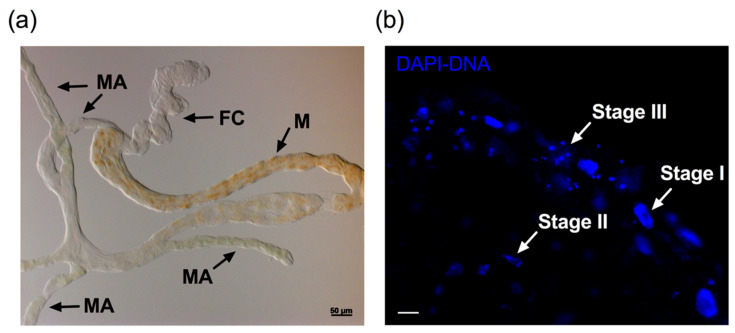
Figure 2.
Nuclear architecture changes after ConA feeding. (a) Light micrograph of the potato psyllid alimentary canal showing characteristic structures. FC: filter chamber, M: midgut, MA: midgut appendages. (b) The nuclear morphology (blue, DAPI staining) in the gut of potato psyllids after feeding in sucrose diets containing ConA for 72 h. Stages I, II, and III represent the three stages of apoptotic nuclei changes. Scale bar is 20 μm.

