Figure 5.
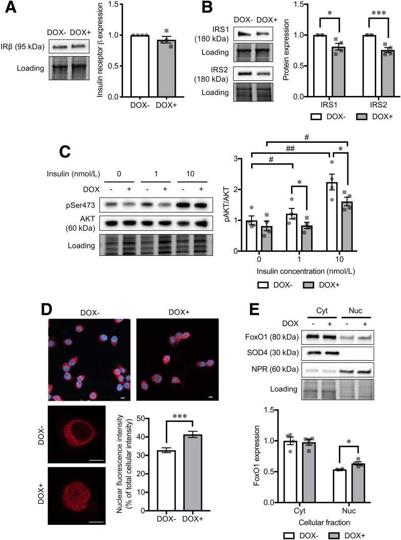
Analysis of insulin-signaling cascade in CD36-overexpressing INS-1 cells. Protein levels of insulin receptor β subunit (IRβ) (A) and IRS1 and IRS2 (B) in INS-1 cells without (DOX−) or with doxycycline-induced CD36 overexpression (DOX+). Normalization factor was used to adjust the target band intensity. N = 4 for each group. C: Phosphorylation levels of AKT Ser473 (pSer473) under a short-term (10-min) stimulation of insulin with various doses (1 and 10 nmol/L). Phosphorylated protein signals were normalized to the total protein levels. N = 4 for each group. D: Immunocytochemical analysis of FoxO1 (red) localization. Nuclei were stained with DAPI (blue). The bar graph shows nuclear FoxO1 fluorescence intensity normalized to the whole-cell intensity. Scale bars, 5 µm. N = 21 cells for each group. E: Western blot analysis of cytoplasmic (Cyt) and nuclear (Nuc) protein levels of FoxO1. N = 4 for each group. The total protein images obtained using the Stain-Free technology are shown as a loading control (Loading). The P values were determined by Student two-tailed t test (unpaired). *P < 0.05, ***P < 0.001 vs. DOX− under the same condition; #P < 0.05, ##P < 0.01 vs. cells without the stimulation of insulin (0 nmol/L) for each group (DOX− or DOX+).

