Abstract
Background
Although several previous studies have examined the association between the platelet to lymphocyte ratio (PLR) and acute appendicitis (AA), findings have been controversial. We aimed to systematically assess the available evidence to elucidate the overall relationship between the PLR and AA.
Methods
Pubmed and Embase databases were searched for all available published literature before August, 2019 by two independent investigators for observational studies reporting the association between the PLR and AA. Random effects models were applied for all meta-analyses. Pooled standardized mean difference (SMD) and 95% confidence interval (CI) were calculated as effect estimates.
Results
Eleven articles met the inclusion criteria and included in this study. Meta-analysis showed that the level of PLR in the AA group was significantly higher than that in the control group (SMD: 1.19, 95% CI: 0.75 to 1.62, P<0.001). A series of subgroup analyses were conducted to investigate the heterogeneity, showing a significant increase in PLV levels in adults with age ≥30 years (SMD: 1.46, 95% CI: 0.89 to 2.02),compared to those in adult <30 years(SMD: 0.58, 95% CI: 0.12 to 1.04) or in children (SMD: 1.03, 95% CI: 0.51 to 1.56). Compared to non-AA controls, a significant increased PLR level was also observed in non-perforated AA (SMD: 1.23, 95% CI: 0.88 to 1.59) and in AA patients during pregnancy (SMD: 0.70, 95% CI: 0.36 to 1.04), while not in perforated AA (SMD: 2.28, 95% CI: -1.72 to 6.28).
Conclusions
A significant increase in PLR level is found in patients with AA, indicating that PLR is a promising biomarker for AA. PLR provides a convenient option for emergency department to quickly screen for clinically or radiologically confirmed AA awaiting appendectomy, especially for pregnant women suspected of having AA. More high-quality evidence is needed to further confirm the diagnostic accuracy of PLR for AA.
Introduction
Acute appendicitis (AA) affects approximately 50000 and 300000 individuals annually who receive appendectomies in the UK and in the US, respectively [1]. It has been proposed that AA is characterized by a series of pathophysiological events including obstruction of the appendix lumen, reduced blood flow to the appendix, destruction of mucosal barrier function, bacterial invasion, inflammatory cell infiltration, tissue hypoxia, necrosis and even perforation [2, 3]. It is reported that perforation may occur in 13–20% of AA patients [4, 5].
Due to the uncertainty of its aetiology, the confirmation or elimination of the diagnosis of appendicitis is the primary concern. If AA is suspected, how to stratify simple and complex appendicitis is also highly significant. Though several diagnostic approaches have been widely applied in clinical practice [6–10], the optimum strategy with non-invasiveness or radiation-free imaging modalities or other laboratory examinations has still not reached consensus, representing a long march for both the patients and surgeons to go.
In the last decades, findings from studies on commonly used blood biomarkers are used to aid the diagnosis of suspected AA patient, especially in children, pregnant women or women of fertile age, and elderly patients. Those inflammatory markers, such as C-reactive protein (CRP), mean platelet volume (MPV), platelet (PLT), platelet distribution width (PDW) and red blood cell distribution width (RDW) or other blood biomarkers, can help identify AA with a certain specificity and sensitivity [11–19].
Recent, increasing evidence suggests that platelet to lymphocyte ratio (PLR) may serve as an inflammatory marker used as a diagnostic or prognostic indicator for various diseases [20–25]. PLR has been shown to be a promising predictive factor for patients with suspected AA [26–28]. A recent study also indicated that the PLR was potent predictor for the differential diagnosis of AA and other disease status [29]. However, the clinical significance of this parameter in patients suspected AA remains unclear. The primary aim of the study was to investigate the relationships between the PLR and AA based on the cumulating evidence of published literature.
Methods
This study was performed and reported according to the Preferred Reporting Items for Systematic Reviews and Meta-analyses (PRISMA) statement [30] and AMSTAR (Assessing the methodological quality of systematic reviews) Guidelines. The protocol for this meta-analysis was registered on PROSPERO (CRD42019146140). The PRISMA checklist is presented in S1 Table.
Literature search
In August, 2019, a comprehensive literature search was performed using online databases by two independent authors, including PubMed, Embase and Cochrane Library. Relevant observational studies were identified that evaluated the association between the PLR level and AA. Search terms including Mesh terms or free text words were listed as follows: “Appendicitis”, “Appendectomy”, “Lymphocyte Count”, “platelet to lymphocyte ratio”, “platelet lymphocyte ratio”, OR “platelet-to-lymphocyte ratio” or “PLR”. Detailed search strategies are provided in S1 File. In addition, manual reference search of eligible literature was screened to further identify potential missing publications.
Study selection
Two independent authors screened the searched relevant studies based on each record’s title or abstract. Full texts were further read after the initial screening process according to the eligibility criteria. Disagreements were resolved by discussion or by a third author by tracing the original article.
Inclusion and exclusion criteria
Studies were consider eligible if they satisfied the following criteria: (1) provided the association between the PLR level and AA; (2) the mean concentration and its standard difference (SD) of the PLR level could be obtained from original studies for both AA patients and controls (healthy controls, healthy pregnant women, outpatient clinic patients without AA, and patients with negative appendectomy) or could be calculated using the indirect methods [31, 32]; (3) provided data for the estimation of the standardized mean difference (SMD) and 95% confidence limit (CI) for the concentrations of PLR level. The exclusion criteria included: (1) reviews, comments, or meta-analysis without original data for meta-analysis; (2) studies without adequate data for abstraction; and (3) irrelevant or duplicated publications.
Data extraction and risk of bias assessment
Pairs of independent authors extracted data from the published articles using a predefined data abstraction form. Variables concerning the study design, population characteristics and laboratory index were investigated including first author, publication year, study country and design, sample size of the study, sex percent, mean age of the participants, the blood index studied, and the type of appendicitis.
Study quality assessment
The quality of each included study was evaluated using the Newcastle–Ottawa Quality Assessment Scale (NOS) [33]. Three domains of this scale regarding study selection, comparability, and outcome were scored with a total of nine points for observational studies. Studies obtaining a score of 7 to 9, 3 to 6, and 0 to 3 points were judged as high, moderate, and low quality, respectively [34].
Statistical analysis
All analyses were performed using the STATA software (version 12.0; Stata Corporation). Data were collected as means ± standard deviation (SD) to estimate the pooled effect estimates. Meta-analysis was carried out using standardized mean difference (SMD) and 95% confidence limit (CI) for assessing the association between the PLR levels and AA. The random effects model, a most common and conservative approach to combine study estimates, was applied for all meta-analyses considering the between-study difference [35]. To test the stability of the meta-analysis results, we also performed the “leave-one-out” sensitivity analyses by omitting one study at each time and examining the influence of each individual study on the summary effect estimate.
Heterogeneity assessment
Cochran’s Q test and I2 statistic were evaluated to assess the inter-study heterogeneity. I2 of 0–25%, 26–50%, 51–75% and 76–100% indicates insignificant heterogeneity, low heterogeneity, moderate heterogeneity, and high heterogeneity, respectively [36]. To further investigate the potential source of inter-study heterogeneity, we also conducted subgroup analyses based on different available variables, including sample size (≥500 vs. <500), study continent (Europe vs. Asia), patient age (adult <30 years vs. ≥30 years vs. children), appendicitis type (perforated AA vs. non-perforated AA vs. AA during pregnancy), NOS score (high quality vs. fair or low quality) and controls (healthy controls vs. healthy pregnant women vs. outpatient clinic patients without AA vs. patients with negative appendectomy).
Publication bias assessment
Publication bias was assessed by visual inspection of the funnel plot symmetry and quantitatively by Egger’s and Begg’s tests, with a P value less than 0.05 indicating significant publication bias [37]. When publication bias existed, the Duvall and Tweedle trim-and-fill analysis would be performed to test the effects of missed study on overall effect estimates [38].
Results
Literature search
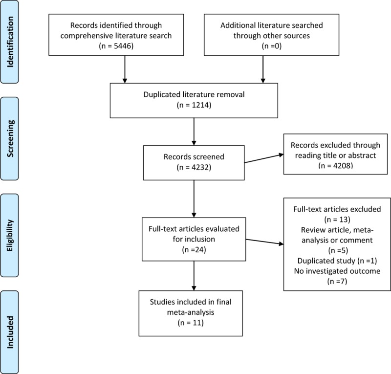
Our initial literature search through databases identified a total of 5446 records. After removing 1214 duplicates, the remaining 4232 records were reviewed based on the title or abstract reading by two independent authors. During this stage, 4208 irrelevant studies were excluded. Twenty-four articles were retrieved and reviewed based on full text. Finally, 11 articles met the inclusion criteria and were included in our meta-analysis [26–28, 39–46]. Fig 1 presenting a flow diagram depicts the results of the study selection process for this meta-analysis.
Fig 1. Flow diagram of the literature search and screening process.

Study characteristics
The baseline characteristics of each included study are shown in Table 1. A total of 11 studies were included in our meta-analysis with their sample size ranging from 63 to 650 subjects. The included studies were published between 2015 and 2019. Mean age of the participants ranged between 9.34 and 40 years. Two studies involved participants of pregnant women. Ten of the 11 studies reported adult patients while one included pediatric patients [28]. Five studies extracted effect estimates using indirect methods proposed by Wan et al. [26,27,40,41,45]. Most of the included studies were conducted in Europe and Asia. All of the studies used a retrospective case-control design. Based on the NOS scoring system, 8 studies ranked as high-quality (NOS score≥7) and the others as were classified as moderate in quality assessment (NOS score<7) (S2 Table).
Table 1. Characteristics of included studies.
| Study | Year | Country | Sample size | Case no. | Controls and no. | Sex (male)/female % | Age(mean), years | Biomarker studied | Appendicitis type |
|---|---|---|---|---|---|---|---|---|---|
| Pehlivanli F | 2019 | Turkey | 558 | 458/14 | Negative appendectomy; 86 | Males 55.2%; Females 44.8% | 34.24 | WBC, PLT, MPV, NLR, PLR | Acute appendicitis, perforated appendicitis |
| Yazar FM | 2018 | Turkey | 640 | 511/54 | Negative appendectomy; 75 | Positive appendectomy (Males 54.7%; Females 45.3%); Negative appendectomy (Males 45.3%; Females 54.7%) | Positve appendectomy 39.23: Negative appendectomy 35.27 | WBC, CRP, NLR, PLR | Acute appendicitis |
| Cinar H | 2018 | Turkey | 94 | 40/7 | Healthy pregnant women under routine pregnancy follow-up at the obstetrics clinics; 47 | Females 100.0% | Group A 27; Group B 25.14; Control group 29.74 | WBC, MPV, NLR, PLR | Acute focal appendicitis, acute suppurated appendicitis, acute perforated appendicitis, and acute gangrenous appendicitis |
| Nazik S | 2017 | Turkey | 63 | 30 | Healthy control subjects; 33 | Males 65.08%; Females 34.92% | 9.34 | ESR,CRP, WBC, MPV, NLR, PLR, Ischemia-modified albumin | Acute appendicitis and perforated appendicitis |
| Kahramanca Ş | 2017 | Turkey | 569 | 475 | Negative appendectomy; 94 | Males 55.94%; Females 44.05% | 40 | PLR | Acute appendicitis |
| Mehmet Ü | 2017 | Turkey | 569 | 455 | Patients with a normal appendix having other complaints in ED excluding abdominal pain; 114 | Males 55.36%; Females 44.64% | 31.97 | WBC, PLT, Neutrophil, Lymphocyte, NLR, PLR, PDW | Acute appendicitis |
| Toktas O | 2017 | Turkey | 60 | 30 | Healthy control subjects; 30 | Males 71.67%; Females 28.33% | 28.5 | Platelet, MPV, RDW, Neutrophil, Lymphocyte, NLR, PLR, Leukocyte | Acute appendicitis |
| Shin DH | 2017 | Korea | 650 | 615 | Negative appendectomy; 35 | Males 51.2%; female 48.8% | 33 | WBC, NLR, LMR, PLR, DNI, CRP | Non-complicated appendicitis, complicated appendicitis |
| Ulukent SC | 2016 | Turkey | 191 | 97 | Healthy control subjects in outpatient clinics; 94 | Appendicitis: males 59.8%; Females 40.2%;Control: Males 63.8%; Females 36.2% | Appendicitis 39; Control 34 | Leukocyte count, neutrophil percentage, NLR, PLR, MPV, RDW, PDW and CRP | Acute appendicitis |
| Acar E | 2016 | Turkey | 476 | 215/200 | Patients without any complaints at the outpatient clinics; 61 | Male 53.5%; female 46.5% | 31.6 | WBC, RDW, MPV, neutrophil, lymphocyte, NLR and PLR. | Acute appendicitis |
| Yazar FM | 2015 | Turkey | 122 | 28/35 | Healthy pregnant control; 29/30 | Females 100.0% | Appendectomy group 26.93; Healthy pregnant control group 29.62 | WBC, CRP, NLR, PLR | Acute appendicitis |
Abbreviations: CRP: C-reactive protein; DNI: Delta neutrophil index; ED, emergency department; ESR: Erythrocyte sedimentation rate; MPV: Mean platelet volume; NLR: Neutrophil to lymphocyte ratio; PDW: Platelet distribution width; PLT: Platelet; PLR: Platelet to lymphocyte ratio; RDW: Red cell distribution of width; WBC: White blood cell.
The association between PLR level and AA
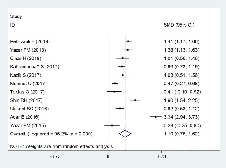
Eleven case-control studies involving 3006 cases and 698 controls examined the association between PLR level and AA. Meta-analysis found that PLR levels were significantly higher in patients with AA (SMD: 1.19, 95% CI: 0.75 to 1.62, P<0.001) than that of the controls, with high inter-study heterogeneity (I2 = 95.2%) (Fig 2).
Fig 2. Forest plot showing the relationship between platelet to lymphocyte ratio and acute appendicitis.
Subgroup analysis and sensitivity analysis
We conducted several preplanned subgroup analyses and sensitivity analyses by several variables. Analyses stratified by study sample size revealed that PLR level was significantly related to AA for studies with both small sample size (<500) (SMD: 1.15; 95% CI: 0.22 to 2.08; P = 0.015) and large sample size (≥500) (SMD: 1.21; 95% CI: 0.76 to 1.67; P <0.001). When we stratified studies by study region, a statistically significant relationship was noted for studies conducted in Europe (SMD: 1.12; 95% CI: 0.66 to 1.57; P <0.001) and in Asia (SMD: 1.90; 95% CI: 1.54 to 2.25; P = 0.003). The associations were also significant in studies with both adult patient age ≥30 years (SMD: 1.46; 95% CI: 0.89 to 2.02; P = 0.004), adult patient age <30 years (SMD: 0.58; 95% CI: 0.12 to 1.04; P = 0.472) and children. Sensitivity analyses revealed significant associations when involving only non-perforated AA (SMD: 1.23; 95% CI: 0.88 to 1.59; P<0.001) or AA during pregnancy (SMD: 0.70; 95% CI: 0.36 to 1.04; P = 0.037), but not perforated AA (SMD: 2.28; 95% CI: -1.72 to 6.28; P = 0.263). The PLR level was not found significantly different among complicated AA compared to that in non-complicated AA. According to the analysis by study quality, a higher PLR level was found in studies with both high quality (NOS score≥7) (SMD: 0.95; 95% CI: 0.57 to 1.32; P <0.001) and moderate or low quality (SMD: 1.84; 95% CI: 0.56 to 3.11; P = 0.005). Based on the analyses by control subjects, significant associations were found among healthy control subjects (SMD: 0.77; 95% CI: 0.46 to 1.07; P <0.001) and control subjects with negative appendectomy (SMD: 1.40; 95% CI: 1.05 to 1.74; P <0.001). The detailed results of subgroup analyses for associations between PLR levels and AA are presented in Table 2.
Table 2. Subgroup analyses results for association between serum levels of platelet to lymphocyte ratio and acute appendicitis.
| Variable | SMD | 95%CI | Degree heterogeneity (I2 statistics; %) | P value | No. of included Studies | P for interaction |
|---|---|---|---|---|---|---|
| Total | 1.19 | 0.75 to 1.62 | 95.2 | <0.001 | 11 | NA |
| Sample size | 0.149 | |||||
| <500 | 1.15 | 0.22 to 2.08 | 96.4 | 0.015 | 6 | |
| ≥500 | 1.21 | 0.76 to 1.67 | 94.0 | <0.001 | 5 | |
| Study region | <0.001 | |||||
| Europe | 1.12 | 0.66 to 1.57 | 95.2 | <0.001 | 10 | |
| Asia | 1.90 | 1.54 to 2.25 | - | 0.003 | 1 | |
| Age (Mean/median) | 0.001 | |||||
| Adult <30 years | 0.58 | 0.12 to 1.04 | 61.7 | 0.472 | 3 | |
| Adult ≥30 years | 1.46 | 0.89 to 2.02 | 96.8 | 0.004 | 7 | |
| Children | 1.03 | 0.51 to 1.56 | - | - | 1 | |
| Appendicitis type | <0.001 | |||||
| Perforated AA | 2.28 | -1.72 to 6.28 | 98.9 | 0.263 | 2 | |
| Non-perforated AA | 1.23 | 0.88 to 1.59 | 75.6 | <0.001 | 2 | |
| AA during pregnancy | 0.70 | 0.36 to 1.04 | 77.1 | 0.037 | 2 | |
| Study quality | <0.001 | |||||
| NOS score≥7 | 0.95 | 0.57 to 1.32 | 90.3 | <0.001 | 8 | |
| NOS score <7 | 1.84 | 0.56 to 3.11 | 98.0 | 0.005 | 3 | |
| Control subject | <0.001 | |||||
| Healthy subjects | 0.77 | 0.46 to 1.07 | 77.1 | <0.001 | 3 | |
| Negative appendectomy | 1.40 | 1.05 to 1.74 | 85.2 | <0.001 | 4 | |
| Healthy pregnant women | 0.66 | -0.06 to 1.37 | 33.3 | 0.074 | 2 | |
| Non-AA outpatients or ED patients | 1.90 | -0.91 to 4.71 | 99.4 | 0.185 | 2 |
Abbreviations: AA, acute appendicitis; CI, confidence interval; ED, emergency department; NA, not available; SMD, standardized mean difference.
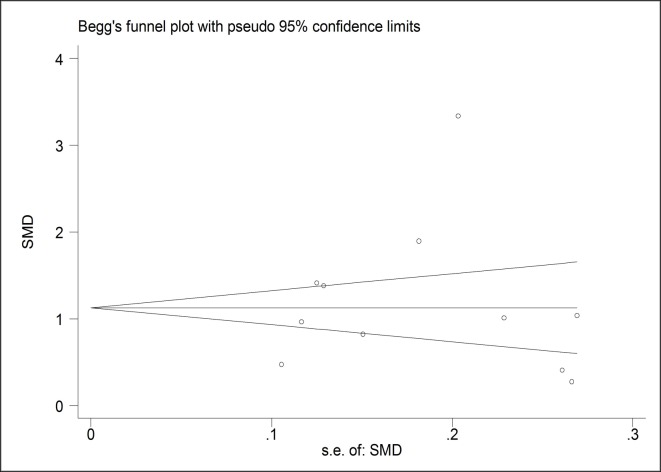
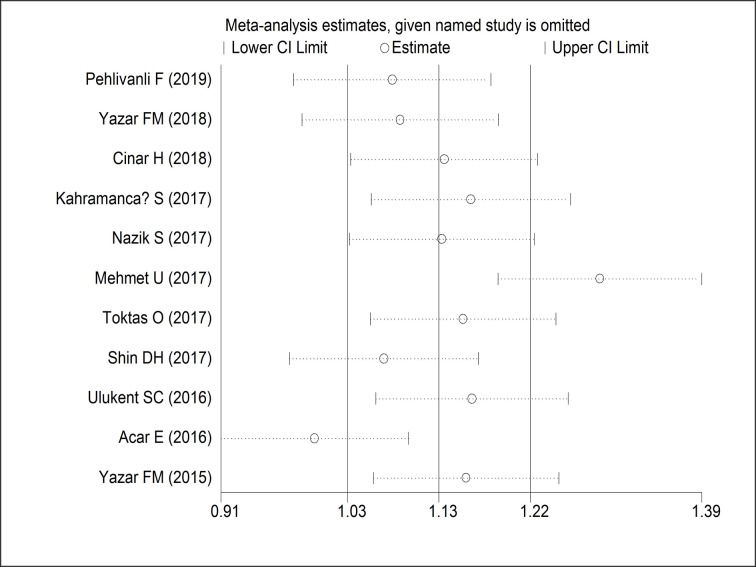
A funnel plot was generated to investigate the potential publication bias of the 11 included studies. Both Egger’s (P = 0.573) and Begg’s (P = 1.000) indicated that no significant publication bias existed in these studies (Fig 3). In addition, no filled studies were input when trim and fill method was applied, yielding the same adjusted summary SMD with the original analysis, further indicating the robustness of the finding. The “leave-one-out” sensitivity analyses by omitting one study at each time and examining the influence of each individual study also confirmed that the results were stable (Fig 4).
Fig 3. A funnel plot analysis of publication bias for association between platelet to lymphocyte ratio and acute appendicitis.
Fig 4. Sensitivity analysis of effect for association between platelet to lymphocyte ratio and acute appendicitis.
Discussion
Principal findings
To the best of our knowledge, this meta-analysis is the first attempt to demonstrate the association between the PLR level and AA. A comprehensive literature search of all observational studies assessing this relationship was performed. The findings indicate that the PLR level is significantly higher in AA individuals compared to the non-AA ones which is in line with previous reports [40, 42, 43]. Moreover, though the results of most subgroups are consistent with the main analysis, findings did not suggest that the PLR levels was significantly correlated with perforated AA, probably because of low statistical power with few included studies. Therefore, further well-designed prospective studies should be advocated to further confirm these relationships.
Potential mechanisms
Numerous reports have revealed that systemic inflammatory response can induce neutrophilia and lymphocytopenia [47, 48], leading to an increase in some inflammatory indices such as PLR and neutrophil to lymphocyte ratio (NLR), some of the inflammatory biomarkers in AA [49–56]. Smith et.al found that some processes of inflammatory response were influenced by the changes in platelet markers [57]. It has been shown that cytokines such as interleukin (IL)-1 and IL-6 may affect the change of mean platelet volume (MPV), which is regarded as a marker of platelet activation inflammation [58, 59]. Studies also found that platelets can serve as key coordinators involved in several inflammatory processes [60, 61] and in the occurrence and development of several inflammatory diseases including inflammatory bowel disease, solid cancers, psoriasis multiple sclerosis and bronchial asthma [62–64]. However, few studies and meta-analyses have demonstrated the association between PLR and AA.
Clinical relevance and implications
This study has important clinical relevance and implications. As a promising diagnostic biomarker, PLR has great potential to help in the diagnosis and decision-making of suspected appendicitis in selected populations and conditions. We found that the diagnostic implication of PLR may play an important role for participants whose age ≥30 years, pregnant women, and AA without progression to perforation. For areas with limited medical resources, due to the limited access to CT or MRI scan at the emergency department, PLR can be used as an effective rapid alternative to auxiliary diagnosis. Our study shows that PLR levels in pregnant women with appendicitis are significantly higher than those without appendicitis, which indicates that the measurement of PLR level provides an important, safe, rapid and radiation-free auxiliary diagnosis method for pregnant women suspected of appendicitis. PLR also has potential value in differentiating perforated appendicitis from nonperforated appendicitis, although large prospective studies are needed further confirmation.
The PLR, like many blood inflammatory indicators, are commonly used, non-invasive and cost-effective blood biomarkers, which can be easily available in the emergency department, even in small hospitals. However, the value of the PLR in AA has only been examined in few studies. A study conducted by Celik et al. [65] showed that the cutoff value of 284 for PLR with a sensitivity of 42% and a specificity of 86% was found to be the best predictive value for the diagnosis of complicated AA. Another study by Mehmet et al. [42] revealed that a higher PLR level was detected in patients with perforated appendicitis than that in normal controls. Pehlivanli et al. [27] assessed 558 patients who underwent appendectomy and found that the PLR appeared to be significantly valuable in the differentiation of normal appendix from AA and in the differentiation of AA from perforated appendicitis.
In this study, the reasons to select SMD as the statistical parameter for this kind of meta-analysis were explained as follows. First, the current literature search did not yield sufficient eligible studies for diagnostic meta-analysis combining the sensitivity and sensitivity of PLR as effect estimates. Instead, 11 studies could be identified providing means and SDs of PLR levels for both AA patients and controls, which could be used as an alternative effect estimate to conduct a meta-analysis. Thus, SMD is a suitable substitute as a statistical parameter to demonstrate the associations between PLR levels (a continuous measure outcome) and AA. Second, a research article published in 2019 [66] has demonstrated that SMD is a commonly used effect estimate to measure continuous outcomes using different scales or units (e.g., blood inflammatory biomarkers with different units). These effect estimates should be standardized before pooling in a meta-analysis. One of the frequently used methods of standardization includes the use of SMD. In our meta-analysis, we also used SMD because units of PLR level in the included studies were different. Finally, the result of our study can be well interpreted. We found that the level of PLR in the AA group was significantly higher than that in the control group by 1.19 standard deviations of PLA levels (SMD: 1.19, 95% CI: 0.75 to 1.62, P<0.001), which indicates that PLR is a promising blood biomarker that can potentially predict AA in patients with clinical suspicion of appendicitis.
Considerable inter-study heterogeneity was detected in our meta-analysis, though we conducted several subgroup analyses. The results of subgroup analyses for relationship between the PLR level and AA indicated that the heterogeneity could partly attribute to variables regarding differences in patient age, appendicitis type and control subjects. However, these variables could not account for all of the heterogeneity. Perhaps further larger well-designed prospective cohort studies or clinical trials are warranted in the future to better demonstrate the relationship between the PLR level and AA.
Strengths and limitations
The present meta-analysis has several advantages. Firstly, this is the first meta-analysis of the relationship between PLR level and AA. From the pooled analysis, we found that patients with AA had significant higher PLR level than the non-AA controls. Moreover, multiple analyses including the “leave-one-out” sensitivity analysis, trim and fill method and subgroup analyses confirmed this association was stable. In addition, the results of subgroup analyses were mostly consistent with those of the main analysis, which also indicated our findings were convincing. Secondly, random-effects model, a more conservative approach, was used when we pooled effect estimates, making the results of our meta-analysis more accurate. Thirdly, publication date and language limits were not restricted for literature search, making the results less possible to leading to publication bias.
This meta-analysis should still be interpreted with caution due to the limitations in several aspects. First, despite that all studies used blood sample analyzer to evaluate PLR levels, the sample collection time and reference values were varied, which can lead to heterogeneous results. Second, besides the lack of diagnostic accuracy studies, the results of this meta-analysis were mainly based on observational studies, mostly case-control studies, which is a limitation of critical importance for demonstrating a causal relationship between PLR level and AA. However, observational study results may also provide significant preliminary data to justify further larger trials and well-designed prospective cohort studies. Third, there seem to be patients mostly from one continent of Europe. The variations on the ethnicity of the population are still under investigation. Because limited number of studies is involved, multi-ethnic cohort studies are warranted to confirm our findings. Fourth, it is known that systemic inflammatory response might induce an increase in blood PLR. It is quite obvious that AA can also lead to the PLR increase. Diagnosis of AA can be challenging due to the fact that a variety of acute gastrointestinal and gynaecologic diseases can mimic the symptoms of AA. Therefore, more high quality evidence should be accumulated before definite conclusions could be drawn. Last but not least, as we extracted estimates using indirect methods in 5 studies, this method might have led to overestimation or underestimation of the sample of SD and thereby causing inaccuracy of the effect estimates.
In conclusion, this meta-analysis shows a significant increase in PLR level in patients with AA, indicating that PLR is a promising biomarker for AA. PLR provides a convenient option for emergency department to quickly screen for clinically or radiologically confirmed AA awaiting appendectomy, especially for pregnant women suspected of having AA. For areas with limited medical resources, due to the limited access to CT or MRI scans in emergency department, PLR can be used as an effective and rapid alternative to auxiliary diagnosis. However, more high-quality evidence is needed to further confirm the diagnostic accuracy of PLR for AA, and whether PLR combined with other blood biomarkers will yield better predictive value needs to be further studied.
Supporting information
Supplementary search strategies.
(DOCX)
(DOC)
(DOCX)
Acknowledgments
We sincerely thank all authors who provided published data for our meta-analysis.
Data Availability
All relevant data are within the manuscript and its Supporting Information files.
Funding Statement
This work was supported by the National Natural Science Foundation of China (grant no. 81774112 to ZM, 81872046 to HW) and a grant from Siming Scholars from Shuguang Hospital (grant no. SGXZ-201913 to ZM). The funders had no role in study design, data collection and analysis, decision to publish, or preparation of the manuscript.
References
- 1.Collaborative NSR. Multicentre observational study of performance variation in provision and outcome of emergency appendicectomy. British Journal of Surgery 2013;100(9):1240–52 10.1002/bjs.9201 [DOI] [PubMed] [Google Scholar]
- 2.Tsuji M, Puri P, Reen D. Characterisation of the local inflammatory response in appendicitis. Journal of pediatric gastroenterology and nutrition 1993;16(1):43–48 10.1097/00005176-199301000-00008 [DOI] [PubMed] [Google Scholar]
- 3.Allister L, Bachur R, Glickman J, et al. Serum Markers in Acute Appendicitis. Journal of Surgical Research 2011;168(1):70–75 [published Online First: Epub Date]|. 10.1016/j.jss.2009.10.029 [DOI] [PubMed] [Google Scholar]
- 4.Andersson R, Hugander A, Thulin A. Diagnostic accuracy and perforation rate in appendicitis: association with age and sex of the patient and with appendicectomy rate. The European journal of surgery = Acta chirurgica 1992;158(1):37–41 [PubMed] [Google Scholar]
- 5.Marudanayagam R, Williams GT, Rees BI. Review of the pathological results of 2660 appendicectomy specimens. Journal of gastroenterology 2006;41(8):745–49 10.1007/s00535-006-1855-5 [DOI] [PubMed] [Google Scholar]
- 6.van Randen A, Bipat S, Zwinderman AH, et al. Acute appendicitis: meta-analysis of diagnostic performance of CT and graded compression US related to prevalence of disease. Radiology 2008;249(1):97–106 10.1148/radiol.2483071652 [DOI] [PubMed] [Google Scholar]
- 7.Shen G, Wang J, Fei F, et al. Bedside ultrasonography for acute appendicitis: An updated diagnostic meta-analysis. International Journal of Surgery 2019;70:1–9 10.1016/j.ijsu.2019.08.009 [DOI] [PubMed] [Google Scholar]
- 8.Repplinger MD, Pickhardt PJ, Robbins JB, et al. Prospective comparison of the diagnostic accuracy of MR imaging versus CT for acute appendicitis. Radiology 2018;288(2):467–75 10.1148/radiol.2018171838 [DOI] [PMC free article] [PubMed] [Google Scholar]
- 9.Kinner S, Pickhardt PJ, Riedesel EL, et al. Diagnostic accuracy of MRI versus CT for the evaluation of acute appendicitis in children and young adults. American Journal of Roentgenology 2017;209(4):911–19 10.2214/AJR.16.17413 [DOI] [PMC free article] [PubMed] [Google Scholar]
- 10.Duke E, Kalb B, Arif-Tiwari H, et al. A systematic review and meta-analysis of diagnostic performance of MRI for evaluation of acute appendicitis. American Journal of Roentgenology 2016;206(3):508–17 10.2214/AJR.15.14544 [DOI] [PubMed] [Google Scholar]
- 11.Zouari M, Jallouli M, Louati H, et al. Predictive value of C-reactive protein, ultrasound and Alvarado score in acute appendicitis: a prospective pediatric cohort. The American journal of emergency medicine 2016;34(2):189–92 10.1016/j.ajem.2015.10.004 [DOI] [PubMed] [Google Scholar]
- 12.Zani A, Teague WJ, Clarke SA, et al. Can common serum biomarkers predict complicated appendicitis in children? Pediatric surgery international 2017;33(7):799–805 10.1007/s00383-017-4088-1 [DOI] [PubMed] [Google Scholar]
- 13.Saxena D, Tandon M, Gedam B. Role of mean platelet volume (MPV) in diagnosis of acute appendicitis. Int J Biomed Res 2015;6(4):235–7 [Google Scholar]
- 14.Msolli MA, Beltaief K, Bouida W, et al. Value of early change of serum C reactive protein combined to modified Alvarado score in the diagnosis of acute appendicitis. BMC emergency medicine 2018;18(1):15 10.1186/s12873-018-0166-5 [DOI] [PMC free article] [PubMed] [Google Scholar]
- 15.Monsalve S, Ellwanger A, Montedonico S. White blood cell count and C-reactive protein together remain useful for diagnosis and staging of acute appendicitis in children. South African Medical Journal 2017;107(9):773–76 10.7196/SAMJ.2017.v107i9.12206 [DOI] [PubMed] [Google Scholar]
- 16.Erdem H, Aktimur R, Cetinkunar S, et al. Evaluation of mean platelet volume as a diagnostic biomarker in acute appendicitis. International journal of clinical and experimental medicine 2015;8(1):1291 [PMC free article] [PubMed] [Google Scholar]
- 17.Dinc B, Oskay A, Dinc SE, et al. New parameter in diagnosis of acute appendicitis: platelet distribution width. World Journal of Gastroenterology: WJG 2015;21(6):1821 10.3748/wjg.v21.i6.1821 [DOI] [PMC free article] [PubMed] [Google Scholar]
- 18.Dayawansa NH, Segan JD, Yao HH, et al. Incidence of normal white cell count and C‐reactive protein in adults with acute appendicitis. ANZ journal of surgery 2018;88(6):E539–E43 10.1111/ans.13760 [DOI] [PubMed] [Google Scholar]
- 19.Bozlu G, Taskinlar H, Unal S, et al. Diagnostic value of red blood cell distribution width in pediatric acute appendicitis. Pediatrics International 2016;58(3):202–05 10.1111/ped.12805 [DOI] [PubMed] [Google Scholar]
- 20.Raungkaewmanee S, Tangjitgamol S, Manusirivithaya S, et al. Platelet to lymphocyte ratio as a prognostic factor for epithelial ovarian cancer. Journal of gynecologic oncology 2012;23(4):265–73 10.3802/jgo.2012.23.4.265 [DOI] [PMC free article] [PubMed] [Google Scholar]
- 21.Gary T, Pichler M, Belaj K, et al. Platelet-to-lymphocyte ratio: a novel marker for critical limb ischemia in peripheral arterial occlusive disease patients. PloS one 2013;8(7):e67688 10.1371/journal.pone.0067688 [DOI] [PMC free article] [PubMed] [Google Scholar]
- 22.Turkmen K, Erdur FM, Ozcicek F, et al. Platelet‐to‐lymphocyte ratio better predicts inflammation than neutrophil‐to‐lymphocyte ratio in end‐stage renal disease patients. Hemodialysis International 2013;17(3):391–96 10.1111/hdi.12040 [DOI] [PubMed] [Google Scholar]
- 23.Balta S, Ozturk C. The platelet-lymphocyte ratio: a simple, inexpensive and rapid prognostic marker for cardiovascular events. Platelets 2015;26(7):680–81 10.3109/09537104.2014.979340 [DOI] [PubMed] [Google Scholar]
- 24.Fu H, Qin B, Hu Z, et al. Neutrophil-and platelet-to-lymphocyte ratios are correlated with disease activity in rheumatoid arthritis. Clin Lab 2015;61(3–4):269–73 10.7754/clin.lab.2014.140927 [DOI] [PubMed] [Google Scholar]
- 25.Uslu AU, Küçük A, Şahin A, et al. Two new inflammatory markers associated with Disease Activity Score‐28 in patients with rheumatoid arthritis: neutrophil‐lymphocyte ratio and platelet‐lymphocyte ratio. International journal of rheumatic diseases 2015;18(7):731–35 10.1111/1756-185X.12582 [DOI] [PubMed] [Google Scholar]
- 26.Shin DH, Cho YS, Cho GC, et al. Delta neutrophil index as an early predictor of acute appendicitis and acute complicated appendicitis in adults. World Journal of Emergency Surgery 2017;12(1):32. [DOI] [PMC free article] [PubMed] [Google Scholar]
- 27.Pehlivanli F, Aydin O. Role of Platelet to Lymphocyte Ratio as a Biomedical Marker for the Pre-Operative Diagnosis of Acute Appendicitis. Surgical infections 2019. 10.1089/sur.2019.042 [published Online First: Epub Date]|. [DOI] [PubMed] [Google Scholar]
- 28.Nazik S, Avci V, Kusku Kiraz Z. Ischemia-modified albumin and other inflammatory markers in the diagnosis of appendicitis in children. Ulusal travma ve acil cerrahi dergisi = Turkish journal of trauma & emergency surgery: TJTES 2017;23(4):317–21 10.5505/tjtes.2016.11823 [published Online First: Epub Date]|. [DOI] [PubMed] [Google Scholar]
- 29.Tuncer AA, Cavus S, Balcioglu A, et al. Can mean platelet volume, Neutrophil-to-Lymphocyte, Lymphocyte-to-Monocyte, Platelet-to-Lymphocyte ratios be favourable predictors for the differential diagnosis of appendicitis? JOURNAL OF THE PAKISTAN MEDICAL ASSOCIATION 2019;69(5):647–53 [PubMed] [Google Scholar]
- 30.Moher D, Liberati A, Tetzlaff J, et al. Preferred reporting items for systematic reviews and meta-analyses: the PRISMA statement. Annals of internal medicine 2009;151(4):264–69 10.7326/0003-4819-151-4-200908180-00135 [DOI] [PubMed] [Google Scholar]
- 31.Wan X, Wang W, Liu J, et al. Estimating the sample mean and standard deviation from the sample size, median, range and/or interquartile range. BMC medical research methodology 2014;14(1):135. [DOI] [PMC free article] [PubMed] [Google Scholar]
- 32.Hozo SP, Djulbegovic B, Hozo I. Estimating the mean and variance from the median, range, and the size of a sample. BMC medical research methodology 2005;5(1):13. [DOI] [PMC free article] [PubMed] [Google Scholar]
- 33.Stang A. Critical evaluation of the Newcastle-Ottawa scale for the assessment of the quality of nonrandomized studies in meta-analyses. European journal of epidemiology 2010;25(9):603–05 10.1007/s10654-010-9491-z [DOI] [PubMed] [Google Scholar]
- 34.Montazeri Z, Nyiraneza C, El-Katerji H, et al. Waterpipe smoking and cancer: systematic review and meta-analysis. Tobacco control 2017;26(1):92–97 10.1136/tobaccocontrol-2015-052758 [DOI] [PubMed] [Google Scholar]
- 35.DerSimonian R, Laird N. Meta-analysis in clinical trials. Controlled clinical trials 1986;7(3):177–88 10.1016/0197-2456(86)90046-2 [DOI] [PubMed] [Google Scholar]
- 36.Higgins JP, Thompson SG, Deeks JJ, et al. Measuring inconsistency in meta-analyses. BMJ (Clinical research ed) 2003;327(7414):557–60 [DOI] [PMC free article] [PubMed] [Google Scholar]
- 37.Egger M, Smith GD, Schneider M, et al. Bias in meta-analysis detected by a simple, graphical test. BMJ (Clinical research ed) 1997;315(7109):629–34 [DOI] [PMC free article] [PubMed] [Google Scholar]
- 38.Duval S, Tweedie R. Trim and fill: a simple funnel‐plot–based method of testing and adjusting for publication bias in meta‐analysis. Biometrics 2000;56(2):455–63 10.1111/j.0006-341x.2000.00455.x [DOI] [PubMed] [Google Scholar]
- 39.Acar E, Ozcan O, Deliktas H, et al. Laboratory markers has many valuable parameters in the discrimination between acute appendicitis and renal colic. Ulusal travma ve acil cerrahi dergisi = Turkish journal of trauma & emergency surgery: TJTES 2016;22(1):17–22 10.5505/tjtes.2015.69488 [published Online First: Epub Date]|. [DOI] [PubMed] [Google Scholar]
- 40.Cinar H, Aygun A, Derebey M, et al. Significance of hemogram on diagnosis of acute appendicitis during pregnancy. Ulusal travma ve acil cerrahi dergisi = Turkish journal of trauma & emergency surgery: TJTES 2018;24(5):423–28 10.5505/tjtes.2018.62753 [published Online First: Epub Date]|. [DOI] [PubMed] [Google Scholar]
- 41.Kahramanca Ş, Özgehan G, Kaya O, et al. Platelet to Lymphocyte Ratio and Acute Appendicitis. Kafkas Tıp Bilimleri Dergisi 2017;7(2):153–57 [Google Scholar]
- 42.Mehmet Ü, Ertuğrul K, Murat O, et al. The role of neutrophils/lymphocyte ratio, platelet/lymphocyte ratio and platelet distribution width values in acute appendicitis diseases. Biomedical Research 2017;28(17):7514–18 [Google Scholar]
- 43.Toktas O, Aslan M. Mean platelet volume, red cell distribution width, neutrophil to lymphocyte ratio and platelet to lymphocyte ratio in the diagnosis of acute appendicitis. Eastern Journal Of Medicine 2017;22(1):5 [Google Scholar]
- 44.Ulukent SC, Sarici IS, Ulutas KT. All CBC parameters in diagnosis of acute appendicitis. International journal of clinical and experimental medicine 2016;9(6):11871–76 [Google Scholar]
- 45.Yazar FM, Bakacak M, Emre A, et al. Predictive role of neutrophil-to-lymphocyte and platelet-to-lymphocyte ratios for diagnosis of acute appendicitis during pregnancy. The Kaohsiung journal of medical sciences 2015;31(11):591–6 10.1016/j.kjms.2015.10.005 [published Online First: Epub Date]|. [DOI] [PMC free article] [PubMed] [Google Scholar]
- 46.Yazar FM, Urfalioglu A, Bakacak M, et al. Efficacy of the Evaluation of Inflammatory Markers for the Reduction of Negative Appendectomy Rates. The Indian journal of surgery 2018;80(1):61–67 10.1007/s12262-016-1558-y [published Online First: Epub Date]|. [DOI] [PMC free article] [PubMed] [Google Scholar]
- 47.Ishizuka M, Shimizu T, Kubota K. Neutrophil-to-lymphocyte ratio has a close association with gangrenous appendicitis in patients undergoing appendectomy. International surgery 2013;97(4):299–304 [DOI] [PMC free article] [PubMed] [Google Scholar]
- 48.Yazici M, Özkisacik S, Öztan MO, et al. Neutrophil/lymphocyte ratio in the diagnosis of childhood appendicitis. The Turkish journal of pediatrics 2010;52(4):400 [PubMed] [Google Scholar]
- 49.Bilici S, Sekmenli T, Goksu M, et al. Mean platelet volume in diagnosis of acute appendicitis in children. African health sciences 2011;11(3):427–32 [PMC free article] [PubMed] [Google Scholar]
- 50.Boshnak N, Boshnaq M, Elgohary H. Evaluation of Platelet Indices and Red Cell Distribution Width as New Biomarkers for the Diagnosis of Acute Appendicitis. Journal of investigative surgery: the official journal of the Academy of Surgical Research 2018;31(2):121–29 10.1080/08941939.2017.1284964 [published Online First: Epub Date]|. [DOI] [PubMed] [Google Scholar]
- 51.Bozkurt S, Kose A, Erdogan S, et al. MPV and other inflammatory markers in diagnosing acute appendicitis. JPMA The Journal of the Pakistan Medical Association 2015;65(6):637–41 [PubMed] [Google Scholar]
- 52.Bozlu G, Taskinlar H, Unal S, et al. Diagnostic value of red blood cell distribution width in pediatric acute appendicitis. Pediatrics international: official journal of the Japan Pediatric Society 2016;58(3):202–5 10.1111/ped.12805 [published Online First: Epub Date]|. [DOI] [PubMed] [Google Scholar]
- 53.Dinc B, Oskay A, Dinc SE, et al. New parameter in diagnosis of acute appendicitis: platelet distribution width. World journal of gastroenterology 2015;21(6):1821–6 10.3748/wjg.v21.i6.1821 [published Online First: Epub Date]|. [DOI] [PMC free article] [PubMed] [Google Scholar]
- 54.Erdem H, Aktimur R, Cetinkunar S, et al. Evaluation of mean platelet volume as a diagnostic biomarker in acute appendicitis. International journal of clinical and experimental medicine 2015;8(1):1291–5 [PMC free article] [PubMed] [Google Scholar]
- 55.Fan Z, Pan J, Zhang Y, et al. Mean Platelet Volume and Platelet Distribution Width as Markers in the Diagnosis of Acute Gangrenous Appendicitis. Disease markers 2015;2015:542013 10.1155/2015/542013 [published Online First: Epub Date]|. [DOI] [PMC free article] [PubMed] [Google Scholar]
- 56.Gu Nes ME, Deniz MM, Yilmaz S. Diagnostic value of platelet indices in acute appendicitis and comparison with histopathology. Annali italiani di chirurgia 2017;88:222–28 [PubMed] [Google Scholar]
- 57.Smith TL, Weyrich AS. Platelets as central mediators of systemic inflammatory responses. Thrombosis research 2011;127(5):391–94 10.1016/j.thromres.2010.10.013 [DOI] [PMC free article] [PubMed] [Google Scholar]
- 58.Kaser A, Brandacher G, Steurer W, et al. Interleukin-6 stimulates thrombopoiesis through thrombopoietin: role in inflammatory thrombocytosis. Blood 2001;98(9):2720–25 10.1182/blood.v98.9.2720 [DOI] [PubMed] [Google Scholar]
- 59.Yuri Gasparyan A, Ayvazyan L, P Mikhailidis D, et al. Mean platelet volume: a link between thrombosis and inflammation? Current pharmaceutical design 2011;17(1):47–58 10.2174/138161211795049804 [DOI] [PubMed] [Google Scholar]
- 60.Blair P, Flaumenhaft R. Platelet α-granules: basic biology and clinical correlates. Blood reviews 2009;23(4):177–89 10.1016/j.blre.2009.04.001 [DOI] [PMC free article] [PubMed] [Google Scholar]
- 61.Duerschmied D, Suidan GL, Demers M, et al. Platelet serotonin promotes the recruitment of neutrophils to sites of acute inflammation in mice. Blood 2013;121(6):1008–15 10.1182/blood-2012-06-437392 [DOI] [PMC free article] [PubMed] [Google Scholar]
- 62.Takeda T, Morita H, Saito H, et al. Recent advances in understanding the roles of blood platelets in the pathogenesis of allergic inflammation and bronchial asthma. Allergology International 2018;67(3):326–33 10.1016/j.alit.2017.11.008 [DOI] [PubMed] [Google Scholar]
- 63.Saluk-Bijak J, Dziedzic A, Bijak M. Pro-Thrombotic Activity of Blood Platelets in Multiple Sclerosis. Cells 2019;8(2):110. [DOI] [PMC free article] [PubMed] [Google Scholar]
- 64.Salamah MF, Ravishankar D, Kodji X, et al. The endogenous antimicrobial cathelicidin LL37 induces platelet activation and augments thrombus formation. Blood advances 2018;2(21):2973–85 10.1182/bloodadvances.2018021758 [DOI] [PMC free article] [PubMed] [Google Scholar]
- 65.Celik B, Nalcacioglu H, Ozcatal M, et al. Role of neutrophil-to-lymphocyte ratio and platelet-to-lymphocyte ratio in identifying complicated appendicitis in the pediatric emergency department. BMC veterinary research 2019;25(3):222–28 10.1186/s12917-019-1945-2 10.5505/tjtes.2018.06709 [published Online First: Epub Date]|. [DOI] [PubMed] [Google Scholar]
- 66.Murad MH,Wang Z,Chu H, et al. When continuous outcomes are measured using different scales: guide for meta-analysis and interpretation.BMJ, 2019, 364: k4817 10.1136/bmj.k4817 [DOI] [PMC free article] [PubMed] [Google Scholar]
Associated Data
This section collects any data citations, data availability statements, or supplementary materials included in this article.
Supplementary Materials
Supplementary search strategies.
(DOCX)
(DOC)
(DOCX)
Data Availability Statement
All relevant data are within the manuscript and its Supporting Information files.


