

Abstract
The great diffusion of the surgical techniques in jaws surgery and the progress of the radiological imagining procedures expressed many interest in clinical anatomy of the mental foramen (MF). The study goal was to determine the precise location of the MF and the surrounding anatomical landmarks. Measurements of the MF position relative to the surgical landmarks and related posterior teeth were made on 20 dry mandibles with complete dentition and intact alveolar bridge obtained from the Institute of Anatomy, School of Medicine, University in Sarajevo. The measurements were made by anthropometric methods on the booth sides of the mandible, and compared with measurement made on the orthopantomogram radiographs of the same mandibles. The most common position of the MF was in line with the longitudinal axis of the second premolar. In the vertical plane on the skulls the MF lays in the midpoint of the distance between the lower border of the mandible and the alveolar margin, however on the orthopantomogram MF appeared slightly bellow the midpoint. In the horizontal plane it lays approximately one third of the distance between the mandibular symphysis and the posterior border of the ramus of the mandible measurement from A-P projection and one quarter of that distance measurement from the profile projection. There were no significant differences between distances MF from posterior border of the ramus of the mandible measurement from A-P and profile projection and the one obtained on orthopantomogram and their ratio is constant value determined as 1,065. The MF was on average 25 mm lateral to the mandibular skeletal midline and symmetrical, and symmetry was preserved on the orthopantomogram. The measurement showed significant differences in distances of MF from superior border mandible measurement on dry mandible and orthopantomogram radiographs while distance bellow the MF was not significantly different. The constant values of MF distance to the posterior border of the ramus of the mandible measured as 1,065 and thedistance to the medial skeletal line of 2,11 made it possible to also determine average angle of 43 degrees stream of the corpus of the mandible behind MF. These values in combination with ratios of MF to the different anatomic landmarks designated as relative horizontal and relative vertical position, would be of importance not only from anatomical but also from practical point of view for estimation of alveolar bridge resorption and preoperative analysis in orthognat postresection or implant surgery in the mandible.
Introduction
Although a lot of research was performed in order to evaluate the appearance and location of anatomical landmarks on panoramic radiographs, there seems to be no systematic study of the appearance of the mental foramen from either, clinical or experimental viewpoint. Knowing the position of the mental foramen is important for clinicians when administering regional anesthesia and performing surgery in the mental region of the mandible. Recent development of the mandibular implants technique and the increasing frequency of orthognathic surgery have increased the possibility of surgical procedures near the mental foramina, also.
Although it is often possible to identify the mental foramen by palpation and radiographically, knowing the normal range of possible locations is essential. Standard anatomic texts contain data collections from dried skulls which are often of unknown origin or from an ethnic group that does not represent the Bosnian population. Sto{i} (1) described anatomical features of mental foramen on 150 dry mandibles and reported that the most common position of the foramen was in the center of the corpus of the mandible in the vertical plane and between the lower premolar. Two different approaches to the study of mental foramina are available: evaluation and analysis of radiographs and procedures carried out on dry skulls. Both approaches are essential for the thorough understanding of the location and appearance of this important anatomical landmark. This article considers both, the analysis of experimental procedures on the dry skulls and their radiograph appearance. The study, in addition to the measurements that define it, review anatomical facts significant for preoperative planning of either, implant procedures of the mandible or reconstructive procedures after mandibular resection surgery and posttraumatic defects. Furthermore, the difference in size and morphology of patient jaws makes the use of absolute measurements from panoramic radiographs of little value. Expressing the ratio of the different anatomic features of the mandible to a measurable distance by using consistent anatomic landmarks is probably a better method. Only a few articles based on this approach appear in the literature. Wical and Swope studied mandibular alveolar bone height in panoramic radiographs of 130 fully dentulous adults with minimal or no evidence of resorbtion using the ratio of the alveolar bone height and distance from the mental foramen. The authors used ratios of measured values with mean value of 2,9 and believed that the calculation of these values would enable classification of the alveolar resorbtion. Other authors studied the radiographic appearance of foramen mental such as Yoshua and LeBrook (3,4) who showed that vertical position of the mental foramen was different in dentelous and edentulous patients as a result of alveolar resorbtion. Alantar at al. (5) compared the appearance of the mental foramen by different radiography techniques using Panorex, Orthopantomograms and Panelipses and concluded that Orthopantomogram and Panorex were more precise for identification (above 80%) than Panelipse (60%)
Aims
The aims of this research were:
To determine the most common position of the mental foramen in Bosnian population in relation to the lower teeth and compare the results with those reported for other populations.
To determining the position of the mental foramen in relation to the mandibular symphysis, the posterior border ramus of the mandible, the lower border of the mandible and the alveolar crest on dry adult dentelous mandibles
To compare the results with those obtained on the orthopantomogram radiographs of mandibles and to establish some standards based on the achieved results in order to expand these studies in some areas of mandible reconstructive surgery in various clinical situations.
Materials And Methods
Twenty sculls obtained from the Institute of Anatomy, School of Medicine, University of Sarajevo with dentelous mandibles and intact alveolar bridges were used in study. The measurements were made by anthropometrical methods on the both sides of every mandible. Every measurement was reported, with its specific variation interval. All measurements were done with high standard and accuracy.
The position of the mental foramen in relation to lower posterior teeth.
The position of the mental foramen with respect to teeth was investigated in a sample of Bosnian population, of unknown age or sex. The position of the mental foramen in relation to the mandibular teeth was recorded as lying in line with long axis of a posterior tooth or their interdental spaces in one of five position as shown in Figure 1. The position was assessed by holding a transparent flexible plastic sheet with a T-mark against the lateral aspect of the occlusal plane and vertical line used to locate the position of the foramen. The relation between the foramen and the body of the mandible in horizontal and vertical planes was determined when the mandible was placed on a horizontal surface and held by vertical pressure on the second molar tooth. This is defined by Morrant (6) as the standard horizontal plane and shown in Figure 2.
Figure 1.
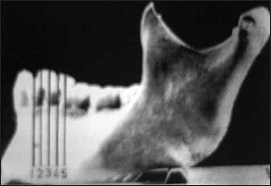
Picture for the numeric expression of the mental foramen position relative to the mandibular teeth. (1. position of FM as lying in line with long axis of the first premolar tooth, 2. interdental spaces between premolars, 3. line with long axis of the second premolar teeth, 4. Interdental spaces between second premolar and first molar, 5. line with long axis of the first molar root)
Figure 2.
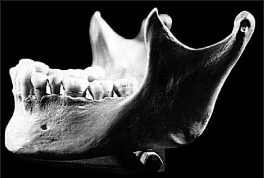
Measurement plane and the relation of the mental foramen on mandibla. Oclusal surface is parallel to the horizontal plane of the floor.
The horizontal position of the mental foramen
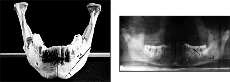
Horizontal positions of the mental foramen on dry skulls were determined in two visual projections, antera-posterior and profile, on drowned horizontal line parallel with oc-elusion surface that went through the center of the mental foramen cutting the mandible in the most anterior and the most distal point as shown in Figure 3. On the projected horizontal line, the distance from the foramen mental to the anterior point on mandibular symphysis was designated as Pd, and the distance from the mental foramen to the posterior border of the ramus of the mandible was designated as Zd. In sagital plane, the distance from the mental foramen to the middle line of the mandible was designated as Ps, and distance from the distal point of the mandible to the middle line was designated as Zs as shown on Figure 4.
Figure 3.
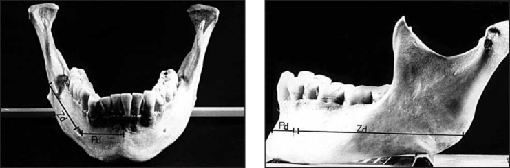
Measurement plane and relationship of the mental foramen to the sumphysis menti (Pd) and posterior border of the ramus of the mandible (Zd).A. Anterioposterior projection; B. Profile projection.
Figure 4.
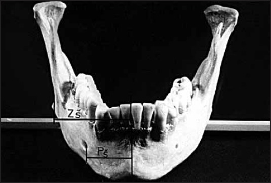
Plane of the measurement and relationship of the mental foramen to the medial skeletal line (Ps), and posterior border of the ramus of the mandible to the medial skeletal line(Zs).
The vertical position of the mental foramen
The technique used for vertical measurement was virtually the same as the Wical and Swope’s. These measurements were done on the drown vertical line perpendicular to the occlusive surface cutting the center of the mental foramen. The distance from the upper edge of the mental foramen to the alveolar crest was designated as Gv, and the distance from the lover edge of the mental foramen to the inferior border of mandible was designated as Dv as shown in Figure 5. These signs were different to those used by Wical and Swoope (2) to designate similar measurements.
Figure 5.
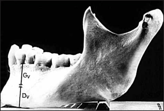
Plane of measurement and relation of the mental foramen to the alveolar crest (Gv) and inferior border of mandible (Dv).
The position of the mental foramen on orthopantomo-gram radiographs
Using this method and distances, appearances and position of the mental foramen from horizontal and vertical planes on the ortopantomogram radiographs was determined. Every mandible was placed in identical position on the orthopantomogram machine, fixed in the machine tube with occlusion surface parallel to the horizontal surface of room floor during recording. Figure 6. shows standard mandible radiographs obtained in this study. Contrast BaSO4 was injected in each mental foramen because of precise identification on X-ray.
Figure 6.
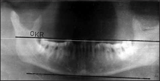
Radiographic images of mandible with oclusal surface parallel with the horizontal surface of the room floor when picture was recorded.
Figure 7. shows measurements done from horizontal position and distance of the mental foramen from the anterior point of corpus mandible (Pd), and distance of the mental foramen from the distal point of the ramus of the mandible (Zd), on determined horizontal line. Figure 8. shows measurements done from vertical position and the distance of the mental foramen from the alveolar crest (Gv), and distance of the mental foramen from the inferi- or mandible border (Dv) on drown vertical line.
Figure 7.

Distances measured on radiographic images of mandibles: (Pd) from foramen mental to the sumphysis,(Zd) from foramen mental to the posterior border of the ramus of the mandible.
Figure 8.

Distances measured on radiographic images of mandibles: (Dv) from inferior border of mandible to lower edge of mental foramen, (Gv) from upper edge of foramen mental to the alveolar crest.
The result obtained on dry skulls and on ortopantomogram radiographs were compared.
Results
For the clinical and diagnostic imaging purposes the medium values of the anatomical measurements can be considered sufficient. For anthropometric, anesthesiological and surgical aims knowing the maximum and the minimum values and the variation interval is also necessary.
The measurements, in this study, were made by anthropo-metric methods and are reported in the tables with mean values and their specific variation intervals.
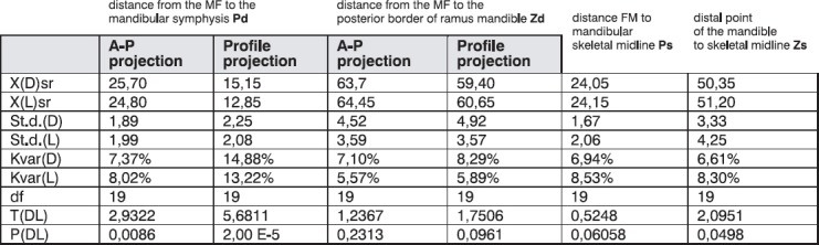
The means and standard deviations for the horizontal position of the mental foramen measurements of dry skulls from different projections are shown in Table 1.
Table 1.
The mean values of horizontal position of the mental foramen (MF) measured on the dry skulls from anteroposterior and profile projections.
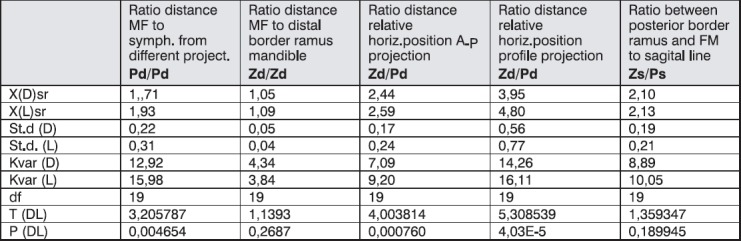
Using these values the ratios between measures from different projections Pd (a-p/profile) and Zd (a-p/profile) and ratios between Zd/Pd from the same projection designated as relative horizontal position were determined.
The means and standard deviations for those ratios and relative horizontal position measures on dry skulls are shown in Table 2.
Table 2.
Mean values of the ratios Pd distances from a-p and profile, Zd distances from a-p and profile and the relative horizontal position designated as ratios Zd/Pd from same projections measured on dry skulls.
The position of the mental foramen in the dry skulls with respect to teeth is shown in Table 3.
Table 3.
Position of the mental foramen in relation to the lower posterior teeth. (I - position of the mental foramen in line of the long axis of the first premolar tooth, II - position to interdental spaces between first and second premolar teeth, III - position of the foramen mental in line of the second premolar teeth, IV - position of the foramen mental to interdental spaces between second premolar and first molar teeth.

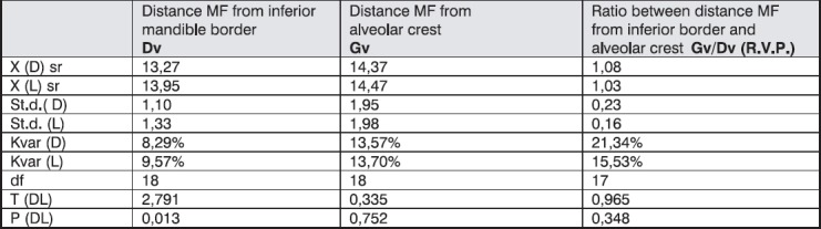
The mean values and standard deviations for the distance of the mental foramen from alveolar crest and inferior border of the mandible and their ratio determined as relative vertical position are shown in Table 4.
Table 4.
The mean values and standards deviation of the vertical position of foramen mental measured on dry skulls.
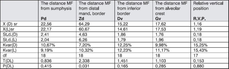
Mean values and standard deviations for the ortopantomogram measurements from horizontal and vertical projections are shown in Table 5.
Table 5.
Mean values of distances and ratios of the position of the mental foramen measured on the orthopantomogram radiographs.
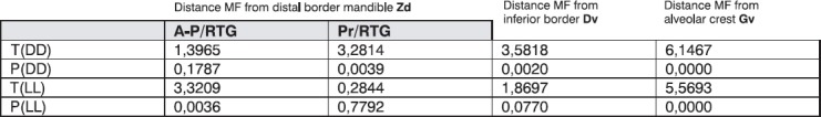
Comparison of mean values of measures on ortopantomogram and dry skulls are shown in Table 6.
Table 6.
Comparison of the mean values of distances of the mental foramen measured on dry skulls and orthopanto-mogram radiographs. No significant differences between distances designated as Zd and Dv, while significant differences were in distances designated as Gv between dry skulls and orthopantomogram radiographs.
Discusion
The present investigation is unique, because it was based on the material from Bosnian ethnic group.
The most common position of the mental foramen was in line with longitudinal axis of the second premolar followed closely by the location between the first and second premolars (Table 3). The mental foramen was not positioned symmetrically. These results concur with the research done by Moiseiwitsch (7) which conclud that foramen was generally found on average between premolars, determined on North American Caucasian population. Similar study was done on other ethnic groups and it yielded different results. Shankland (8) showed, in his study, that the mental foramen on Asian Indians mandibles was located in 75,36% of 138 total mandibular sides directly bellow the second premolar. Oguz (9), in his research, showed that the mental foramen was positioned in 61,7% of 34 of total Turkish adults on dry skulls below the root of the second premolar on the right and in 50 % on the left. Mbajiorgu (10) presented the most common position of the mental foramen between the lower premolars on Black Zimbabwean populations. These results, as other previous similar studies indicated ethnic and racial differences in most common positions of the mental foramen. In white population the mental foramen was positioned more distal than in black or Asian population. The results in the study confirmed these conclusions. Santini (11) compared positions of the mental foramen in Chinese and British population, and found that modal position of the mental foramen was relative to the standing mandible teeth in the British sample and lay in line with the longitudinal axis of the second premolar tooth whereas the modal position in the Chinese sample was in line between the first and second premolar teeth. The standard anatomy textbook notion that the foramen always lies between the first and second premolars cannot be accepted now. Racial differences occur and it is clear that in Asian and Chinese people, the most common position is between first and second premolar teeth whereas the result of this study also agree that in white Bosnian population the mental foramen was most commonly positioned in the long axis of the second premolar tooth. Clinicians should expect to find the mental foramen mostly in line between premolar in black or Asian population and more distal in white population.
According to the anatomy texts by Sicher (12) and Shiller (13), the mental foramen lies midway between the alveolar border and the inferior border of the mandible or closer to the latter. As can be seen in this study (Table 4), the mental foramen is positioned in the middle of the distance from the inferior border of the mandible to the alveolar crest on projected perpendicular line. These values are accordingly compared with the results of Oguz (9) that show that the distance of the mental foramen to the inferior border of the mandible is 14,61 mm and 14,29 mm to the right and left, then to the superior border was 13,62 mm to the right and 14,62 mm to the left. Mbajiorgu (10) showed that in the Black Zimbabweans population, in vertical plane, the mental foramen lays slightly below the midpoint of the distance between the lover border of the mandible and the alveolar margin in 41% of 32 mandibles right and 45,5% for the left sides. When these results are compared with the results of our study done on white Bosnian population no ethnic differences in the position of the mental foramen in the vertical plane are found.
Although the true vertical position of the mental foramen as measured on skulls is approximately centered in the alveolar bone, the majority of ortopantomogram radiographs showed the mental foramen significantly closer to the inferior border of the mandible than it actually is (Table 5). The distance of the mental foramen from the inferior mandible border was not significantly different compared with the measurements on dry skulls. The higher mean values of the relative vertical position in orthopan-tomogram of 1,19-0,20 right and 1,19-0,23 left confirm the lower positioned foramina on the radiographs and we conclude that orthopantomogram changes true values in vertical plane. Packota at al. (14) compared absolute radiographic measurements of marginal bone loss on ortho-pantomograms and intraoral radiographs with actual measurements on dry skulls. They concluded that all radiographic measurements overestimated the amount of bony support present and that in assessing marginal bone loss pantomographs gave results that deviated less from actual measurements on dry skulls than those obtained from intraoral radiographs. In studies of the mandible the mental foramen was used as a constant anatomic landmark based on the relative constancy of the relation of the foramen to the inferior border of the mandible in spite of resorbtion of the edentulous alveolar process (14). Wical and Swoope (2) believed that the lower edge of the mental foramen was used as a reference mark in panoramic radiographs for estimating the amount of alveolar bone loss.
Alveolar ridge height determined in their study was expressed as ratio instead of absolute measurements, because differences in patient size and the position may make absolute measurements less meaningful for clinical use. The basic method used in this investigation for the mandibular analyses was virtually the same as the one used by Wical and Swoope. Studies of the mandible in present study have used the mental foramen as a constant anatomical landmark based on the relative constancy of the relation of the foramen to the inferior border of the mandible in spite of resorption of the alveolar process. We believed that the lower edge of the mental foramen is to be used as a reference mark in orthopantomogram radiographs for estimating the amount of alveolar bone loss. We rationalized that the bone below the foramen consists of predictable proportion of the total bone height in most of normal patients and is not significantly affected by resorption until extreme atrophy occurs. One of the criteria used in our study was the position of the mandible in Orthopantomogram machine and clearly visible mental foramen (Figure 9).
Figure 9.
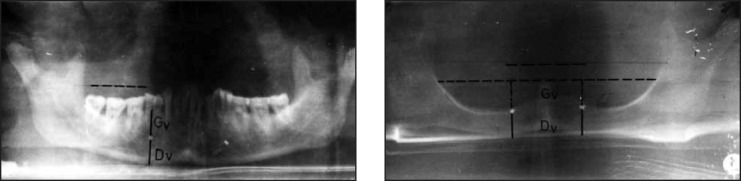
Relative vertical position (R.V.P.) and Dv as constant value, method for estimation of alveolar resorption. (A. radiograph mandible with intact alveolar bridge B. radiograph mandible with total resorbtion of alveolar bridge and R.V.P. ? 0)
The distance of the mental foramen from symphysis measured from A-P projection were 25,7 mm for right side and 24,8 mm for left side which coincides with research of Santini A., who compared the position of the mental foramen in different ethnic groups and showed dependence of the position of the mental foramen from ethnic and racial groups. The author showed distally positioned mental foramen in Chinese population compared to the British. The mean distance of the foramen from symphysis in the British was 25,2 mm and not significantly different when compared with our study. In Chinese that distance was 27,6 mm. The result of our study confirm that the mental foramen in Asian population is positioned more distally than in the white one. The ratio between the distances of the mental foramen from anterior point Pd and the distances of the mental foramen from distal point of the ramus of the mandible Zd designated as relative horizontal position were mean 2,44 right and 2,66 left for A-P projection. From that profile the values were right 4,06 and left 4,83 (Table 2). Comparing mean values between distances of the mental foramen from symphysis of the mandible (Pd) from different projections, we showed significant differences. The mean distances of the mental foramen from the distal point of the ramus of the mandibles showed no significant differences (Table 2), and their ratio had constant value of 1,065. Mean value of distances of the mental foramen from the middle line of the mandible was symmetrical and distances of the distal point of the mandible from the middle line was symmetrical also. Using the results in this study ratio of these distances has a constant value of 2,115 (Table 2). Using these results it was possible to determine the average angle corpus of the mandible behind the mental foramen in relation with the middle line. Using that ratio and the ratio above pointed that the corpus of the mandible behind the foramen is always a straight line that spreads out under the angle of 43 degrees to the middle line in normal mandible. (Figure 10, 11). It was possible to affirm that the ortopanthomogram twisted normal angle corpus of the mandible behind the mental foramen, 47 degrees to the normal mandible (Figure 12).
Figure 10.
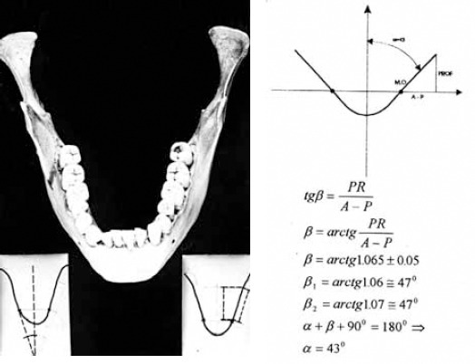
Scheme for the assessment of average angle strength of the corpus of mandible behind mental foramen under the angle of 43 degrees to the skeletal middle line using values ratios Zd / Pd = 2,115., and ratios Zd from AP and profile projection Zd/Zd 1,065.
Figure 11.
A) Average angle of strength of the mandible corpus behind foramen mental 43 degree. B) Average angle deformation strength of the mandible corpus behind the mental foramen on the orthopanthmogram of normal mandibles. It was possible to affirm that surrender in normal average values measurements on orthopantomograms radiograph images; R.H.P. (2,83 - 2,73), R.V.P. (1,16 - 1,19) and angle of 47 degrees suggested an anomaly.
Figure 12.
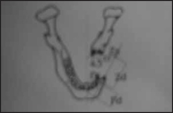
The scheme for adequate preoperative reconstruction of corpus mandible defect using values designated in this study; Relative horizontal position designated as ratio Zd/Pd, relationship of distal point of mandible and foramen mental in accordance with medial skeletal midline Zs/Ps determined as 2,1, and angle of corpus mandible behind mental foramen is 43 degree
These results using the mental foramen as a constant anatomic landmark and its relation with other parts of the mandible may be used in preoperative schedules and planning of reconstructive procedures on the mandible after resection, surgery or posttraumatic defects.
Knowledge of the distances from surgically encountered anatomic landmarks may be of assistance in locating these important maxillofacial neurologic structures during many procedures. This information may play an even more important role in new techniques for minimally invasive surgery that are developed. Understanding the location of these foramina will also assist the clinicians in performing local anesthetic blocks procedures.
Conclusions
- The most common position of the mental foramen in relation to the mandibular teeth was in line with the second premolar teeth in 47,06 % right and 64,71% left, and in the line between first and second premolar teeth in 41,18 % right 29,41 % left.
- In the sagital plane, mean value of the distance from the mental foramen to the middle line of the mandible was 24,05 mm right and 24,15 mm left, and the mean distance from the distal point of the mandible to the middle line was 50,35 mm right and 51,20 mm left.
- The mean distance in the horizontal plane on the skulls from the foramen mental to the anterior point on the mandibular symphysis from A-P projection was 29,41 mm right, 24,80 mm left and 15,15 mm right and 12,85 mm left from the profile projection.
- The mean distance from the mental foramen to the posterior border of the ramus of the mandible was 63,7 mm right and 64,45 mm left measured from A-P projection, and 59,40 mm right and 60,65 mm left, measured from the profile projection.
- On the ortopantomogram mean distance measured from the foramen mental to the medial point of the corpus mandible was 22,56 mm right and 22,17 mm left and was significantly different when compared with the values determined on the skulls
- The mean distance from the foramen mental to the posterior border of the ramus of the mandible was 64,29 mm right and 60,67 mm left measured on the ortopantomogram and there were no significant differences compared with the measurements determined on the skulls.
- The mean distance from the upper edge of the mental foramen to the alveolar crest measured on the skulls was 14,37 mm right and 14,47 mm left and the mean distance from the lover edge of the mental foramen to the inferior border of mandible was 13,27 mm right and 13,95 mm left.
- The mean distance from the upper edge of the mental foramen to the alveolar crest measured on the orto-pantomogram was 17,62 mm right and 17,53 mm left and was significantly different when compared with this values determined on the skulls
- The mean distance from the lover edge of the mental foramen to the inferior border of mandible on the or-topantomogram was 15,22 mm right and 14,61 mm left and there were no significant differences compared with the measurements determined on the skulls.
- Ratio of these values determined as relative vertical position and relative horizontal position measured on the ortopantomogram may be used in preoperative planning of reconstructive procedures on the mandible
References
- (1).Sto{i} S. Varijacije bradnog otvora, stomatoloski glasnik Srbije. 1978;3:203–208. [PubMed] [Google Scholar]
- (2).Wickal K.E, Swoope C.C. Studies of residual ridge resorbtion. Part I. Use of panoramic radiograph for evaluation and classification of mandibular resorption. J. Prost. Dent. 1974;32:7–12. doi: 10.1016/0022-3913(74)90093-6. [DOI] [PubMed] [Google Scholar]
- (3).Takashi Y, Sharon L.B. The appearance of the mental foramina on panoramic radiographs. I. Evaluation of the patients. Oral Surg, Oral Med, Oral Path. 1989;68:360–364. doi: 10.1016/0030-4220(89)90224-7. [DOI] [PubMed] [Google Scholar]
- (4).Takashi Y, Sharon L.B. The appearance of the mental foramina on panoramic and periapic radiographs. II. Evaluation of the patients. Oral Surg. Oral Med. Oral Path. 1989;68:488–492. [Google Scholar]
- (5).Alantar S.A, Collent W, Ronald A, Baughman L. A survey of panoramic radiographs for evaluation of normal and pathologic findings. Oral Surgery, Oral. Med, Oral Path. 1988;50:472–478. doi: 10.1016/s0030-4220(80)80017-x. [DOI] [PubMed] [Google Scholar]
- (6).Morrant G.M. A biometric study of the human mandible. Biometrika. 1936;28:84–122. [Google Scholar]
- (7).Moiseiwitsch J.R. Position of the mental foramen in a North American, white population. Oral Surg Oral Med Oral Pathol Oral Radiol Endod. 1998;85:457–460. doi: 10.1016/s1079-2104(98)90074-9. [DOI] [PubMed] [Google Scholar]
- (8).Shankland W.E. The position of the mental foramen in Asian Indians. J Oral Impiantai. 1994;20:118–123. [PubMed] [Google Scholar]
- (9).Oguz 0, Bozkir M.G. Evaluation of location of mandibular and mental foramina in dry, young, adult human male, dentulous mandibles. West Indian Med. J. 2002;51:14–16. [PubMed] [Google Scholar]
- (10).Mbajiorgu E.F, Mawera G, Asala S.A, @ivanovi} S. Position of the mental foramen in adult black Zimbabwean mandibles: a clinical anatomical study. Cent. Afr. J. Med. 1998;44:24–30. [PubMed] [Google Scholar]
- (11).Santini A, Moira L. A comparison of the Position of the mental foramen in Chinese and British mandibles. Acta Anat. 1990;137:208–212. doi: 10.1159/000146822. [DOI] [PubMed] [Google Scholar]
- (12).Sicher L.L, Du Bral E. Oral Anatomy. 6th ed. St Louis: The CVMosby Co; 1975. pp. 424–430. 450-451. 1954; 118: 387-390. [Google Scholar]
- (13).Shiller W.R. The mental foramina of the mandible. Anatomical Record. 1954;118:387–390. doi: 10.1002/ar.1091190308. [DOI] [PubMed] [Google Scholar]
- (14).Packota G.V, Hoover J.N, Neufeld B.D. A study of the height of intact alveolar bone on panoramic and intraoral radiographs of adult patients. J. Prosthetic Dentistry. 1988;60:504–510. doi: 10.1016/0022-3913(88)90258-2. [DOI] [PubMed] [Google Scholar]