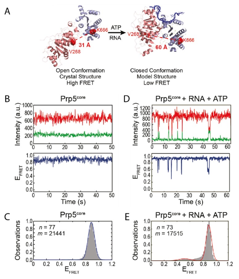
Figure 1.
Conformational changes within Prp5. (A) Crystal structure of Prp5 in the twisted open conformation and modelled structure of closed conformation. The two RecA-like domains of Prp5 are shown in pink and purple and the RNA in the closed conformation in blue. The residues mutated for fluorophore introduction are indicated in red. (B) Exemplary Förster resonance energy transfer (FRET) trajectory of Prp5 alone and (C) the corresponding FRET histogram fitted with one peak (blue line). (D) FRET behavior of Prp5 in the presence of poly (A) RNA and ATP and (E) the corresponding FRET histogram fitted with two peaks (red line). This Figure was taken from Beier et al. [18] and is used under Creative Commons Non-Commercial License (CC-BY NC).

