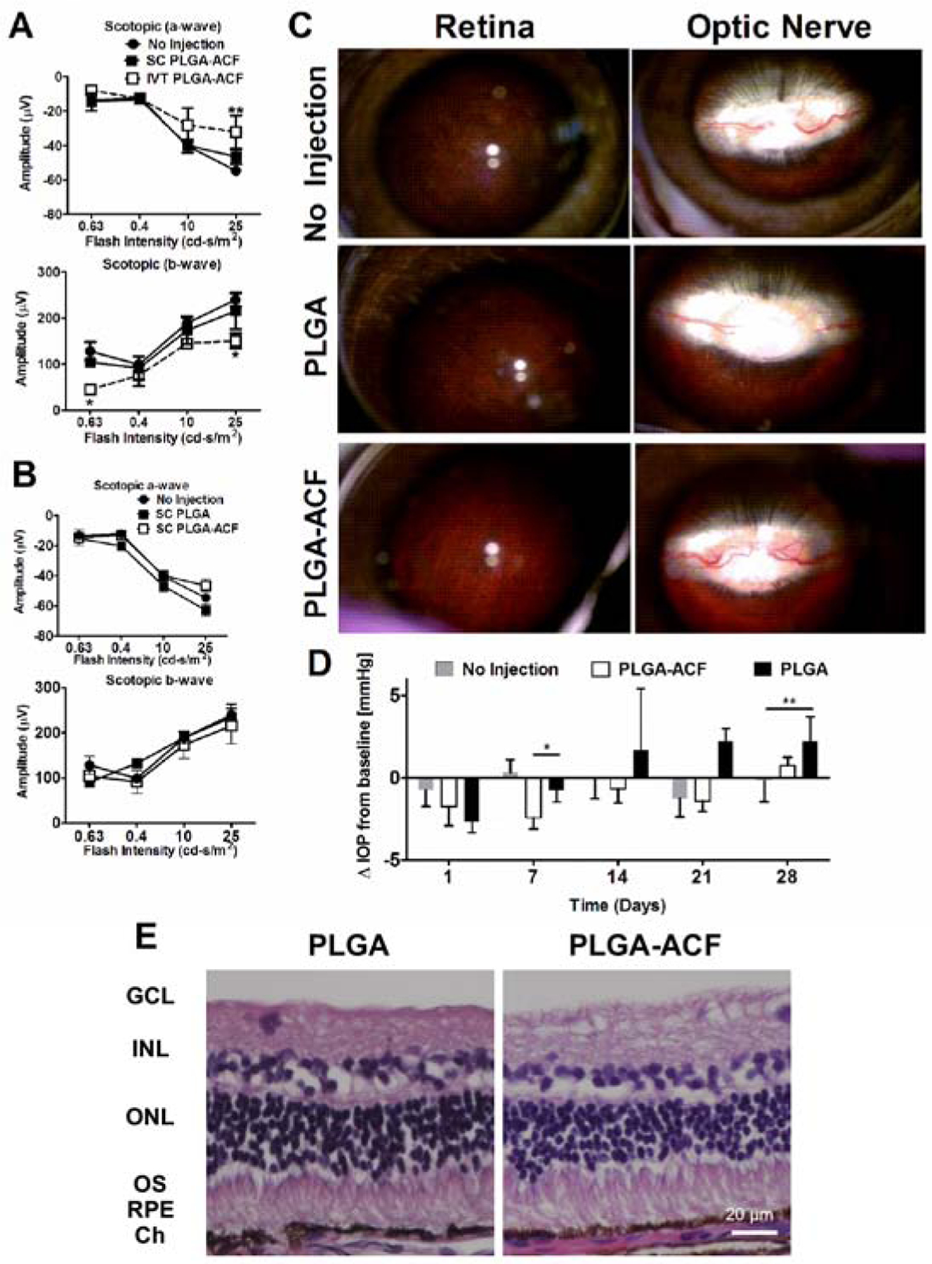
Figure 4. Suprachoroidal Injection (SC), but not Intravitreous Injection (IVT) of PLGA-Acriflavine (PLGA-ACF) Microparticles (MPs) in Rabbits is Safe.

Dutch belted rabbits were given a suprachoroidal (SC) or intravitreous injection of 50 μl containing 10 mg of PLGA-ACF MPs (38 μg of ACF), 10 mg of empty PLGA MPs, or no injection (n=3–4). (A) Scotopic electroretinograms 28 days after injection showed that compared to eyes with no injection, there was no significant difference in mean (±SEM) a-wave or b-wave amplitude in eyes given a SC injection of PLGA-ACF, but there was a significant reduction in mean a-wave amplitude at the highest stimulus intensity and in b-wave amplitude at the highest and lowest stimulus intensity in eyes given a an IVT injection of PLGA-ACF (*p<0.05; **p<0.01 by ANOVA with Bonferroni correction for multiple comparisons). (B) Compared with eyes that received no injection, there was no significant reduction in a-wave or b-wave amplitudes 28 days after SC injection 50 μl containing 10 mg of PLGA-ACF MPs (38 μg of ACF) or 10 mg of empty PLGA MPs (n=3–4). (C) Fundus photographs showed normal appearing peripheral retina (first column) and optic nerve (second column) 28 days after SC injection of 50 μl containing 10 mg of PLGA-ACF MPs (38 μg of ACF) or 10 mg of empty PLGA MPs, indistinguishable from eyes that had no injection. (D) Mean (± SEM) changes from baseline intraocular pressure (ΔIOP) were small and not clinically significant through 28 days after SC injections of PLGA-ACF MPs or PLGA MPs, but there were some statistically significant differences between groups. At day 7, there was a significant reduction in the PLGA-ACF group compared with the others (*p<0.05 by ANOVA with Bonferroni correction for multiple comparisons) and at day 28, there was a significant increase in the PLGA group compared with no injection (**p<0.01). (E) Histopathology of the retina was normal 28 days after SC injection of PLGA-ACF or PLGA MPs (n=3).
