

Abstract
Mycologists have always been curious about the elaborate morphotypes and shapes of species of the genus Helvella. The small, black, cupulate Helvella specimens have mostly been assigned to Helvella corium, a broadly defined morpho-species. Recent phylogenetic analyses, however, have revealed an aggregate of species hidden under this name. We performed a multispecies coalescent analysis to re-assess species limits and evolutionary relationships of the Helvella corium species aggregate in the Nordic countries. To achieve this, we used morphology and phylogenetic evidence from five loci – heat shock protein 90 (hsp), translation elongation factor 1-alpha (tef), RNA polymerase II (rpb2), and the 5.8S and large subunit (LSU) of the nuclear ribosomal DNA. All specimens under the name Helvella corium in the larger university fungaria of Norway, Sweden and Denmark were examined and barcoded, using partial hsp and/or rpb2 as the preferential secondary barcodes in Helvella. Additional fresh specimens were collected in three years (2015–2018) to obtain in vivo morphological data to aid in species discrimination. The H. corium species aggregate consists of seven phylogenetically distinct species, nested in three divergent lineages, i.e. H. corium, H. alpina and H. pseudoalpina sp. nov. in the /alpina-corium lineage, H. alpestris, H. macrosperma and H. nannfeldtii in the /alpestris-nannfeldtii lineage, and H. alpicola as a weakly supported sister to the /alpestris-nannfeldtii lineage. Among the seven species, the ribosomal loci expressed substantial variation in evolutionary rates, suggesting care in the use of these regions alone in delimitation of Helvella species. Altogether, 469 out of 496 available fungarium specimens were successfully barcoded.
Keywords: molecular phylogeny, new taxon, Pezizales, Stacey
INTRODUCTION
Helvella is a species-rich genus of ascomycetous fungi (Pezizales, Helvellaceae) probably consisting of more than 100 species worldwide. Recently, the broad concept of Helvella was restricted, i.e. to exclude some para- and polyphyletic groups of species now referred to the segregate genera Balsamia, Dissingia, Pindara and Wynnella (Hansen et al. 2019). Originally, Helvella included only stipitate-capitate (i.e. non-cupulate) taxa, but cupulate species, which are today included in Helvella, were assigned to Peziza and/or Acetabula (Pezizaceae) (Fries 1822, Fuckel 1870). Quélet (1886), and later Nannfeldt (1937) and Dissing (Dissing 1966a, b), introduced a broad generic concept that includes both stipitate-capitate (saddle-shaped) and stipitate-cupulate taxa in Helvella (sensu lato). They considered apothecial gross morphology too variable and phenotypically plastic to be useful in genus level delineation. Instead, microanatomy of excipular tissues was introduced as more useful for this purpose. Species with similar microanatomy, the capitate as well as the cupulate, were thus included in Helvella. Still, apothecial gross morphology was retained as an important characteristic in delineation of subgroups (sections) within the genus (Boudier 1907, Dissing 1964, 1966b).
Helvella corium, first described as Peziza corium by Weberbauer (1873), is a medium-sized, stipitate-cupulate black species. It has seen many revisions over time. It was referred to as Cyathipodia and Leptopodia by Boudier (1907, 1910) until it was accommodated in Helvella (Massée 1895). Over the years, a few additional species have been introduced as closely related to H. corium. Helvella alpestris was described from alpine areas of France (Boudier & Fischer 1894, Boudier 1895), H. arctica (Nannfeldt 1937) from Abisko in Northern Sweden and H. arctica var. macrosperma (Favre 1955) from the Swiss Alps. The latter variety was later given specific rank by Fellner & Landa (1991), as has been followed by subsequent authors (Van Vooren 2014, 2015, Skrede et al. 2017). Contrary, in his monumental work on Helvella, Dissing (1966b) included H. alpestris, H. arctica and H. arctica var. macrosperma as part of his broad concept of H. corium.
Traditionally, fungal species have been recognised based on morphological characters, ecology and/or mating behavioural traits. Since many morphological traits in fungi show a high degree of phenotypic plasticity (West-Eberhard 1989), it has often been challenging for taxonomists to interpret and understand patterns of fungal relationships. Today, with the availability of molecular data, the concept of genealogical species recognition [i.e. recognition of species based on congruent gene trees across multiple unlinked loci (Taylor et al. 2000, Dettman et al. 2003)] has completely out-performed traditional morphological species concepts in this group of organisms.
Molecular markers used to infer phylogenies and erect phylogenetic species in fungi are diverse, including both RNA-coding, non-coding and protein coding loci. Several studies have pointed out that single-copy, protein-coding markers are preferred and may perform better than multi-copy ribosomal genes in delimiting fungal species (Raja et al. 2011, Stielow et al. 2015, Hansen & Olariaga 2015). For species delineation the commonly used protein-coding loci include β-tubulin (tub2), the translation elongation factor 1-α (tef) and the DNA directed RNA polymerase II core subunits (rpb1 and rpb2). The latter locus has proven useful in resolution of deep as well as shallow clades of ascomycetes (Spatafora et al. 2006, Hofstetter et al. 2007, Schoch et al. 2009). Recently, the minichromosome maintenance complex component 7 (mcm7) and the ribosome biogenesis protein tsr1 were added to the list of valuable markers for resolving high- as well as low level taxonomic units in Ascomycota (Aguileta et al. 2008, Raja et al. 2011). In Helvella a portion of the heat shock protein 90 (hsp), in combination with rpb2, were shown to have strong discriminating power at the species level (Skrede et al. 2017). Hsp was also shown to be successfully amplified from fungaria specimens. This is probably due to the short sequence length (i.e. 272 bp). It was introduced as a most useful secondary barcode marker in Helvella (Skrede et al. 2017).
In the study by Skrede et al. (2017), species limits, phylogeny and taxonomy within Helvella were assessed, using a multilocus genealogical approach. They found that the morphospecies concept of H. corium in fact represented a pseudo-cryptic species aggregate that comprised five phylogenetic species nested in two divergent evolutionary lineages: the /alpina-corium lineage with H. corium and H. alpina, and the /alpestris-nannfeldtii lineage with H. alpestris, H. macrosperma and H. nannfeldtii. It was suggested as well that a sixth species, i.e. H. alpicola, might belong to this species aggregate, but its phylogenetic placement within the genus was unresolved (Skrede et al. 2017). The phylogenetic species of this species aggregate are morphologically very similar and therefore difficult to distinguish in the field. They also occupy overlapping habitats of the alpine zone, often in close proximity to Salix spp. (Weidemann 1998).
It is against this background that we aimed to get a better resolution of the species-level relationships within the H. corium species aggregate, using a broader set of genetic markers. In addition, we wanted to re-evaluate morphological characters used in species discrimination.
MATERIALS AND METHODS
The study is based on a set of newly collected specimens of the Helvella corium aggregate in the years 2015 to 2017 and samples deposited as H. corium and associated species in the Nordic University fungaria of O, TRH, BG, TROM, S, UPS, GB, UME, and C (in total 496 specimens). All specimens were identified by molecular barcoding (Tables 1, S1).
Table 1.
Helvella specimens included in the phylogenetic analyses of this study. Specimens included in the final analysis are marked with an asterisk. All specimens are marked with sample ID and fungaria ID. Type specimens are written in bold. GenBank accession numbers are added for all obtained sequences. X indicates sequence was not long enough for publishing in GenBank.
| Species | Sample and fungarium ID | Locality | Collection year | GenBank accession number | ||||
|---|---|---|---|---|---|---|---|---|
| hsp | rpb2 | tef | LSU | ITS | ||||
| H. corium | H2184, O-255756* | Svalbard. Longyearbyen | 2017 | MN692370 | MN692314 | MN658196 | MN655873 | MN656185 |
| H547, O-255757* | France. Savoie | 1992 | MN692363 | MN692309 | MN658197 | MN655870 | MN656165 | |
| H436, O-253281 | Svalbard. Spitsbergen. Kongsfjorden | 1988 | KY784528 | KY772771 | – | KY773163 | – | |
| H950, TROM-F-610059 | Norway. Nordland. Saltdal. Junkerdalen | 2016 | MN692364 | MN692312 | – | MN655880 | – | |
| H956, O-255753 | Norway. Hedmark. Folldal. Einunndalen | 2016 | MN692365 | MN692313 | – | MN655875 | – | |
| H957, O-255754 | Norway. Sør-Trøndelag. Oppdal. Vinstradalen | 2016 | MN692366 | MN692310 | – | MN655871 | – | |
| H1958, TROM-F-610014 | Norway. Troms. Salangen | 2017 | MN692369 | – | – | MN655876 | MN656172 | |
| H248, O-253277 | Norway. Hordaland. Ulvik. Finse | 1996 | KY784366 | KY772614 | – | KY773075 | – | |
| H1088, C-F-86904 | Greenland. Thule Airbase | 1994 | MN692367 | – | – | MN655872 | – | |
| H1089, C-F-63828 | Greenland. Mestervig. Ochenpas | 1968 | MN692368 | – | – | MN655874 | – | |
| H294, C-F-16568 | Russia. North Ural Mountaint | 1990 | KY784407 | KY772654 | – | KY773100 | – | |
| H. alpina | H2106, O-255751* | Norway. Oppland. Dovre. Grimdalen | 2017 | MN692362 | MN692307 | MN689291 | MN655865 | MN656181 |
| H1124, C-F-55730* | Greenland | 1987 | MN692361 | MN692306 | MN658193 | MN655863 | MN656171 | |
| H223, O-253228* | France. Savoie | 1992 | KY784343 | KY772593 | MN658192 | KY773054 | – | |
| H336, O-253227 | Canada. British Colombia. Whistler | 1994 | KY784439 | KY772690 | – | KY773116 | – | |
| H1159, C-F-54601 | Norway. Nordland. Rana. Virvassdalen | 1979 | MN692359 | X | – | MN655864 | – | |
| H540, C-F-34420 | Russia. Khatanga airport | NA | MN692358 | MN692305 | – | MN655862 | – | |
| H1095, C-F-50287 | Greenland. Sdr. Strømfjord | 1982 | MN692360 | – | – | – | – | |
| H. pseudoalpina | H498, O-255748* | Svalbard. Longyearbyen | 2015 | MN692354 | MN692302 | MN689292 | MN655869 | MN656164 |
| H1965, TROM-F-610048* | Norway. Troms. Balsjord | 2017 | MN692355 | MN692303 | MN658194 | MN655866 | MN656173 | |
| H1966, TROM-F-610049* | Norway. Troms. Balsjord | 2017 | MN692356 | MN692304 | MN658195 | MN655868 | – | |
| H2278, TRH-F-20631 | Norway. Nordland, Saltdal, Junkerdalen. Bibeldalen | 1988 | MN692357 | MN692308 | – | MN655867 | – | |
| H. alpicola | H231, O-253226* | Switzerland. Graubunden | 1984 | KY784349 | KY772598 | MN689298 | KY773061 | – |
| H952, TROM-F-610061* | Norway. Nordland. Saltdal. Junkerdalen | 2016 | MN692344 | MN692296 | MN658206 | MN655847 | – | |
| H953, TROM-F-610062* | Norway. Nordland. Saltdal. Junkerdalen | 2016 | MN692345 | MN692297 | MN658207 | MN655848 | – | |
| H1439, C-F-53856 | Norway. Nordland. Rana | 1972 | MN692346 | – | – | – | – | |
| H1534, C-F-103009 | Norway. Nordland. Rana | 1971 | X | X | – | – | – | |
| H. nannfeldtii | H1972, TROM-F-610034* | Norway. Troms. Kåfjord | 2017 | MN692330 | MN692291 | MN658199 | MN655861 | MN656174 |
| H954, O-255744* | Norway. Hedmark. Folldal | 2016 | MN692328 | MN692290 | MN689293 | MN655860 | MN656167 | |
| H2104, O-255745* | Norway. Oppland. Dovre. Grimsdalen | 2017 | MN692327 | MN692293 | MN658200 | MN655859 | – | |
| H027, O-253338* | Norway. Oppland. Dovre. Grimsdalen | 2009 | KY784203 | KY772447 | KY772836 | KY772919 | X | |
| H1994, TROM-F-610036* | Norway. Troms. Målselv | 2017 | MN692326 | MN692292 | MN689294 | MN655856 | – | |
| H212, O-253332* | France. Savoie. Val d'Isere | 1992 | MN692323 | KY772585 | KY772888 | KY773044 | – | |
| H216, O-253333* | France. Savoie. Bon Valle | 1992 | KY784337 | KY772589 | KY772891 | KY773048 | – | |
| H545, O-255740 | Austria. Tirol. Obergurgl | – | MN692324 | MN692288 | – | MN655858 | – | |
| H564, O-255741 | France. Savoie. Val d'Isere | 1992 | MN692325 | MN692289 | – | MN655857 | – | |
| H1105, C-F-86947 | Greenland. Narssarssuaq. | 1991 | MN692329 | – | – | – | – | |
| Helvalla sp | H1995, O-255738* | Norway. Troms. Målselv | 2017 | MN692339 | MN692283 | MN689299 | MN655845 | MN656178 |
| H1996, O-255739* | Norway. Troms. Målselv | 2017 | MN692340 | MN692284 | MN689300 | MN655846 | MN656179 | |
| H. alpestris | H460, ex DAOM574891* | Canada. Nuvanut | 2014 | KY784542 | KY772789 | MN658202 | MN655837 | MN656163 |
| H548, O-255736* | France. Savoie. Val d'Isere | 1992 | MN692332 | MN692276 | MN658203 | MN655835 | MN656166 | |
| H2111, O-255737* | Norway. Oppland. Dovre. Grimsdalen | 2017 | MN692341 | MN692280 | MN658205 | MN655834 | MN656183 | |
| H865, S-F-122351* | Norway. Oppland. Dovre. Grimsdalen | 1985 | MN692334 | MN692277 | MN658204 | MN655838 | – | |
| H1098, C-F-50667 | Greenland. Ella Island. St. Elvdal E of the house. | 1982 | MN692342 | MN692279 | – | MN655839 | – | |
| H719, UPS-F-145393 | Sweden. Torne Lappmark. Jukkasjärvi | 1946 | MN692333 | X | – | MN655836 | – | |
| H916, TROM-F-11410 | Norway. Nordland. Fauske. Blåmannsisen | 1967 | MN692335 | MN692278 | – | – | – | |
| H928, TROM-F-11403 | Norway. Troms. Tromsø. Tromsø museum | 1980 | MN692336 | MN692311 | – | MN655833 | – | |
| H1103, C-F-86999 | Greenland. Std. Strømfjord, 4km E of airport | 1988 | MN692337 | – | – | – | – | |
| H1115, C-F-86617 | Greenland. Jameson Land. Valley W of Nathorts fjeld | 1989 | MN692338 | – | – | MN655834 | – | |
| H1190, C-F-45351 | Norway. Nordland. Fauske. Blåmannsisen | 1967 | MN692343 | – | – | – | – | |
| H. macrosperma | H2146, O-F-285169* | Norway. Oppland. Lom | 2007 | MN692322 | MN692286 | MN658201 | MN655844 | MN656184 |
| H2100, TROM-F-610004* | Norway. Troms. Balsfjord | 2017 | MN692321 | MN692285 | MN658198 | MN655843 | MN656180 | |
| H947, TROM-F-610056* | Norway. Nordland. Junkerdalen | 2016 | MN692318 | MN692281 | MN689295 | MN655841 | – | |
| H029, O-253328* | Norway. Oppland. Dovre. Grimsdalen | 2007 | KY784205 | KY772449 | KY772838 | KY772921 | MN656159 | |
| H047, O-253329* | Norway. Oppland. Dovre. Grimsdalen | 2009 | KY784217 | KY772462 | KY772850 | KY772936 | – | |
| H1982, TROM-F-610001* | Norway. Troms. Målselv | 2017 | MN692319 | MN692282 | MN689297 | MN655842 | MN656176 | |
| H050, O-253330* | Norway. Oppland. Dovre. Grimsdalen | 2009 | MN692316 | KY772464 | KY772852 | KY772938 | – | |
| H053, O-253331* | Norway. Oppland. Dovre. Grimsdalen | 2009 | MN692317 | KY772466 | MN689296 | KY772939 | – | |
| H1997, TROM-F-610017 | Norway. Troms. Balsfjord. Lakselvbukt | 2017 | MN692320 | MN692287 | – | MN655840 | – | |
| H. arctoalpina | H033, O-253237* | Norway. Oppland. Dovre. Grimsdalen | 2009 | KY784207 | KY772453 | KY772841 | KY772924 | – |
| H. acetabulum | H225, O-253212* | Norway. Oppland. Dovre. Grimsdalen | 1984 | KY784344 | KY772594 | KY772894 | KY773055 | MN656162 |
| H. subilica | H148, O-70080* | Norway. Akershus. Asker | 1994 | KY784281 | KY772531 | KY772880 | KY772997 | MN656160 |
| H. rivularis | H1978, O-255764* | Norway. Troms. Balsfjord | 2017 | MN692371 | MN692295 | MN689306 | MN655850 | MN656175 |
| H. fallax | H018, O-253351* | Norway. Oppland. Dovre. Grimsdalen | 2009 | KY784195 | KY772439 | KY772830 | KY772913 | – |
| H. pezizoides | H061, O-253366* | Sweden. Halmstad | 2009 | KY784225 | KY772471 | KY772854 | KY772945 | – |
| H. scyphoides | H140, O-65348* | Norway. Hedmark. Åmot | 2002 | KY784273 | KY772523 | KY772879 | KY772989 | – |
| Helvalla sp2 | H1983, O-255763* | Norway. Troms. Målselv | 2017 | MN692351 | MN692294 | MN689305 | MN655849 | MN656177 |
| H. macropus | H238, O-291425* | Norway. Rogaland | 2009 | KY784356 | KY772605 | KY772896 | KY773067 | – |
| H. fibrosa | H240, O-291352* | Norway. Sør-Trøndelag | 2008 | KY784358 | KY772607 | KY772898 | KY773069 | – |
| H. lacunosa | H1041, O-255761* | Norway. Nordland. Saltdal. Junkerdalen | 2016 | MN692347 | MN692299 | MN689302 | MN655855 | MN656169 |
| H. atra | H1055, O-255762* | Norway. Hedmark. Kvikne | 2016 | MN692348 | MN692300 | MN689304 | MN655852 | MN656170 |
| H. philonotis | H2110, O-255760* | Norway. Oppland. Dovre. Grimsdalen | 2017 | MN692353 | MN692301 | MN689303 | MN655853 | MN656182 |
| H. calycina | H022, O-253255* | Norway. Oppland. Dovre. Grimsdalen. | 2009 | KY784198 | KY772442 | KY772833 | KY772915 | MN656158 |
| H. costifera | H247, O-253283* | Norway. Oppland. Vågå | 1998 | KY784365 | KY772613 | KY772900 | KY773074 | – |
| H. crispa | H235, O-360158 | Norway. Nordland. Andøy | 2005 | KY784353 | KY772602 | – | KY773065 | – |
| H. hyperborea | H1309, C-F-55004 | Norway. Nordland. Rana | 1981 | MN692349 | X | MN689301 | – | – |
| H. hypocrateriformis | H275, C-F-57126 | Switzerland. Graubünden | 1982 | KY784390 | KY772638 | – | – | – |
| H. pulla | H149, O-069282 | Norway. Møre og Romsdal. Nesset | 2008 | KY784282 | KY772532 | – | KY772998 | MN656161 |
| H. bicolor | H1033, O-255759 | Norway. Nordland. Saltdal. Junkerdalen | 2016 | MN692352 | MN692298 | – | MN655851 | MN656168 |
| H. capucina | H1051, O-255758 | Norway. Hedmark. Kvikne. Innerdalsvatnet | 2016 | MN692350 | MN692315 | – | MN655854 | – |
DNA extraction and PCR
A small (sesame seed sized) piece of the stipe of each fungarium specimen was put in CTAB Lysis buffer (BioChemica, Panreac AppliChem) for DNA extraction. For the specimens that were newly collected for this study, fresh pieces of ascomata were put directly in CTAB buffer before drying the whole ascocarp at 30 °C for 24 h.
Prior to DNA extraction, one tungsten bead was added to each sample, i.e. specimens in CTAB Lysis buffer (AppliChem Panreac). Samples were first frozen, then incubated at 65 °C on a heating block for 30 min, and subsequently vortexed thoroughly. Samples were frozen a second time, and allowed to thaw at room temperature before DNA extraction. DNA was extracted using the E.Z.N.A.®HP Fungal DNA Kit (Omega Biotek D3195), following a slightly modified version of the manufacturer's protocol for dried samples, i.e. the optional step of adding 10 μL 2-mercaptoethanol was undertaken, and the column was equilibrated with NaOH. The DNA was eluted in 50 μL elution buffer to increase DNA concentration.
For barcoding purposes, efforts were made to PCR amplify a partial segment of the heat shock protein 90 (hsp) for all sampled specimens. In addition, a subset of 80 specimens, of which 21 represented outgroup taxa, were selected to infer multilocus phylogenies (Table 1). Within the species aggregate an effort was made to include broad taxon sampling. For some species, this proved difficult due to lack of collections (i.e. H. alpicola, H. macrosperma). Five loci were targeted: a 272 bp region of the protein-coding gene hsp, a 356 bp region of the protein-coding gene rpb2, a 571 bp region of the protein-coding gene tef, a 743 bp region of the large ribosomal subunit LSU (including the D1 and D2 regions), and the whole internal transcribed spacer region (ITS1, 5.8S, ITS2). Only the highly conserved 160 bp region of the 5.8S ribosomal RNA region was used in the phylogenetic analyses since the complete ITS region is difficult to align across the whole species aggregate. The primers used are shown in Table 2.
Table 2.
PCR and sequencing primers used to amplify the Helvella corium species aggregate and relevant outgroup taxa in the study.
| Locus1 | Forward primer sequence (5ʹ-3ʹ) | Reverse primer sequence (5ʹ-3ʹ) |
|---|---|---|
| hsp | H_hspf4: CRGGCATCCGGGTGACGTAAT | H_hspr4: AGGGKGTTGTCGACTCCGAGG |
| rpb2 | H_rpb2r24: TCCACAATCTGCATCCCGATTC | H_rpb2f4: CCAGACATGGACAGAAGGTTGAG |
| tef | EF595F3: CGTGACTTCATCAAGAACATG | EF1160R3: CCGATCTTGTAGACGTCCTG |
| LSU | H_LSUf12: AGCGGAGGAAAGAAACCAAC | H_LSUr22: TCCCAACAGCTATGCTCCTAC |
| ITS | ITS55: GGAAGTAAAAGTCGTAACAAGG | ITS45: TCCTCCGCTTATTGATATGC |
| ITS2 | ITS35: GCATCGATGAAGAACGCAGC | ITS45: TCCTCCGCTTATTGATATGC |
1LSU: 28S large subunit ribosomal RNA, domains D1/D2; rpb2: RNA polymerase II, second largest subunit; hsp: heat shock protein 90; tef: translation elongation factor 1-alpha; ITS: The internal transcribed spacer region (ITS1, 5.8S and ITS2).
2From Landeros et al. (2015), modified in Skrede et al. (2017).
3From (Kauserud & Schumacher 2001).
4From Skrede et al. (2017).
5From White et al. (1990).
For all PCR reactions, PuReTaq Ready-To-Go PCR Beads (GEhealthcare, Waukesha, WI) were used in 25 μL reactions. The following PCR protocols were used to amplify the five loci: 4 min at 95 °C, 40 (50 for LSU) cycles of 25 s (30 s for hsp and LSU) at 95 °C, 30 s at 53 °C (58 °C for hsp, 52°C for LSU) and 60 s at 72 °C, followed by a 10 min extension at 72 °C and an indefinite hold at 10 °C. Amplified PCR products were visualized with electrophoresis on 1 % agarose gels. For PCR reactions that yielded product, 5 μL PCR product was purified with 0.2 μL ExoSAP-IT (GEhealthcare) and 1.8 μL H2O. Samples were then run on a thermocycler at 37 °C for 15 min, followed by 80 °C for 15 min. Cleaned PCR product was diluted with 45 μL water per sample. 5 μL PCR product and 5 μL primer were added to clean tubes and labelled before sequencing. Sanger sequencing was performed by GATC Biotech (Constance, Germany).
Phylogenetic inference and species delimitation
Sequence assembly and editing was done using Geneious v. 9.1.6 (http://www.geneious.com, Kearse et al. 2012). All sequences were manually inspected and edited. Preliminary alignments were made using MAFFT v. 7.309 (Katoh & Standley 2013) within Geneious v. 9.1.6, under default parameter settings. All alignments were inspected and manually adjusted when necessary.
Substitution models for each locus were determined based on the AICc model selection criterion (small-sample-size corrected version of Akaike information criterion) as implemented in PartitionFinder v. 1.1.1 (Lanfear et al. 2017). Search was set to "greedy" and branch lengths set to "linked".
In recent years, new methods and software for delimiting species based on multilocus data have become available [e.g. BPP (Yang & Rannala 2010), Structurama (Huelsenbeck et al. 2011), PTP (Zhang et al. 2013), and DISSECT (Jones et al. 2015)]. Based on DISSECT, STACEY (Jones 2017) was introduced, as a package implemented in BEAST2 (Bouckaert et al. 2014).
This analytical tool is based on multispecies coalescent theory (Rannala & Yang 2003, Degnan & Rosenberg 2009, Yang & Rannala 2010), and utilizes multilocus data and Bayesian inference to estimates gene trees, the species tree and species delimitations simultaneously. In this study, molecular species delimitation was performed using STACEY. All possible species combinations were assessed, treating each individual as a hypothetical species (Heled & Drummond 2010, Jones et al. 2015). In the resulting maximum clade credibility tree, each cluster represents a putative species under the multispecies coalescent model.
Input xml files were prepared in BEAUti v. 2.4.7 (Bouckaert et al. 2014) and the corresponding substitution models were set according to results from PartitionFinder. Bayesian posterior probabilities of different species scenarios were estimated using a strict clock model for all loci. The following priors were adjusted: PopPriorScale was given a lognormal distribution with M = -7 and S = 2, and the CollapseWeight a beta distribution with alpha = 1 and beta = 1. The collapse height was set to 0.00001. All other priors were accepted as the defaults in BEAUti. The analyses were run for 115 M generations and sampled at every 5 000th. The output file was inspected in Tracer v. 1.6.0 (Rambaut et al. 2018) to ensure convergence of the MCMC chains (ESS values > 200). Output trees were processed in TreeAnnotator v. 2.4.7 (supplied with the BEAST package), where burnin was set to 10 %, and a maximum clade credibility tree was produced. The tree was displayed with FigTree v. 1.4.3 (tree.bio.ed.ac.uk/software/figtree/). The final visualization was done using iTOL (Letunic & Bork 2016) and edited in Adobe Photoshop.
Cluster analyses were performed using SpeciesDelimitationAnalyser (Jones et al. 2015), where burn-in was set to 10 %, collapseheight to 0.001 and simcutoff to 0.95. Visualization was done in RStudio (RStudio Team 2016) using PlotSimMatrix [script provided in the supplementary information for DISSECT (Jones et al. 2015)].
In addition,maximum likelihood (ML) analyses were performed using RAxML v. 7.2.8 (Stamatakis 2006, 2014) as implemented in Geneious. For these analyses, Rapid Bootstrapping and search for best-scoring ML tree algorithms were applied. Bootstrap analyses were performed with 1 000 pseudo-replicates.
Morphological examinations
Macromorphological traits of cup and stipe as well as ecological traits were noted on site during fieldwork. Microanatomical examinations and morphological descriptions were done after molecular identification of specimens. Detailed microscopical examination of selected specimens was done on dried material (number of specimens examined in parentheses): Helvella corium (4), H. alpina (3), H. pseudoalpina (3), H. alpestris (4), H. macrosperma (4), H. nannfeldtii (4), and H. alpicola (3). Manually cut sections and squash preparations of rehydrated apothecia were studied and observed in distilled water and lactophenol cotton blue (LCB). For each specimen, 20 ascospores were measured, and measurements of other character states were measured in 10 examples. The terminology of microanatomical terms follows Korf (1973).
RESULTS
Sequence amplification
Out of 469 barcoded specimens, 281 were assigned to Helvella corium s. str., 12 specimens to H. alpina, nine specimens to H. pseudoalpina sp. nov., nine specimens to H. alpicola, 69 specimens to H. nannfeldtii, 37 specimens to H. alpestris, and five specimens to H. macrosperma. Based on barcode sequences obtained, 47 specimens were referred to a wide range of species outside the H. corium species aggregate (e.g. H. arctoalpina, H. solitaria, H. rivularis and H. philonotis).
Amplicons were not produced for all individuals originally targeted for multilocus phylogenies; from 80 initial individuals, 80 hsp, 71 rpb2, 48 tef, 73 LSU and 29 5.8S sequences were obtained (Table 1). The tef and ITS regions proved especially difficult to amplify from fungarium specimens. The protein-coding genes MCM7 and TSR1 were targeted in initial studies, but the available universal primers failed to amplify these regions in the Helvella corium species aggregate.
Phylogenetic inference and species delimitation
Sequences for four loci (hsp, rpb2, tef and LSU) were obtained from 47 out of 80 specimens. The multispecies coalescent analysis using STACEY was thus based on 47 accessions (32 individuals of the Helvella corium species aggregate plus 15 individuals representing outgroup taxa). Twenty-nine 5.8S sequences were included a posteriori to the data set. The run converged with ESS values > 200 for all parameters. The resulting maximum clade credibility tree (based on 21 600 sampled trees, 2 400 trees were discarded as burn-in) is shown in Fig. 1. Bayesian posterior probabilities (BBP) are shown above branches. For simplicity, maximum likelihood RAxML bootstrap values (MLB) were also added to the tree, since the topologies resulting from the two multilocus analyses were congruent.
Fig. 1.

Maximum clade credibility tree of the Helvella corium species aggregate, along with outgroup taxa, resulting from the multilocus coalescent analyses in STACEY (Beast2). The analysis is based on partial sequences of the heat shock protein 90 (hsp), the nuclear ribosomal large subunit (LSU, including D1/D2 domains), the second largest subunit of RNA polymerase II (rpb2), the translation elongation factor 1-α (tef) and the complete 5.8S ribosomal RNA. RAxML maximum likelihood bootstrap values (added manually) > 60 % and Bayesian posterior probability (BPP) > 0.8 are shown above nodes. Selected morphological character states are mapped on the tree, as explained in detail in the results section. The scale bar reflects the number of substitutions per site. Drawings: S.B. Løken.
The multispecies coalescent analysis supported seven distinct clades of the Helvella corium species aggregate, distributed in three evolutionary lineages: (1) the /alpinacorium lineage (1.0 BBP, 100 % MLB) with H. corium, H. alpina and H. pseudoalpina (2) the /alpestris-nannfeldtii lineage (1.0 BBP, 100 % MLB) with H. alpestris, H. macrosperma and H. nannfeldtii; and (3) H. alpicola (Fig. 1). Helvella pseudoalpina was strongly supported as sister to H. alpina (1.0 BPP, 97 % MLB) and H. corium as sister to this clade (1.0 BPP, 100 % MLB). Helvella alpestris and H. macrosperma were supported as sister species (1.0 BPP, 100 % MLB) and H. nannfeldtii as sister to this subclade (1.0 BPP, 100 % MLB). Two specimens (denoted "H. sp") were separated as a distinct clade from H. alpestris (0.99 BBP, 100 % MLB), yet with a short branch length. These two individuals had rpb2 sequences similar to H. macrosperma and hsp sequences similar to H. alpestris. Helvella alpicola was recovered as sister to the /alpestris-nannfeldtii lineage with moderate support (0.89 BBP, 60 % MLB). The average distance between sister species varied from 0.005 to 0.015 substitutions per site. According to the inferred phylogeny, the H. corium species aggregate represents a paraphyletic group, although with low support for basal nodes (Fig. 1). A run based on the full dataset (i.e. 80 individuals, with missing data) produced the same topology and delimitation of species.
For the seven supported clades, Bayesian inference with STACEY and ML analyses showed a high degree of congruence. Resolution was poor outside the identified clades, and the majority of the basal nodes had very low posterior probabilities (as well as bootstrap support). The placement of the lineages within the genus Helvella was therefore difficult. STACEY showed topological congruence among all gene trees, but gene trees produced by the ML analysis showed incongruence regarding the basal nodes as well as the placement of the two specimens denoted "H. sp". In the hsp gene tree these specimens were nested within H. alpestris and in the rpb2 gene tree within H. macrosperma. For the other two markers, maximum likelihood analyses placed them as sister to H. alpestris.
The cluster analysis performed with SpeciesDelimitation Analyser produced a similarity matrix separated into seven distinct clusters with strong support within (red colour) and low support in-between (white colour) (Fig. 2). The individuals within the clusters had zero posterior probability of belonging to a different cluster, as indicated by the white colour that separates the clusters. The in-between singletons represent Helvella outgroup taxa outside the species aggregate under study. Some clusters exhibited considerable intraspecific genetic structure. The specimens denoted "H. sp." displays some degree of separation from H. alpestris, yet not significant enough for STACEY to delimit them as a separate species. Helvella nannfeldtii exhibited extensive intraspecific variation, but the STACEY analysis still accepted all included individuals as belonging to the same species.
Fig. 2.

Similarity matrix showing Bayesian posterior probabilities (BPP) for pairs of individuals from the Helvella corium species aggregate and outgroup taxa belonging to the same cluster from the STACEY analysis. Red means 1.0 BPP, white means 0.0 BBP. For this analysis, collapseheight was set to 0.001 and simcutoff to 0.95.
Performance of molecular markers
The three protein-coding loci (hsp, rpb2 and tef) provided good phylogenetic signal and showed low levels of intraspecific genetic variation. The LSU marker also clearly separated six of the seven species, but displayed slightly more intraspecific genetic variation compared to the protein-coding loci. The ITS region was found to be highly variable, and an objective alignment of this region across the two divergent lineages of the H. corium aggregate was not achievable. The conserved part of this region, i.e. the 5.8S gene, however, provided valuable phylogenetic signal and was therefore included as a fifth locus in the phylogenetic analyses. We obtained complete sequences from the ITS region (ITS1, 5.8S and ITS2) from six of the species, while for the seventh species, i.e. H. alpicola, we only succeeded in obtaining ITS2 and parts of 5.8S. The pairwise similarity identity across the ITS1 and ITS2 regions of the H. corium species aggregate is presented in Table 3.
Table 3.
Pairwise percent identity in the ITS-region (ITS1 and ITS2) between species of the Helvella corium species aggregate. Individuals of H. nannfeldtii included in table represent one of the genetic groupings within the species. NA indicates no obtained sequence.
| H. corium | H. alpina | H. pseudoalpina | H. alpicola | H. nannfeldtii | H. alpestris | H. macrosperma | |
|---|---|---|---|---|---|---|---|
| H. corium | 100/100 | 86/81–84 | 85/80–82 | NA/67 | 57–60/61 | 61–63/68–71 | 59–62/65–71 |
| H. alpina | 86/81–84 | 96–100/96–100 | 91–92/92–93 | NA/61–64 | 59–60/56–60 | 62–63/68 | 60–62/65–69 |
| H. pseudoalpina | 85/80–82 | 91–92/92–93 | 100/100 | NA/63 | 58/58–61 | 60/68–71 | 60/67–71 |
| H. alpicola | NA/67 | NA/61–64 | NA/63 | NA/100 | NA/76 | NA/83–85 | NA/78–84 |
| H. nannfeldtii | 57–60/61 | 59–60/56–60 | 58/58–61 | NA/76 | 100/100 | 81–82/83 | 80–81/81–85 |
| H. alpestris | 61–63/68–71 | 62–63/67–69 | 60/68–71 | NA/83–85 | 81–82/82–84 | 100/100 | 94/97–98 |
| H. macrosperma | 59–62/65–71 | 60–62/65–69 | 60/67–71 | NA/78–84 | 80–81/81–85 | 94/97–98 | 100/100 |
Little or no intraspecific variation was found in LSU and the ITS region of H. corium, H. alpina, H. pseudoalpina, H. alpicola, H. alpestris and H. macrosperma. This contrasted the high levels of intraspecific genetic variation observed in the ribosomal gene clusters (LSU and ITS) of H. nannfeldtii. Two distinct genetic groups of LSU (H1972 and H954 vs. the rest of individuals of H. nannfeldtii included in the phylogeny) showed a considerable number of substitutions between them. Some within-group variation was also observed. From two to 43 LSU substitutions separated the specimens of H. nannfeldtii included in this dataset. The two LSU groupings also largely corresponded to genetic groups observed in the ITS region of this species. For comparison, 25 LSU substitutions separated specimens of H. corium from specimens of H. alpina. Seven LSU substitutions separated specimens of H. macrosperma from specimens of H. alpestris.
Morphology and taxonomy
Due to the large degree of morphological similarity of species recognized as parts of the H. corium aggregate, a conclusive identification of the pertinent species in the field proved challenging. Still, after more careful examinations, we observed that morphological characters largely corresponded to the seven supported clades obtained from the STACEY analysis (Fig. 1). The seven clusters are therefore acknowledged here as "good" species. In H. nannfeldtii, the genetic subgrouping observed in STACEY was not observed in distribution of morphological characters. The two specimens annotated as Helvella sp. showed some degree of macro-morphological differentiation, but the sampled material was insufficient for recognition as a new species at this point.
A synoptic key, as well as emended taxonomic descriptions of the inferred phylogenetic species of the Helvella corium species aggregate, is presented below.
1. ............................................................................................................................................................................................... H. corium
2. ................................................................................................................................................................................................ H. alpina
3. .................................................................................................................................................................................... H. pseudoalpina
4. ............................................................................................................................................................................................. H. alpicola
5. ........................................................................................................................................................................................ H. nannfeldtii
6. ............................................................................................................................................................................................ H. alpestris
7. ................................................................................................................................................................................... H. macrosperma
Apothecium
a. Receptacle and hymenium blackish to black all over ..................................................................................................... 1, 2, 3, 5, 6, 7
b. Receptacle brownish, hymenium blackish ......................................................................................................................................... 4
c. Cup up to 3.5 cm diam ....................................................................................................................................................................... 1
d. Cup always less than 3.5 cm diam ................................................................................................................................. 2, 3, 4, 5, 6, 7
e. Receptacle surface little to moderately pubescent, with scattered, short-celled, hyphoid hairs .................................................. 2, 3
f. Receptacle surface moderately pubescent, with tufts of hyphoid hairs usually less than 250 μm in length ............................ 1, 4, 6
g. Receptacle surface heavily pubescent, with tufts of hyphoid hairs reaching 250–500 μm in length towards the margin ............ 5, 7
Stipe
a. Slender, more than two times longer than apothecium width ...................................................................................................... 2, 3
b. Thick, equal to or less than apothecium width .................................................................................................................. 1, 4, 5, 6, 7
c. Brown-blackish above, whitish below ........................................................................................................................................... 4, 5
d. Black above, whitish only below ground ........................................................................................................................... 1, 6, 2, 3, 7
e. Terete (cylindrical) without groove ........................................................................................................................................ 4, 5, 6, 7
f. +/– Grooved ................................................................................................................................................................................ 1, 2, 3
Ascus type
a. Aporhynchous ............................................................................................................................................................................ 1, 2, 3
b. Pleurorhynchous................................................................................................................................................................... 4, 5, 6, 7
Ascospores
a. Predominantly exceeding 20 μm in length ................................................................................................................................ 1, 4, 6
b. Predominantly less than 20 μm in length .............................................................................................................................. 2, 3, 5, 7
c. With one large internal guttule, sometimes so large that the spore appears empty .................................................... 1, 3, 4, 5, 6, 7
d. With one large internal guttule and several small droplets towards the poles ................................................................................. 2
Description of species
Helvella corium (O. Weberb.) Massee, Brit. Fung. Fl. 4: 463. 1895. Fig. 3A–C, E.
Fig. 3.
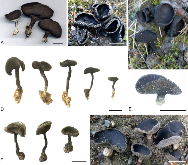
Photos of fresh (A–D, G) and dried (E–F) apothecia of the /alpina-corium lineage and H. alpicola of the Helvella corium species aggregate. A. H. corium. B. H. corium (H2184). C. H. corium (H1958). D. H. alpina (H336). E. H. corium (H1998). F. H. pseudoalpina (H2278). G. H. alpicola (H552). Scale bars = 1 cm. Photos: B–E: S.B. Løken; A, G: T. Schumacher.
Synonym: Helvella arctica Nannf., Svensk Bot. Tidskr. 31: 60. 1937.
Detailed list of synonyms in Skrede et al. (2017).
Apothecium ± regularly stipitate-cupulate, black all over, cup up to 3.5 cm wide. Stipe uniformly black, above ground portions black, predominantly with longitudinal grooves that do not continue onto receptacle. Receptacle surface densely pubescent with brown-walled hyphoid hairs forming fascicles 150–360 μm long, fascicles gradually increasing in length towards margin where they form distinct triangular tufts; in specimens from arctic-alpine areas often with white crystals, individual hair cells up to 20 μm broad. Medullary excipulum of densely interwoven textura intricata, hyphae 3–7 μm broad, septate. Outer excipulum of textura angularis, innermost cells irregular in shape and size, walls pale brown, outermost cells cylindrical or club-shaped, with subhyaline or brownish walls, individual cells 10–25 × 8–30 μm, arranged in tufts of fascicled hyphae. Asci aporhynchous, 210–340 × 11.3–18.8 μm. Ascospores ellipsoid, with one large internal guttule, 16.3–22.5 × 10–13.8 μm. Paraphyses septate, cells with brownish walls along the whole length, gradually increasing in pigmentation towards the tips, 2.5 μm broad below, gradually enlarged to 3.8–7.0 μm at the subcapitate tips.
Included specimens for macro- and microanatomical examinations: France, Savoie, T. Schumacher [H547] (O-255757). Sweden, Torne Lappmark, Jukkasjärvi, E of Abiskojok, Aug. to Sep. 1928, J.A. Nannfeldt [H292] (C-F-92111-Fung. Exs. Suec. 369 isotype of H. arctica Nannf.). Norway, Troms, Salangen, 8 Aug. 2017, S.B. Løken & T. Schumacher [H1958] (TROM-F-610014); Finnmark, Alta, Talvik, 12 Aug. 2017, S.B. Løken & T. Schumacher [H1970] (TROM-F-610015); Troms, Balsfjord, Lakselvbukt, 20 Aug. 2017, S.B. Løken & T. Schumacher [H1998, H1999, H2101] (TROM-F-610004, TROM-F-610017, TROM-F-610040); Nordland, Saltdal, Junkerdalen, Aug. 2016, S.B. Løken & T. Schumacher [H950] (TROM-F-610059); Hedmark, Folldal, Einunndalen, Aug. 2016, S.B. Løken & T. Schumacher [H955] (O-255766); Sør-Trøndelag, Oppdal, Vinstradalen, Aug. 2016, S.B. Løken & T. Schumacher [H957] (O-255754). Svalbard, Longyeardalen, Longyearbyen, 20 Jul. 2017, S.B. Løken & B.A. Granbo [H2184] (O-255756).
Notes: Helvella corium forms a well-supported lineage with H. alpina and H. pseudoalpina. Eleven hsp, 10 rpb2, 18 tef, 25 LSU, zero 5.8S and 96–99 ITS substitutions separate H. corium and H. alpina. Ten hsp, nine rpb2, 16 tef, 26 LSU, zero 5.8S and 111–112 ITS substitutions separate H. corium and H. pseudoalpina. Helvella corium is by far the most common species of the H. corium species aggregate and is also the only species that thrives in temperate as well as in the boreal and arctic-alpine biomes. The holotype of H. corium [Poland, Georgenberg Landeck, May 1870, Weberbauer (WRSL)] has not been examined. Due to its old age it is presumably unsuitable for molecular identification. We base our concept of Helvella corium on the epitype designated by Skrede et al. (2017) [Denmark, Mid Zealand, Kirke Hvalsø, Brødlesgård, 2 Jul. 1984, U. Søchting (C-F-71638)].
Helvella alpina Skrede et al., Persoonia 39: 19. 2017. Fig. 3D.
Apothecium regularly stipitate-cupulate, black all over, occasionally with white crystals at margin, cup 0.8–2.0 cm wide. Stipe slender, solid to hollow, above ground portions black, occasionally with a few longitudinal grooves, 0.2–0.3 cm thick, 1.0–3.5 cm long. Medullary excipulum of textura intricata, hyphae 2–5 μm wide, hyaline. Outer excipulum of textura angularis, cells 10–25 μm diam, intermixed with subhyaline to brown-walled hyphae, turning out perpendicularly to receptacle surface. Receptacle surface subpubescent, with scattered, brown-walled hyphoid hairs, not tufted, individual hairs 60–200 (occasionally up to 350) μm long, composed of ovoid to subglobose cells, up to 20 μm broad. Asci aporhynchous, 230–340 × 12.5–15 μm. Ascospores ellipsoid, with one large internal guttule and several smaller ones towards the poles, 16.3–21.3 × 10–13.8 μm. Paraphyses 2.0–2.8 μm broad below, septate, cells with brownish walls along the whole length, gradually enlarged to 4.0–6.5 μm at the subcapitate tips.
Included specimens for macro- and microanatomical examinations: Canada, British Columbia, Whistler National Park, 13 Aug. 1994, T. Schumacher [H336] (O-253227). France, Savoie, Plan des Evettes, 26 Aug. 1992, T. Schumacher [H223] (O-253226 holotype). Russia, Khatanga Air Port, H.F. Gøtzsche [H540] (C-F-34420). Sweden, Torne Lappmark, Jukkasjärvi, 1945, G. Degelius [H711] (UPS-F-145392). Norway, Nordland, Ballangen, Langvatn, 8 Aug. 1970, O. Skifte [H921] (TROM-F-41055); Oppland, Dovre, Grimsdalen, 23 Aug. 2017 [H2106] (O-255751); Troms, Tromsø, ca. 2 km W of Breivikeidet, 29 Aug. 1954, F.E. Eckblad [H2162] (O-F-174772).
Notes: Helvella alpina is sister species to H. pseudoalpina from which it diverges in one substitution in hsp, one substitution in rpb2, six substitutions in tef, 10 substitutions in LSU, zero substitutions in 5.8S and 52–61 substitutions in ITS.
Helvella pseudoalpina S.B. Løken, Skrede & T. Schumach. sp. nov. MycoBank MB833241. Fig. 3F.
Etymology: From Greek "false" and Latin "occurring in mountainous regions", referring to morphological resemblance to H. alpina.
Typus: Norway, Nordland, Saltdal, Junkerdalen, Bibeldalen, 28 Aug. 1988, L. Ryvarden [H2278] (holotype TRH-F-20631).
Apothecium regularly stipitate-cupulate, black all over, cup 0.8–1.5 cm wide. Stipe slender, solid, above ground portions black, occasionally with a few longitudinal grooves, 0.2–0.3 cm thick, 1.0–3.5 cm long. Receptacle surface subpubescent, with scattered, brown-walled hyphoid hairs, 90–250 μm long. Medullary excipulum of textura intricata, hyphae septate, hyaline to subhyaline, generally 3–6 μm broad, intermixed with swollen cells up to 15 μm broad. Outer excipulum of textura angularis, cells 20–45 μm diam, brown-walled, intermixed with subhyaline to brown-walled hyphae, cells in rows turning out perpendicularly to receptacle surface, individual cells 12–30 × 7–25 μm, outermost cells subcapitate to capitate in shape., Asci aporhynchous, 250–330 × 12.5–17.5 μm. Ascospores ellipsoid, with one large internal guttule, 15–21.3 × 10–13.8 μm. Paraphyses 2.5 μm broad below, septate, walls light brownish along the whole length, gradually enlarged to 5.0–8.8 μm at the subcapitate tips.
Included specimens for macro- and microanatomical examinations: Greenland, Qeqertarsuaq (Godhavn), 11 Aug. 1977, P.M. Petersen [H349] (C-F-63820). Norway, Troms, Tromsø, Tromsdalen, 27 Aug. 1961, O. Skifte [H941, H942, H943] (TROM-F-11412, TROM-F-11405, TROM-F-11404); Troms, Balsfjord, Mestervik, 9 Aug. 2017, S.B. Løken & T. Schumacher [H1965, H1966, H1967] (TROM-F-610048, TROM-F-610049, TROM-F-610050); Nordland, Saltdal, Junkerdalen, Bibeldalen, 28 Aug. 1988, L. Ryvarden [H2278] (TRH-F-20631 holotype). Svalbard, Longyeardalen, Longyearbyen, Aug. 2015, S. Svantesson [H498] (O-255748).
Notes: Helvella pseudoalpina is sister species to H. alpina from which it diverges in one substitution in hsp, one substitution in rpb2, six substitutions in tef, 10 substitutions in LSU, zero substitutions in 5.8S and 52 to 61 substitutions in ITS.
Helvella alpicola Skrede et al., Persoonia 39: 19. 2017. Fig. 3G.
Apothecium regularly stipitate-cupulate, cup 0.5–1.5 cm wide, hymenium greyish black, receptacle dark greyish. Stipe terete, greyish to whitish below, 0.2–0.3 cm broad, 0.5–1.8 cm high, with 2–3 shallow grooves at base. Receptacle surface densely pubescent, covered with multiseptate, subhyaline, hyphoid hairs forming fascicles near margin, fascicles 60–350 μm long, individual hair cells 10–40 × 7.5–25 μm, with conspicuous brown pigments at septa. Medullary excipulum of loose textura intricata, hyphae 2–5 μm broad, hyaline. Outer excipulum of textura angularis, cells 10–27 μm diam. Asci pleurorhynchous, 250–400 × 12.5–18 μm. Ascospores ellipsoid, with one large internal guttule, 18–22.5 × 11–15 μm. Paraphyses 2.5–3.8 μm below, walls light brownish along the whole length, septate, gradually enlarged to 5–10 μm at the clavate tips.
Included specimens for macro- and microanatomical examination: Switzerland, Graubünden,, Inn at Resgia, 26 Aug. 1984, H. Dissing [H231] (O-253226). Norway, Nordland, Saltdal, Junkerdalsura. 27 Aug. 1988, A.E. Torkelsen [H175] (O-185924 holotype); Nordland, Saltdal, Junkerdalsura, 11 Aug. 2016, S.B. Løken & T. Schumacher [H552, H553, H554, H952, H953] (TROM-F-610052, TROM-F-610053, TROM-F-610054, TROM-F-610061, TROM-F-610062).
Notes: Helvella alpicola is nested in a divergent sister lineage to the /alpestris-nannfeldtii lineage (Fig. 1). It diverges from H. nannfeldtii in four substitution in hsp, 11 substitutions in rpb2, 12 substitutions in tef and 52 substitutions in LSU.
Helvella nannfeldtii Skrede et al., Persoonia 39: 33. 2017. Fig. 4A–C.
Fig. 4.
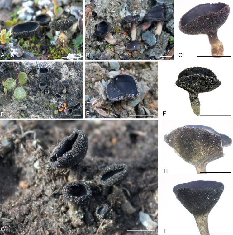
Photos of fresh apothecia from the /alpestris-nannfeldtii lineage of the Helvella corium species aggregate. A. Helvella nannfeldtii (H1971). B. H. nannfeldtii (H2104). C. H. nannfeldtii (H1971). D. H. alpestris (H2111). E. H. alpestris (H2102). F. H. alpestris (H2111). G. H. macrosperma (H1982). H. H. macrosperma (H2100). I. Helvella sp. (H1996). Scale bars = 1 cm. Photos: S.B. Løken.
Apothecium stipitate-cupulate, 1–3 cm wide, hymenium black, receptacle dark brown to black, rarely with white crystals at margin. Stipe short, terete, solid, greyish black above, whitish below in above ground portions, 0.2–0.3 cm wide, 0.5–2 cm long. Receptacle surface pubescent, covered with subhyaline to light brown-walled hyphoid hairs forming hyphal fascicles, individual hairs 190–500 μm long, irregularly shaped and often much constricted at septa, toward the margin increasing in length and forming distinct triangular tufts. Medullary excipulum of loosely interwoven textura intricata, hyphae 3–5 μm broad. Outermost excipulum of textura angularis, thick-walled, cell walls dark brown, 15–30 μm diam, intermixed with broad short-segmented brownish hyphae forming a textura intricata. Asci pleurorhynchous, 250–340 × 13.75–20 μm. Ascospores ellipsoid, with one large internal guttule, 15–22.5 × 10⁻¹⁵ μm. Paraphyses 2.5–3.8 μm broad below, septate, walls dark brown along upper two thirds, gradually increasing in pigmentation towards tips, gradually enlarged to 5–8.8 μm at the subcapitate tips.
Included specimens for macro- and microanatomical examinations: France, Savoie, Val d'Isere, Gorges du Mal, 31 Aug. 1992, T. Schumacher [H212] (O-253332); Savoie, Bon Valle, Sur Arc, 2 Sep. 1992, T. Schumacher [H216] (O-253333). Norway, Oppland, Dovre, Grimsdalen, Veslegrimsa, 8 Aug. 2009, T. Carlsen, T. Schumacher & I. Skrede [H027] (O-253338 holotype); Hedmark, Folldal, Einunndalen, Aug. 2016, S.B. Løken & T. Schumacher [H954] (O-255744); Troms, Kåfjord, Guolasjärvi, 13 Aug. 2017, S.B. Løken & T. Schumacher [H1971, H1972] (TROM-F-610033, TROM-F-610034); Troms, Målselv, Frøkentindelva, 16 Aug. 2017, S.B. Løken & T. Schumacher [H1984, H1985] (TROM-F-610031, TROM-F-610032); Troms, Målselv, Iselvdalen, 19 Aug. 2017, S.B. Løken & T. Schumacher [H1991, H1992, H1993, H1994] (TROM-F-610020, TROM-F-610002, TROM-F-610005, TROM-F-610036); Oppland, Dovre, Grimsdalen. 23 Aug. 2017, S.B. Løken & T. Schumacher [H2103, H2104] (O-255765, O-255745).
Notes: Helvella nannfeldtii forms a well-supported lineage with H. alpestris and H. macrosperma. Four hsp, two rpb2, seven tef, 47 LSU, two 5.8S and 138–139 ITS substitutions separate H. nannfeldtii and H. alpestris. Three hsp, one rpb2, five tef, 43 LSU, 3 5.8S and 139 ITS substitutions separate H. nannfeldtii and H. macrosperma.
Helvella alpestris Boud., Bull. Soc. Bot. France 41: CCXL. 1894. Fig. 4D–F.
Apothecium stipitate-cupulate to almost plane, black all over, predominantly with white crystals at margin, cup 0.5–1.8 cm wide. Stipe terete, solid, above ground portions black, 0.2–0.4 cm broad, 0.4–1.5 cm high. Medullary excipulum of textura intricata, hyphae 2–5 μm wide. Outer excipulum of brown-walled globose to angular cells, 10–20 μm diam. Receptacle surface densely pubescent, covered with dark brown-walled hyphoid hairs forming fascicles, hairs 50–380 μm long, gradually increasing in length towards the margin where they form distinct triangular tufts, individual hair cells 10–40 × 7.5–17.5 μm. Asci pleurorhynchous, 230–350 × 12–17.5 μm. Ascospores ellipsoid, with one large internal guttule, 16–22.5 × 10–13.8 μm. Paraphyses septate, 2.5–3.75 μm broad below, walls brownish along the whole length, gradually increasing in pigmentation towards apex, enlarged to 4.5–8.8 μm at the gnarled, clavate tips.
Included specimens for macro- and microanatomical examinations: Canada, Nunavut, Kitikmeot Region, above Bloody Falls, 19 Jul. 2014, J.M. Saarela, P.C. Sokoloff & R.D. Bull [H460] (O ex DAOM-574891). France, Savoie, Val'dIsere, T. Schumacher [H548] (O-255736). Norway, Troms, Tromsø, Tromsø museum, 10 Jul. 1980, S. Sivertsen & O. Skifte [H928] (TROM-F-11403); Oppland, Dovre, Grimsdalen, Jegerhøi, 23 Aug. 2017, S.B. Løken & T. Schumacher [H2111] (O-255737); Finnmark, Porsanger, Lakselv, 16 Jul. 1961, F.E. Eckblad [H2154] (O-F-174783).
Notes: Helvella alpestris is sister species to H. macrosperma from which it diverges in one substitution in hsp, one substitution in rpb2, four substitutions in tef, seven substitutions in LSU, one substitution in 5.8S and 42–43 substitutions in ITS. Specimens are found on bare silt or slate grounds, usually in close proximity to Salix reticulata. One of the genotypes for 5.8S and ITS2 (i.e. S1) from root tips of S. reticulata, obtained in the work by Weidemann (1998), proved identical to the 5.8S/ITS2 genotypes of H. alpestris provided in the present study.
Helvella macrosperma (J. Favre) R. Fellner & Landa, Česka Mykol. 45: 35. 1991. Fig. 4G–H.
Apothecium regularly stipitate-cupulate, black all over, rarely with white crystals at the margin, 0.5–2 cm wide. Stipe short, terete, solid, above ground portions black, 0.4–1.2 cm high, 0.2–0.4 cm broad. Receptacle and stipe surface densely pubescent, covered with dark, brown-walled hyphoid hairs, forming fascicles, hairs 180–500 μm long, gradually increasing in length towards margin where they form distinct triangular tufts; individual hair cells 10–40 × 10–20 μm, constricted at septa and with conspicuous incrusted pigments on the interior of the cell walls. Medullary excipulum of loosely interwoven textura intricata, hyphae 3–5 μm broad, short-celled; outer excipulum of brown-walled globose to angular cells, 10–20 μm diam, turning out perpendicularly to the receptacle surface. Asci pleurorhynchous, 240–330 × 10–20 μm. Ascospores broadly ellipsoid, with one large internal guttule, 17.5–22.5 × 11.3–13.8 μm. Paraphyses septate, 2.5–3.75 μm broad below, walls brownish along the whole length, gradually increasing in pigmentation towards tip, enlarged to 5–7.5 μm at the gnarled, clavate tips.
Included specimens for macro- and microanatomical examinations: Norway, Nordland, Saltdal, Junkerdalen, Aug. 2016, S.B. Løken & T. Schumacher [H947] (TROM-F-610056); Troms, Målselv, Håkåfjellet, 16 Aug. 2017, S.B. Løken & T. Schumacher [H1982] (TROM-F-610001); Troms, Balsfjord, Lakselvbukt, 20 Aug. 2017, S.B. Løken & T. Schumacher [H1997, H2100] (TROM-F-610017, TROM-F-610004); Oppland, Lom, Høyrokampen at Bøvertun, 31 Aug. 2007, A.K. Wollan [H2146] (O285169); Oppland, Dovre, Grimsdalen, Veslegrimsa, 2007, Master Field Course [H029] (O-253328).
Notes: Helvella macrosperma is sister species to H. alpestris from which it diverges in one substitution in hsp, one substitution in rpb2, four substitutions in tef, seven substitutions in LSU, one substitution in 5.8S and 42–43 substitutions in ITS.
DISCUSSION
Phylogenies based on a few, phylogenetically highly informative loci are still preferred when delimiting species and inferring relationships among species of fungi. Indeed, the larger amount of sequence data produced by next generation sequencing techniques is not necessarily associated with better phylogenetic signal and resolution (Lemmon & Lemmon 2013). Multilocus phylogenies of phylogenomic data often include extensive non-phylogenetic signal and may produce conflicting, yet highly supported, phylogenetic trees (Philippe et al. 2011). Thus, the choice and number of genetic markers necessary to infer reliable evolutionary histories of species is still an important matter of debate in phylogenetics (Rokas et al. 2003, Aguileta et al. 2008, Balasundaram et al. 2015, Stielow et al. 2015), and should be evaluated individually for each group under study. The present study confirmed the high informativeness of selected single-copy protein-coding loci, in combination with ribosomal markers, in delimiting species. Altogether, five markers (hsp, rpb2, tef, LSU and 5.8S) were sufficient to get a stable support for species recognition as well as shallow clades in our phylogeny. Not unexpectedly, this election of markers was not sufficient to infer a stable topology of the deeper branches of the tree (Fig. 1). The inclusion of highly conserved loci, e.g. SSU, mcm7 and/or tsr1, may have improved the support for basal nodes. We initially attempted to include mcm7 and tsr1, which had been shown to reveal higher-level relationships in Helvella and other ascomycete genera (Raja et al. 2011, Zhao et al. 2015, Mark 2016), however, PCR-amplification using general primers for these regions failed in our study. It was concluded that the design of new, specific Helvella primers for these loci are needed if these markers are to be used in future phylogenetic studies of this genus. The ITS region also proved difficult to amplify with universal ITS primers, as also demonstrated in previous studies (Landvik et al. 1999, Skrede et al. 2017). Nevertheless, we succeeded in obtaining complete ITS sequences for six out of seven species of the H. corium species aggregate. In general, the ITS region displayed extremely high levels of interspecific variability across the Helvella species under study, making it impossible to construct objective alignments across the /alpestris-nannfeldtii and /alpina-corium lineages jointly. ITS is thus considered unsuitable as a phylogenetic marker across these lineages, as also demonstrated for the genetically divergent Helvella genus as a whole (Landvik et al. 1999, Skrede et al. 2017).
Nevertheless, the ribosomal loci, i.e. LSU and ITS, successfully delimited six out of seven species in the Helvella corium species aggregate. The seventh species, i.e. H. nannfeldtii, showed remarkably high levels of intraspecific divergence in these ribosomal regions. The high level of intraspecific variability in H. nannfeldtii observed in Fig. 2 can be explained by its variability in the LSU region, as no other locus displayed comparable levels of intraspecific divergence. Conflicting gene trees, as observed in H. nannfeldtii, may result from intraspecific recombination, which implies that the two genetic clusters observed are not reproductively isolated (Taylor et al. 2000). Based on the observed inconsistency, these two divergent clusters are not considered to represent two cryptic species. This is also demonstrated by the output from the STACEY analysis (Fig. 2), where all individuals of H. nannfeldtii were regarded as belonging to the same species.
The LSU and ITS nrDNA regions are present in numerous copies in fungal genomes. These copies form gene families that are expected to be subject to concerted evolution (Arnheim et al. 1980, Strausbaugh 2001), where homogenization of paralogous genes is achieved through homologous recombination. However, exceptions do occur, resulting in heterogeneous copies within a genome and apparent heterozygosity (Selosse et al. 2016). Substantial intragenomic heterozygosity of ribosomal DNA is thought to be infrequent (Thiéry et al. 2016), but is still observed in the fungal kingdom (Stensrud et al. 2007, Nilsson et al. 2008, Harder et al. 2013, Lindner et al. 2013). Thus, if the primers used in this study amplified paralogs of LSU and ITS in H. nannfeldtii, we might have been comparing variable non-homologous sequences. As the same level of intraspecific variation is not observed in any other species of the H. corium species aggregate, we conclude that the ribosomal DNA of H. nannfeldtii is evolving in a different manner and at a different rate compared to even its closest relatives.
The STACEY analysis inferred seven well-supported clusters in the present species aggregate (Figs 1, 2). Though, when a limited number of individuals and loci are used, STACEY may over-estimate the number of clusters (Toprak et al. 2016, Sukumaran & Knowles 2017). However, as morphological examinations supported the seven clusters, we feel confident they represent well-defined species. The STACEY analysis is based on the multispecies coalescent model and assumes random mating and that no gene flow occurs after speciation (Leaché et al. 2014, Xu & Yang 2016). While restricted gene flow between sister species will not severely effect species tree topology (Heled & Drummond 2010, Toprak et al. 2016), gene flow between non-sisters will lead to incongruence between gene trees and species trees (Taylor et al. 2000, Leaché & Fujita 2010). Speciation is seldom instantaneous in nature, thus violations to the implied evolutionary model might have occurred in our dataset. In this study, the Bayesian inference shows no topological incongruence across loci or between gene trees (except for within H. nannfeldtii) and species trees, but the ML analyses raises uncertainty regarding the placement of the possibly new but unnamed species of Helvella sp. It is not fully understood how well STACEY handles the inclusion of hybrid individuals (Wagner et al. 2017). Still, if Helvella sp. represents hybrid individuals, i.e. as a result of hybridisation between the sister species H. alpestris and H. macrosperma, it will not have severe effect on the overall tree topology.
In this study, using a molecular genealogical approach as basis for classification, allowed us to get a better understanding of the taxonomical value of morphological characters in the Helvella corium species aggregate. Although previous authors have recognized the morphological variation in H. corium s. lat. (Boudier 1907, Nannfeldt 1937, Favre 1955, Dissing 1964, 1966a, b), an accurate delimitation of species within this aggregate has not existed before molecular data were introduced (Skrede et al. 2017). In Dissing's (1966) comprehensive review of Helvella in Europe, the new section Macropodes was proposed to include all Helvella species with cupulate apothecia and a pubescent to villose receptacle surface, a solid stipe with or without furrows and ribs, and ribs not extending from the stipe onto the receptacle. The section Macropodes then included Helvella corium, H. macropus, H. villosa (= H. fibrosa) H. cupuliformis (= H. hypocrateriformis) and H. queletii (= H. solitaria). As already shown by Skrede et al. (2017), and further supported by this study, these macro-morphological characters are useless in recognizing an infrageneric phylogenetic classification. Indeed, the combination of stipitate-cupulate and pubescent Helvella species is found scattered across many lineages in the genus.
Our study of the /alpina-corium and /alpestris-nannfeldtii evolutionary lineages of Helvella further elaborates on the morphological differentiation between species. We identified several informative characters that are useful in discriminating among lineages and species. The ascus development, whether aporhynchous or pleurorhynchous (Chadefaud 1943, Berthet 1964), is considered valuable for defining lineages and sections of Helvella (Weber 1972, Landeros et al. 2015), and even genera of the family Helvellaceae (Hansen et al. 2019). Aporhynchous asci seem important to delineate the /alpinacorium lineage (Häffner 1987), while pleurorhynchous asci characterise the /alpestris-nannfeldtii lineage (Van Vooren 2014, 2015, Skrede et al. 2017). Indeed, aporhynchous asci represent a synapomorphy for the /alpina-corium lineage (Fig. 1). Skrede et al. (2017) also acknowledged the diagnostic value of arrangement of hyphoid hairs on the receptacle surface, also confirmed in this study: hairs are scattered in H. alpina and H. pseudoalpina, and primarily tufted (in fascicles) in H. corium, H. nannfeldtii, H. alpestris, H macrosperma and H. alpicola. Additional taxonomically informative morphological characters in this species aggregate include: (1) colour of stipe base (white in above ground portions in H. nannfeldtii and H. alpicola); (2) hair length (hairs approaching 500 μm near the margin in H. nannfeldtii and H. macrosperma); (3) ascospore size (predominantly more than 20 μm in length in H. corium, H. alpicola and H. alpestris vs. less than 20 μm in H. alpina, H. pseudoalpina, H. nannfeldtii and H. macrosperma); (4) ascospore morphology (one large guttule in H. corium, H. pseudoalpina, H. alpicola, H. alpestris, H. macrosperma, and H. nannfeldtii vs. one large guttule and several smaller ones at the poles in H. alpina); and (5) shape of paraphysis tips (clavate in the /alpestris-nannfeldtii lineage vs. subcapitate in the /alpinacorium lineage).
In describing Helvella arctica, Nannfeldt (1937) paid a great deal of attention to the external, white crystals observed on the marginal receptacle hairs in this species. This trait played a major role in distinguishing it against H. corium. Later, Dissing (1966) concluded that this trait represented a mere adaptation to alpine environments, and synonymized H. arctica with H. corium. This latter disposition also had molecular support in the work by Skrede et al. (2017), who corroborated the synonymy of the two taxa. In fact, in the present study it was found that white crystalline deposits on receptacle hairs are frequently observed under alpine conditions of all species of the Helvella corium species aggregate, however, being most prominent in H. alpestris and H. corium.
Evidently, considerable genetic differentiation has occurred independently of morphological differentiation among species that belong to the Helvella corium species aggregate, resulting in the species sharing many morphological traits. This phenotypical similarity may possibly represent morphological stasis. It has been argued, generally speaking, that morphological stasis may result from strong stabilizing selection for adaptation to harsh environments (Nevo 2001, Lumbsch & Leavitt 2011). In fact, many cryptic and pseudo-cryptic plant species are found in harsh alpine and Arctic regions (Grundt et al. 2006, Skrede et al. 2008, Brochmann & Brysting 2010), and morphological similarity in non-sister species may be due to strong adaptive value of certain morphological traits (Bickford et al. 2007). This may also apply to the genetic divergent, but pseudo-cryptic nature of the H. corium species aggregate. In addition, it should be noted that the species under study show a high degree of niche conservatism in favouring calcareous soils and growth in close proximity to Dryas octopetala and/or Salix spp. These morphologically similar, yet genetically divergent species appear in sympatry in alpine regions of the northern hemisphere. Thus, we hope that the species delimitations made in this study will provide a valuable basis for future investigations of the specific ecological roles of such species in the natural systems they may occupy.
ACKNOWLEDGEMENTS
We acknowledge all the collectors of the Helvella corium aggregate who deposited specimens in fungaria of O, TRH, BG, TROM, S, UPS, GB, UME, and C for their dedicated efforts in collecting valuable material, as well as the curators and staff for supplying material. We thank Karen Hansen for allowing us to use some of her personal collections. We thank Cecilie Mathiesen for all the assistance in the molecular laboratory. We acknowledge the Norwegian Biodiversity Information Centre and the University of Oslo for funding.
Conflict of interest: The authors declare that there is no conflict of interest.
Supplementary Material: http://fuse-journal.org/
Table S1. Specimens of the Helvella corium species aggregate in this study. Specimen ID as well as geographical origin (with coordinate data) is shown for each specimen. “NA” means non annotated data. Type specimens are written in bold.
REFERENCES
- Aguileta G, Marthey S, Chiapello H, et al. (2008). Assessing the performance of single-copy genes for recovering robust phylogenies. Systematic Biology 57: 613–627. [DOI] [PubMed] [Google Scholar]
- Arnheim N, Krystal M, Schmickel R, et al. (1980). Molecular evidence for genetic exchanges among ribosomal genes on nonhomologous chromosomes in man and apes. Proceedings of the National Academy of Sciences 77: 7323–7327. [DOI] [PMC free article] [PubMed] [Google Scholar]
- Balasundaram SV, Engh IB, Skrede I, et al. (2015). How many DNA markers are needed to reveal cryptic fungal species? Fungal Biology 119: 940–945. [DOI] [PubMed] [Google Scholar]
- Berthet P. (1964). Essai biotaxinomique sur les Discomycètes. Thesis, Lyon. [Google Scholar]
- Bickford D, Lohman DJ, Sodhi NS, et al. (2007). Cryptic species as a window on diversity and conservation. Trends in Ecology & Evolution 22: 148–155. [DOI] [PubMed] [Google Scholar]
- Bouckaert R, Heled J, Kühnert D, et al. (2014). BEAST 2: A Software Platform for Bayesian Evolutionary Analysis. PLoS Computational Biology 10(4): e1003537. [DOI] [PMC free article] [PubMed] [Google Scholar]
- Boudier J, Fischer ED. (1894). Rapport sur les espèces de champignons trouvées pendant l’assemblée a Genève et les excursions faites en Valais, par les Sociétés de Botanique de France et de Suisse, du 5 au 15 Aout 1894. Bulletin de la Société Botanique de France 41: 7. [Google Scholar]
- Boudier J. (1895). Description de quelques espèces récoltées en août 1894 dans les regions élevées des Alpes du Valais. Bulletin de la Société Botanique de France 11: 27–30. [Google Scholar]
- Boudier J. (1907). Histoire et classification des Discomycetes d’Europe. Librarie des Sciences Naturelles, Paris, France. [Google Scholar]
- Boudier J. (1910). Icones mycologicae ou iconographie des champignons de France. Librairie des Sciences Naturelles, Paris, France. [Google Scholar]
- Brochmann C, Brysting AK. (2010). The Arctic – an evolutionary freezer? Plant Ecology & Diversity 1: 181–195. [Google Scholar]
- Chadefaud M. (1943). Sur les divers types d’elements dangeardiens chez les Ascomycetes. Revue Scientifique 81: 77–80. [Google Scholar]
- Degnan JH, Rosenberg NA. (2009). Gene tree discordance, phylogenetic inference and the multispecies coalescent. Trends in Ecology & Evolution 24: 332–340. [DOI] [PubMed] [Google Scholar]
- Dettman JR, Jacobson DJ, Taylor JW. (2003). A multilocus genealogical approach to phylogenetic species recognition in the model eukaryote Neurospora. Evolution 57: 2703–2720. [DOI] [PubMed] [Google Scholar]
- Dissing H. (1964). Studies in Arctic and Subarctic Discomycetes. I. The genus Helvella. Botanisk Tidsskrift 60: 108–128. [Google Scholar]
- Dissing H. (1966a). A revision of collections of the genus Helvella L. ex St-Amans emend. Nannf. in the Boudier Herbarium. Revue de Mycologie 31: 189–224. [Google Scholar]
- Dissing H. (1966b). The genus Helvella in Europe with special emphasis on species found in Norden. Dansk Botanisk Arkiv 25: 1–172. [Google Scholar]
- Favre J. (1955). Les champignons supérieurs de la zone alpine du Parc National Suisse. Ergebnisse der wissenschaftligen Untersuchungen des schweizerischen Nationalparks 33: 1–112. [Google Scholar]
- Fellner R, Landa J. (1991). Arctic and alpine fungi in Czechoslovakia. Česka Mykol 45: 35. [Google Scholar]
- Fries EM. (1822). Systema Mycologicum. Vol. 2 (1). Officina Berlingiana, Lund, Sweden. [Google Scholar]
- Fuckel L. (1870). Symbolae mycologicae. Beiträge zur Kenntnis der rheinischen Pilze. Jahrbucher des Nassauischen Vereins fur Naturkunde 23–24: 1–459. [Google Scholar]
- Grundt HH, Kjølner S, Borgen L, et al. (2006). High biological species diversity in the arctic flora. Proceedings of the National Academy of Sciences USA 103: 972–975. [DOI] [PMC free article] [PubMed] [Google Scholar]
- Hansen K, Schumacher T, Skrede I, et al. (2019). Pindara revisited – evolution and generic limits in Helvellaceae. Persoonia 42: 186–204. [DOI] [PMC free article] [PubMed] [Google Scholar]
- Hansen K, Olariaga I. (2015). Species limits and relationships within Otidea inferred from multiple gene phylogenies. Persoonia 35: 148–165. [DOI] [PMC free article] [PubMed] [Google Scholar]
- Harder CB, Læssøe T, Frøslev TG, et al. (2013). A three-gene phylogeny of the Mycena pura complex reveals 11 phylogenetic species and shows ITS to be unreliable for species identification. Fungal Biology 117: 764–775. [DOI] [PubMed] [Google Scholar]
- Häffner J. (1987). Die Gattung Helvella, Morphologie und Taxonomie. Beihefte zur Zeitschrift für Mykologie 7: 1–165. [Google Scholar]
- Heled J, Drummond AJ. (2010). Bayesian inference of species trees from multilocus data. Molecular Biology and Evolution 27: 570–580. [DOI] [PMC free article] [PubMed] [Google Scholar]
- Hofstetter V, Miądlikowska J, Kauff F, et al. (2007). Phylogenetic comparison of protein-coding versus ribosomal RNA-coding sequence data: A case study of the Lecanoromycetes (Ascomycota). Molecular Phylogenetics and Evolution 44: 412–426. [DOI] [PubMed] [Google Scholar]
- Huelsenbeck JP, Andolfatto P, Huelsenbeck ET. (2011). Structurama: bayesian inference of population structure. Evolutionary Bioinformatics Online 7: 55–59. [DOI] [PMC free article] [PubMed] [Google Scholar]
- Jones G. 2017. Algorithmic improvements to species delimitation and phylogeny estimation under the multispecies coalescent. Journal of Mathematical Biology 74: 447–467. [DOI] [PubMed] [Google Scholar]
- Jones G, Aydin Z, Oxelman B. (2015). DISSECT: an assignment-free Bayesian discovery method for species delimitation under the multispecies coalescent. Bioinformatics 31: 991–998. [DOI] [PubMed] [Google Scholar]
- Katoh K, Standley DM. (2013). MAFFT Multiple Sequence Alignment Software Version 7: improvements in performance and usability. Molecular Biology and Evolution 30: 772–780. [DOI] [PMC free article] [PubMed] [Google Scholar]
- Kauserud H, Schumacher T. (2001). Outcrossing or inbreeding: DNA markers provide evidence for type of reproductive mode in Phellinus nigrolimitatus (Basidiomycota). Mycological Research 105: 676–683. [Google Scholar]
- Kearse M, Moir R, Wilson A, et al. (2012). Geneious Basic: an integrated and extendable desktop software platform for the organization and analysis of sequence data. Bioinformatics 28: 1647–1649. [DOI] [PMC free article] [PubMed] [Google Scholar]
- Korf RP. (1973). Sparassoid ascocarps in Pezizales and Tuberales. Reports of the Tottori Mycological Institute 10: 389–403. [Google Scholar]
- Landeros F, Iturriaga T, Rodríguez A, et al. (2015). Advances in the phylogeny of Helvella (Fungi: Ascomycota), inferred from nuclear ribosomal LSU sequences and morphological data. Revista Mexicana de Biodiversidad 86: 856–871. [Google Scholar]
- Landvik S, Kristiansen R, Schumacher T. (1999). Pindara: a miniature Helvella. Mycologia 91: 278. [Google Scholar]
- Lanfear R, Frandsen PB, Wright AM, et al. (2017). PartitionFinder 2: new methods for selecting partitioned models of evolution for molecular and morphological phylogenetic analyses. Molecular Biology and Evolution 34: 772–773. [DOI] [PubMed] [Google Scholar]
- Leaché AD, Fujita MK. (2010). Bayesian species delimitation in West African forest geckos (Hemidactylus fasciatus). Proceedings of Biological Sciences 277: 3071–3077. [DOI] [PMC free article] [PubMed] [Google Scholar]
- Leaché AD, Harris RB, Rannala B, et al. (2014). The influence of gene flow on species tree estimation: a simulation study. Systematic Biology 63: 17–30. [DOI] [PubMed] [Google Scholar]
- Lemmon EM, Lemmon AR. (2013). High-throughput genomic data in systematics and phylogenetics. Annual Review of Ecology, Evolution, and Systematics 44: 99–121. [Google Scholar]
- Letunic I, Bork P. (2016). Interactive tree of life (iTOL) v3: an online tool for the display and annotation of phylogenetic and other trees. Nucleic Acids Research 44: 242–5. [DOI] [PMC free article] [PubMed] [Google Scholar]
- Lindner DL, Carlsen T, Nilsson HR, et al. (2013). Employing 454 amplicon pyrosequencing to reveal intragenomic divergence in the internal transcribed spacer rDNA region in fungi. Ecology and Evolution 3: 1751–1764. [DOI] [PMC free article] [PubMed] [Google Scholar]
- Lumbsch HT, Leavitt SD. (2011). Goodbye morphology? A paradigm shift in the delimitation of species in lichenized fungi. Fungal Diversity 50: 59–72. [Google Scholar]
- Mark K. (2016). Diversification and species delimitation of lichenized fungi in selected groups of the family Parmeliaceae (Ascomycota). Thesis, University of Tartu Press. [Google Scholar]
- Massée G. (1895). British Fungus-Flora 4. Bell & sons, London, Great Britain. [Google Scholar]
- Nannfeldt J. (1937). Contributions to the mycoflora of Sweden. 4. On some species of Helvella, together with a discussion of the natural affinities within Helvellaceae and Pezizaceae trib. Acetabuleae. Svensk Botanisk Tidsskrift 31: 47–66. [Google Scholar]
- Nevo E. (2001). Evolution of genome-phenome diversity under environmental stress. Proceedings of the National Academy of Sciences USA 98: 6233–6240. [DOI] [PMC free article] [PubMed] [Google Scholar]
- Nilsson RH, Kristiansson E, Ryberg M, et al. (2008). Intraspecific ITS variability in the kingdom fungi as expressed in the international sequence databases and its implications for molecular species identification. Evolutionary Bioinformatics Online 4: 193–201. [DOI] [PMC free article] [PubMed] [Google Scholar]
- Philippe H, Brinkmann H, Lavrov DV, et al. (2011). Resolving difficult phylogenetic questions: why more sequences are not enough. PLoS Biology 9: e1000602. [DOI] [PMC free article] [PubMed] [Google Scholar]
- Quélet L. (1886). Enchiridion Fungorum in Europa media et praesertim in Gallia vigentium. Lutetiae, France. [Google Scholar]
- Raja H, Schoch CL, Hustad V, et al. (2011). Testing the phylogenetic utility of MCM7 in the Ascomycota. MycoKeys 1: 63–94. [Google Scholar]
- Rannala B, Yang Z. (2003). Bayes estimation of species divergence times and ancestral population sizes using DNA sequences from multiple loci. Genetics 164: 1645–1656 [DOI] [PMC free article] [PubMed] [Google Scholar]
- Rambaut A, Drummond AJ, Xie D, et al. (2018). Posterior summarisation in Bayesian phylogenetics using Tracer 1.7. Systematic Biology 22: 341. [DOI] [PMC free article] [PubMed] [Google Scholar]
- Rokas A, Williams BL, King N, et al. (2003). Genome-scale approaches to resolving incongruence in molecular phylogenies. Nature 425: 798–804. [DOI] [PubMed] [Google Scholar]
- RStudio Team (2016). RStudio: Integrated development for R. RStudio. Inc., Boston, MA: http://www.rstudio.com/. [Google Scholar]
- Schoch CL, Sung G-H, López-Giráldez F, et al. (2009). The Ascomycota Tree of Life: A phylum-wide phylogeny clarifies the origin and evolution of fundamental reproductive and ecological traits. Systematic Biology 58: 224–239. [DOI] [PubMed] [Google Scholar]
- Selosse M-A, Vincenot L, Öpik M. (2016). Data processing can mask biology: towards better reporting of fungal barcoding data? The New Phytologist 210: 1159–1164. [DOI] [PubMed] [Google Scholar]
- Skrede I, Borgen L, Brochmann C. (2008). Genetic structuring in three closely related circumpolar plant species: AFLP versus microsatellite markers and high-arctic versus arctic–alpine distributions. Heredity 102: 293–302. [DOI] [PubMed] [Google Scholar]
- Skrede I, Carlsen T, Schumacher T. (2017). A synopsis of the saddle fungi (Helvella: Ascomycota) in Europe – species delimitation, taxonomy and typification. Persoonia 39: 201–253. [DOI] [PMC free article] [PubMed] [Google Scholar]
- Spatafora JW, Sung GH, Johnson D, et al. (2006). A five-gene phylogeny of Pezizomycotina. Mycologia 98: 1018–1028. [DOI] [PubMed] [Google Scholar]
- Stamatakis A. (2006). RAxML-VI-HPC: maximum likelihood-based phylogenetic analyses with thousands of taxa and mixed models. Bioinformatics 22: 2688–2690. [DOI] [PubMed] [Google Scholar]
- Stamatakis A. (2014). RAxML version 8: a tool for phylogenetic analysis and post-analysis of large phylogenies. Bioinformatics 30: 1312–1313. [DOI] [PMC free article] [PubMed] [Google Scholar]
- Stensrud Ø, Schumacher T, Shalchian-Tabrizi K, et al. (2007). Accelerated nrDNA evolution and profound AT bias in the medical fungus Cordyceps sinensis. Mycological Research 111: 409–415. [DOI] [PubMed] [Google Scholar]
- Stielow JB, Lévesque CA, Seifert KA, et al. (2015). One fungus, which genes? Development and assessment of universal primers for potential secondary fungal DNA barcodes. Persoonia 35: 242–263. [DOI] [PMC free article] [PubMed] [Google Scholar]
- Strausbaugh LD. (2001). Concerted Evolution. In: Encyclopedia of Genetics. Elsevier, Amsterdam, Netherlands: 436–441. [Google Scholar]
- Sukumaran J, Knowles LL. (2017). Multispecies coalescent delimits structure, not species. Proceedings of the National Academy of Sciences USA 114: 1607–1612. [DOI] [PMC free article] [PubMed] [Google Scholar]
- Taylor JW, Jacobson DJ, Kroken S, et al. (2000). Phylogenetic species recognition and species concepts in fungi. Fungal Genetics and Biology 31: 21–32. [DOI] [PubMed] [Google Scholar]
- Thiéry O, Vasar M, Jairus T, et al. (2016). Sequence variation in nuclear ribosomal small subunit, internal transcribed spacer and large subunit regions of Rhizophagus irregularis and Gigaspora margarita is high and isolate dependent. Molecular Ecology 25: 2816–2832. [DOI] [PubMed] [Google Scholar]
- Toprak Z, Pfeil BE, Jones G, et al. (2016). Species delimitation without prior knowledge: DISSECT reveals extensive cryptic speciation in the Silene aegyptiaca complex (Caryophyllaceae). Molecular Phylogenetics and Evolution 102: 1–8. [DOI] [PubMed] [Google Scholar]
- Valladares F, Gianoli E, Gómez JM. (2007). Ecological limits to plant phenotypic plasticity. The New Phytologist 176: 749–763. [DOI] [PubMed] [Google Scholar]
- Van Vooren N. (2014). Notes sur le genre Helvella L. (Ascomycota, Pezizales). 2. Les sous-genres Cupuliformes et Macropodes. Bulletin Mycologique et Botanique Dauphiné-Savoie 212: 29–47. [Google Scholar]
- Van Vooren N. (2015). Quelques Pézizales intéressantes recoltées en Vanoise. Bulletin Mycologique et Botanique Dauphiné-Savoie 218: 23–29. [Google Scholar]
- Wagner F, Härtl S, Vogt R, et al. (2017). “Fix Me Another Marguerite!”: Species delimitation in a group of intensively hybridizing lineagesof ox-eye daisies (Leucanthemum Mill., Compositae-Anthemideae). Molecular Ecology 26: 4260–4283. [DOI] [PubMed] [Google Scholar]
- Weber N. (1972). The genus Helvella in Michigan. The Michigan Botanist 11: 147–201. [Google Scholar]
- Weberbauer O. (1873). Die pilze Norddeutschlands mit besonderer berücksichtigung Schlesiens. J. U. Kern’s verlag, Wrocław, Poland. Weidemann HM (1998). Påvisning av Helvella ektomykorrhiza hos Dryas og Salix ved hjelp av taxon-selektive nrDNA baserte Helvella primere. Thesis, Department of Biology, University of Oslo, Norway. [Google Scholar]
- West-Eberhard MJ. (1989). Phenotypic plasticity and the origins of diversity. Annual Review of Ecology and Systematics 20: 249–278. [Google Scholar]
- White TJ, Bruns T, Lee S, et al. (1990). Amplification and direct sequencing of fungal ribosomal RNA for phylogenetics. In: PCR protocols: a guide to methods and applications (Innis MA, Gelfand DH, Sninsky JJ, et al., eds). Academic Press Inc., New York, USA: 315–322. [Google Scholar]
- Yang Z, Rannala B. (2010). Bayesian species delimitation using multilocus sequence data. Proceedings of the National Academy of Sciences USA 107: 9264–9269. [DOI] [PMC free article] [PubMed] [Google Scholar]
- Zhang J, Kapli P, Pavlidis P, et al. (2013). A general species delimitation method with applications to phylogenetic placements. Bioinformatics 29: 2869–2876. [DOI] [PMC free article] [PubMed] [Google Scholar]
- Zhao Q, Tolgor B, Zhao YC, et al. (2015). Species diversity within the Helvella crispa group (Ascomycota: Helvellaceae) in China. Phytotaxa 239: 130–142. [Google Scholar]
Associated Data
This section collects any data citations, data availability statements, or supplementary materials included in this article.
Supplementary Materials
Table S1. Specimens of the Helvella corium species aggregate in this study. Specimen ID as well as geographical origin (with coordinate data) is shown for each specimen. “NA” means non annotated data. Type specimens are written in bold.