

Neonatal lupus syndrome occurs in babies of mothers who are anti-SSA and/or anti-SSB positive; most mothers are often asymptomatic. It can also occur in neonates born to mothers with Sjogren’s syndrome, lupus, or undifferentiated connective tissue disease (1). The child can present with erythematous rashes in the peri-orbital region called as raccoon eyes, congenital heart block, thrombocytopenia, and transaminitis. It is often termed as cutaneous neonatal lupus syndrome if the presentation is limited to skin alone.
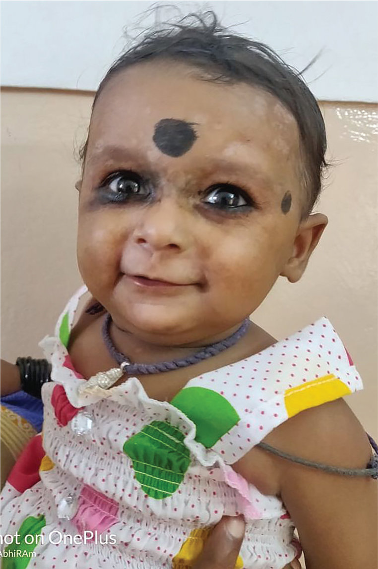
A 4-week infant was referred from a pediatrician to a rheumatologist for ruling out lupus considering skin rashes on the face. The infant was examined and found to have erythematous rashes over the face; she was active, and her milestones were appropriate for the age (Figure 1). She was delivered through a normal vaginal delivery. Her mother was asymptomatic and denied treatment for any rheumatological illness; this was her first pregnancy. The child was diagnosed with raccoon eyes, suggestive of neonatal lupus syndrome.
Figure 1.

Erythematous lesions in the periorbital region - Raccoon eyes.
Investigations done on the infant before referral showed a normal blood count; indirect immunofluorescence showed antinuclear antibodies (ANA) positive at 1:80 dilution with a speckled pattern. ECHO and ECG performed on the infant showed normal results; her C3 and C4 levels were 115 mg/dL and 25 mg/dL, respectively. We advised the mother to undergo immunological workup; mother had ANA positivity at 1:160 dilution with a speckled pattern. On further workup, she had a high positive anti-SSA (Ro 52 and Ro 60) and anti-SSB. The infant was followed up regularly and advised mild low-potency steroid application along with limitation of sun exposure. At the end of 9 months, we observed complete resolution of the erythematous lesions with mild hypopigmentation, and her repeat ANA was negative (Figure 2).
Figure 2.
Complete resolution of cutaneous lesions leaving behind mild atrophic scars.
This infant presented only with cutaneous manifestation and did not have any internal organ involvement. It is ideal to look for anti-SSA/SSB antibodies in mothers since fetal monitoring during the next pregnancy becomes an absolute necessity. ANA, since it is of Ig G type, can be transferred from mother to fetus, and ANA positivity in this infant could be attributed to such a passive transmission. Repeat ANA testing is recommended at 6–9 months of age.
Most infants born to anti-SSA positive mothers are born without any major abnormalities. Complete heart block, the most dreaded complication of neonatal lupus syndrome happens in 2% of anti-Ro-positive pregnancies; the recurrence of which rises to 20% in subsequent pregnancies. Fetal monitoring starting at week 16 becomes mandatory to detect early conduction abnormalities, some of which may be reversible (2).
The cutaneous manifestations of neonatal lupus syndrome are usually reversible, occur more frequently, and resemble those of subacute cutaneous lupus erythematosus lesions. They occur in the first 8 weeks of birth, though rarely they can be observed at birth itself. They resolve without scarring at around 6 months, often coinciding with the disappearance of maternal anti-Ro and -La antibodies from the infant (3).
Footnotes
Content of this journal is licensed under a Creative Commons Attribution-NonCommercial 4.0 International License.
Informed Consent: Written informed consent was obtained from the patient’s parent for publication of this manuscript and accompanying images (Date: November 20, 2018).
Peer-review: Externally peer-reviewed.
Author Contributions: Concept - K.M., A.G.; Design - A.G.; Supervision - A.G.; Resources - K.M.; Materials - A.G.; Data Collection and/or Processing - K.M.; Analysis and/or Interpretation - K.M.; Literature Search - K.M.; Writing Manuscript - K.M.; Critical Review - K.M.
Conflict of Interest: The authors have no conflict of interest to declare.
Financial Disclosure: The authors declared that this study has received no financial support.
References
- 1.Brito-Zerón P, Izmirly PM, Ramos-Casals M, Buyon JP, Khamashta MA. The clinical spectrum of autoimmune congenital heart block. Nat Rev Rheumatol. 2015;11:301–12. doi: 10.1038/nrrheum.2015.29. [DOI] [PMC free article] [PubMed] [Google Scholar]
- 2.Vanoni F, Lava SAG, Fossali EF, Cavalli R, Simonetti GD, Bianchetti MG, et al. Neonatal Systemic Lupus Erythematosus Syndrome: a Comprehensive Review. Clin Rev Allergy Immunol. 2017;53:469–76. doi: 10.1007/s12016-017-8653-0. [DOI] [PubMed] [Google Scholar]
- 3.Klauninger R, Skog A, Horvath L, Winqvist O, Edner A, Bremme K, et al. Serologic follow-up of childrenborn to mothers with Ro/SSA autoantibodies. Lupus. 2009;18:792–8. doi: 10.1177/0961203309103188. [DOI] [PubMed] [Google Scholar]