Abstract
Purpose of Review
Hip arthroscopy has seen increasing utilization over the last decade. This is largely related to increased recognition and improved techniques for treating femoroacetabilar impingement (FAI). Though hip arthroscopy generally yields favorable outcomes, there are a subset of patients who have residual or recurrent symptoms that require reoperation. The current review discusses an algorithmic approach to evaluating patients following a failed hip arthroscopy including a framework for clinical and radiographic assessment, available treatment options, and associated outcomes in revision surgery.
Recent Findings
Residual FAI has been demonstrated to be the most common indication for revision arthroscopy. Other indications include residual or recurrent labral pathology, gross instability, microinstability, or adhesions. Appropriate history and imaging are important to determine the cause for residual symptoms. Novel techniques including labral and capsular reconstruction, and modified remplissage procedures have been developed to deal with complex revision cases. Though studies have shown improved outcomes after revision surgery, they have been shown to result in inferior outcomes compared to a matched cohort following primary hip arthroscopy.
Summary
Management of a failed hip arthroscopy remains a complex problem. Focused history, cross-sectional imaging, and revision hip arthroscopy with novel techniques can improve outcomes, albeit to a lesser extent than patients undergoing successful primary hip arthroscopy. The information provided here can help guide treatment and set appropriate patient expectations for revision surgery.
Keywords: Revision, Hip, Arthroscopy, Femoroacetabular impingement, Microinstability, Labral pathology
Introduction
Arthroscopic surgery of the hip has had a significant increase in popularity during the last two decades [1–3]. Improving disease awareness, investigations, and surgical techniques have helped to expand the indications for this less invasive approach to the hip. Population-based studies and those evaluating trends of recently trained surgeons have demonstrated tremendous increases in international procedural use. In the USA alone, there was a 600% increase in hip arthroscopy from 2006 to 2010 [3]. Similarly, in England, there was a 727% increase between 2002 and 2013. It is projected that these trends will continue in the years to come, with a proposed 1388% increase by 2023 [4].
Though favorable outcomes are reported for primary hip arthroscopy, there are a proportion of patients that experience inferior outcomes with several of these patients going on to require revision surgery. A 2013 systematic review of 92 studies and over 6000 patients reported an overall reoperation rate of 6.3%. Of those patients that required reoperation approximately 30% underwent revision arthroscopy, the remainder underwent open procedures including conversion to total hip arthroplasty (THA) [5]. Similar results were seen in a study in 2019 which demonstrated a 4% rate of revision hip arthroscopy (72 of 1807 hip arthroscopies) [6•]. Of the revision procedures, 43% occurred within 6 months after the index procedure, and 86% occurred within 18 months.
Several studies have indicated residual impingement is the most common reason for revision arthroscopy [7–9, 10••]. A study of revision hip arthroscopy reported that residual impingement was noted in 40% of patients requiring reoperation. This study also noted that patients had an average of 4 diagnoses at the time of revision surgery, indicating that the reason for revision surgery is often multifactorial. The most common diagnoses noted in this study were chondral lesions (both femoral and acetabular), labral tearing, and residual impingement. Other diagnoses noted included labral calcification, heterotopic ossification, adhesions, loose bodies, synovitis, iliopsoas pathology, and trochanteric bursitis [8].
Due to the multitude of factors that could contribute to clinical failure following a primary hip arthroscopy, it is imperative to have an algorithmic approach to evaluating and treating recurrent or residual hip pain following an unsuccessful arthroscopy. The current review aims to discuss revision hip arthroscopy including a framework for clinical and radiographic assessment, treatment options, and outcomes in revision surgery.
History and Physical Examination
Patients presenting with pain following hip arthroscopy should be carefully assessed to clarify symptomology and guide future investigations or treatment. It is important to distinguish between residual symptoms (lack of improvement from surgery), which is most commonly related to residual impingement, and recurrent symptoms (period of relief followed by recurrence), which could be attributed to new labral pathology including labral calcification or failed repair, iatrogenic instability, chondral lesions, or adhesions [11].
Impingement
Patients with residual bony impingement often complain of similar symptoms to those presenting with primary symptomatic FAI, including activity-related groin pain and an inability to perform activities that include higher degrees of hip flexion with or without combined internal rotation, or periods of prolonged sitting [12]. A physical examination can often reveal a reduced range of motion and pain with flexion, adduction, and internal rotation in the case of a residual cam deformity. Pain may also be exacerbated with direct in-line hip flexion, suggestive of subspinous impingement, or focal acetabular retroversion.
Labral Pathology
Recurrent labral pathology may also present with groin pain, although the location of pain can vary based on the location of the pathology. Conventionally, anterosuperior labral tears present similarly to residual impingement with anterior groin pain. Pain can also be reported in the lateral groin or deep posterior buttock [12, 13]. Anterior hip pain is more consistent with an anterior labral tear, while posterior or buttock pain is consistent with a posterior labral tear, although atypical presentations can occur [14]. Symptomatic labral tears may also cause mechanical symptoms such as painful clicking or locking [12, 15]. On physical examination, patients will generally have a positive impingement test, and may also demonstrate pain with a labral shear test.
Instability
Instability can also occur after primary hip arthroscopy, and can range from macroinstability, with a history of frank dislocation, or microinstability, with pain attributed to adverse physiologic motion secondary to soft-tissue deficiencies. Rarely, patients may experience frank subluxation or dislocation of the affected hip, which is more common in those patients with generalized ligamentous laxity [16]. Instead, “microinstability” is of greater concern, as this may present vaguely with deep groin pain, or a sensation of apprehension or giving way with certain activities [17••]. Activities that produce external rotation with an axial load or hyperextension are most frequently to blame. Microinstability can be due to iatrogenic injury to the hip joint capsule. However, it may also be related to labral insufficiency following debridement, or over-resection of cam deformity disrupting the hip joint suction seal. On physical examination, excessive hip internal or external rotation are most commonly reported. An additional test has been described, where the leg is positioned in flexion, abduction, and external rotation (FABER) or the figure-of-4 position and if the lateral aspect of the knee joint is less than 3 in. from the table it may be suggestive of instability. [17••]. A 2017 study examined three different special tests for instability, including the abduction-hyperextension-external rotation test (AB-HEER), prone instability test, and hyperextension-external rotation test (HEER) [18]. The AB-HEER test is performed with the patient in the lateral decubitus position with the affected hip placed upward. The hip then is abducted to 30° to 45°, extended, and externally rotated, while an anteriorly directed force is applied to the posterior greater trochanter. The test finding is positive if pain is reproduced [19]. The prone instability test is performed with the patient in the prone position. The hip is externally rotated, while the examiner applies a downward force on the posterior greater trochanter. The reproduction of anterior hip pain is consistent with a positive test result [20]. The HEER test is performed with the patient supine at the foot of the table with the legs hanging free. An anteriorly directed force is applied at the hip, the contralateral hip is flexed, while the ipsilateral hip is hyperextended and externally rotated. Reproducible pain indicates a positive test result [21]. All three tests showed excellent specificity for microinstability (89.4%, 97.9%, and 85.1%, respectively. The AB-HEER and HEER tests also had good sensitivity. The prone instability test, however, had low sensitivity (33.9%). The authors therefore recommended this test be used to rule in instability rather than to rule it out. [18]. If all three tests are positive, there is a 95% likelihood that the patient will demonstrate instability intraoperatively [17••, 18].
Miscellaneous
Adhesions and chondral injuries have also been listed as causes for residual symptoms after primary hip arthroscopy. The capsulolabral junction has an abundant blood supply which predisposes it to adhesion formation with manipulation as well as the rim osteochondroplasty and placement of suture anchors for labral repair [22, 23]. Adhesions present with pain and limited range of motion [22, 23]. Patients may report pain with straight hip flexion and not with adduction and internal rotation, differentiating it from the anterior impingement sign [23]. Risks for the development of adhesions include age < 30, modifies Harris hip score (mHHS) < 50, and lack of circumduction exercises in the rehab protocol [24].
Pure chondral injuries may not present with much pain or symptoms as cartilage does not have nociceptive receptors. They may have mechanical symptoms of locking or catching if loose bodies are present. Additionally, synovial irritation from chondral damage can present as a painful hip [25]. There are no specific maneuvers to test for chondral lesions. A full hip examination should be completed to rule out other etiologies of hip pain.
Imaging
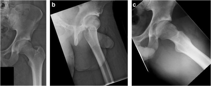
Repeat imaging in the form of plain radiographs should be completed first while assessing the symptomatic hip after primary hip arthroscopy. Anterior-posterior (AP) pelvis and 45° Dunn lateral views are the most effective in assessing for residual cam or pincer lesions (Fig. 1). A 2017 study compared multiple radiographic views of the hip (AP pelvis, cross-table lateral, 90° Dunn, 45° Dunn, and modified 45° Dunn views) to radial cuts of MRI. An alpha angle on the 45° Dunn lateral radiograph had the highest correlation to cuts on MRI (0.81) [26]. Another study compared to 2 views of the hip (AP pelvis and 45° Dunn lateral) versus a 5-view series (AP pelvis and AP, 45° Dunn lateral, frog lateral, and false profile of the affected him) in evaluation of cam morphology. This study demonstrated the 2-view series to be non-inferior to the 5-view series for cam identification (P value = 0.010). There was no difference in sensitivity or specificity between the 2-view and 5-view series [27].
Fig. 1.
A 21-year-old male with residual pain 7 months post left hip arthroscopy. Postoperative AP pelvis a and cross-table lateral b with no obvious residual deformity seen. Postoperative 45° Dunn lateral c showing residual cam lesion resulting in continued symptoms
CT imaging can be used to further assess for cam morphology and assess acetabular version [28]. Three-dimensional CT images are often useful in characterizing the extent of morphological changes and location of residual impingement [29]. Low-dose CT scans have been shown to reduce radiation exposure by 90% compared to traditional CT scans while not compromising image quality and are an effective means for evaluating for residual structural impingement or over-resection [30••].
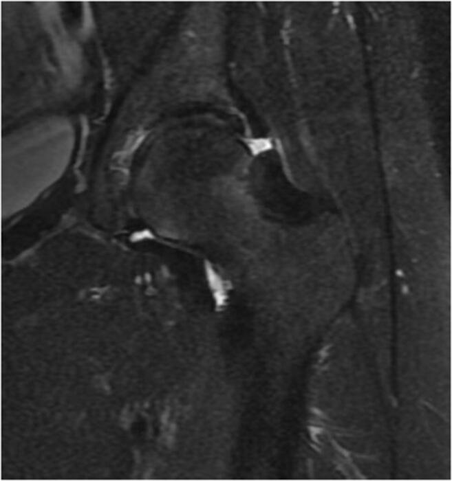
MR arthrogram can be used to assess for soft tissue pathology including recurrent or residual labral tears, chondral pathology, capsular defects, or adhesions (Fig. 2). A study in 2013 examined the sensitivity, specificity, positive predictive value, and negative predictive value of MR arthrogram in revision hip arthroscopy [31]. The study included 70 hips undergoing revision hip arthroscopy and looked at the correlation between preoperative MR arthrogram and intraoperative findings. For labral pathology, MR arthrogram showed a sensitivity of 82%, specificity of 70%, PPV of 94%, and NPV of 39%. For chondral pathology, sensitivity was 65%, specificity was 90%, PPV was 94%, and NPV was 50%. MR arthrography was shown to be superior at ruling in, rather than ruling out, labral lesions, and cartilage lesions [31].
Fig. 2.

A 44-year-old female with symptoms of new onset of deep groin pain 3 months after hip arthroscopy following a postoperative fall. MRI shows disruption of her interportal capsulotomy
Treatment Options and Associated Outcomes
Overall, revision arthroscopy has been shown to improve patient-reported outcomes. A systematic review and meta-analysis in 2019 examined outcomes in revision hip arthroscopy [32••]. The study indicated that there was an improvement in patient-reported outcomes after revision surgery. The modified Harris hip score (mHHS) was reported in 10 studies and had an increase of 17.20 (p < 0.001), the hip outcome score for activities of daily living (HOS-ADL) was reported in 5 studies with a mean difference of 13.98 (p < 0.001) and non-arthritic hip score (NAHS) was reported in 2 studies with a mean difference of 25.30 (p = 0.0162). Though this shows statistically significant improvements, they are inferior to primary hip arthroscopy. The same systematic review showed the pooled mean score after primary hip arthroscopy was 82.77 (95% CI, 81.52–84.03) based on 3 studies versus 74.61 (95% CI, 71.16–78.06). Similarly, the HOS-ADL and NAHS had scores which were significantly higher in the primary hip arthroscopy cohort compared to the revision cohort [32••].
A study in 2018 examined the minimal clinically important difference (MCID) and substantial clinical benefit (SCB) in revision hip arthroscopy [33]. The MCID is defined as the smallest change in outcome that the patient is able to appreciate. The SCB is defined as the improvement in outcome or absolute postoperative health state that the patient considers to be a substantial improvement. The authors showed that MCID values ranged from 7.9 on the mHHS and the HOS-ADL to 13.1 on the HOS sports. SCB net change for the mHHS, HOS activities of daily living (ADL), HOS sports, and iHOT-33 were 23.1, 16.2, 25.0, and 25.5, respectively. Previously, the same authors had reported MCID and SCB values in primary hip arthroscopy of 8.2 and 19.8 for the mHHS, 8.3 and 10.0 for the HOS-ADL, 14.5 and 29.9 for the HOS sports, and 12.1 and 24.5 for the iHOT-33, respectively [34]. This indicates that MCID and SCB values are similar for primary hip arthroscopy and revision hip arthroscopy. Similarly, the results of this study, along with those reporting MCID following arthroscopy for primary symptomatic femoroacetabular impingement syndrome (FAIS), demonstrate that a comparable proportion of patients achieve MCID between these revision (74.3% and primary (73%) cohorts [35].
Residual Impingement
In the case of residual impingement, repeat arthroscopy with revision osteochondroplasty should be completed. Intraoperative fluoroscopy alone gives a limited picture of impingement; thus, careful examination of preoperative images including three-dimensional CT is imperative to avoid missing areas of deformity [11]. A 2014 study of residual deformity requiring revision arthroscopy showed that the most common area for missed cam pathology was at 1:15 o’clock [36]. In the same review study by Nwachukwu et al., they found that a higher proportion of patients with a diagnosis of residual impingement achieved MCID (74.3%) compared with those who carried other diagnoses or reasons for revision (57.1%). This suggests that those with residual impingement may experience the greatest improvement of all patients undergoing revision arthroscopy.
Labral Pathology
Multiple issues with the labrum have been documented at the time of revision arthroscopy. Techniques to address labral pathology depend on the extent of damage and include revision labral repair, labral augmentation, or labral reconstruction. In 2013, Philippon described an algorithm for the treatment of labral tears [37]. For tears in a large labrum (>7 mm), Philippon described performing a repair with rim trimming of the acetabulum and labral re-fixation. Debridement can be performed only if there is sufficient residual tissue. In a small labrum (<7 mm), repair with or without augmentation is suggested [37]. Augmentation can range from local tissue (indirect head of the rectus femoris) to augmentive reconstruction using allograft. Labral reconstruction is also an option in cases where the labrum is irreparable, has calcified, or failed to heal following a prior repair. A systematic review in 2019 stated that the most common indication for labral reconstruction was in young patients who have undergone previous hip surgery or present with an irreparable, hypotrophic (<2 mm) or hypertrophic (>8 mm) non-functional or calcified labrum [38]. Grafts used for reconstruction included iliotibial band allograft, gracilis autograft, semitendinosus autograft or allograft, peroneus brevis allograft, and indirect head of rectus femoris autograft. Labral allografts were also used. Labral reconstruction has shown short- and intermediate-term improvement in patient-reported outcomes and functional scores postoperatively; however, long-term outcome studies are presently lacking [38].
Capsular Pathology
Capsular repair or plication can be used to treat capsular defects and symptomatic instability. Capsular plication aims to imbricate and shift the capsule to augment the screw home mechanism of the capsuloligamentous structures and improve joint stability [39]. Multiple techniques have been described for capsular plication [39–42]. A recent cadaveric study compared two techniques for capsular plication. The techniques used were a T-capsulotomy plication and interportal capsular shift. Plication was completed using 5-mm bites on either side of the capsulotomy. Mean intra-articular volumes were measure before and after plication. Both groups demonstrated statistically significant reductions in capsular volume with an average reduction of 10.0 ml for T-capsulotomy plication and 15.6 ml for the interportal capsular shift. The capsular plication had a relatively greater volumetric reduction, but this difference was not statistically significant, and the clinical significance is not known [40]. Capsular reconstruction has been described in cases of deficient capsular tissue. Iliotibial band (ITB) and dermal allograft can be used to reconstruct the capsuloligamentous complex [43–46]. A study in 2019 examined capsular reconstruction with ITB or dermal allograft in patients undergoing revision hip arthroscopy for symptomatic instability. Eighteen patients underwent reconstruction with ITB allograft and 18 patients underwent reconstruction with dermal allograft. At a mean follow-up of 25 months, the HOS-ADL, Western Ontario & McMaster Universities Osteoarthritis Index (WOMAC), and SF-12 physical component scores significantly improved after surgery in both groups (p < 0.01). At the final follow-up, the ITB allograft group had a higher quality of life scores compared to the dermal the allogaft group [43]. Though this study showed positive outcomes, there were 4 patients in each group (22% overall) that were considered to go on to failure and required subsequent surgery (4 revision arthroscopy, 3 THA, 1 periacetabular osteotomy). Longitudinal follow-up is required to determine long-term outcomes associated with these novel techniques.
Over-resection
As with labral deficiency, cam over-resection can lead to issues with hip biomechanics and stability, primarily due to a disruption of the suction-seal of the labrum. A “remplissage” procedure has been described to address over-resection at the femoral head-neck junction [47, 48]. In this technique, an iliotibial band (ITB) allograft is used to fill in the area of over-resection, similar to the way a Hill-Sachs lesion is addressed in the shoulder [47]. Filling the defect helps to re-establish the seal between femoral head and labrum. This technique requires a careful dynamic examination of the hip intraoperatively to determine the size of the defect. Over-filling of the defect can lead to recreation of the initial impingement syndrome [22]. While this represents a potential salvage technique, clinical outcome data is lacking and should be a focus of future studies.
Miscellaneous
Adhesions can be treated with arthroscopic lysis of adhesions with or without the placement of a capsulolabral spacer graft [22, 23]. Release of adhesions should be performed carefully to avoid damage to healthy labral tissues. Once adhesions have been removed an ITB allograft spacer can be placed in the capsulolabral recess and secured with suture anchors [22, 23]. Circumduction exercises should be the focus of exercises postoperatively to prevent reformation of adhesions. As above, this is a novel procedural with limited outcome data to support routine utilization and further study is required before widespread adoption of this technique.
Chondral injuries can be addressed with microfracture or chondroplasty depending on the size and thickness of the lesion. A review in 2010 described an algorithm for the treatment of chondral injuries. The authors suggested chondroplasty for partial-thickness injuries with careful use of a shaver blade. In the case of small full-thickness injuries, debridement and microfracture are completed to induce a fibrocartilage healing response [25]. The lesions should be contained and less than 400 mm2. In larger lesions, scaffold augmentation has been proposed to enhance properties of repair tissue in microfracture. A 2018 study examined the effects of using a chitosan-based scaffold in the treatment of chondral lesions of the hip [49]. Twenty-three patients with chondral lesions > 2 cm2 were included in the study. Significant improvement occurred comparing the preoperative to the 1-year postoperative patient-reported outcomes including the NAHS (p = 0.0001), iHOT33 (p = 0.0001), HOS-ADL (p = 0.0005), and HOS-SSS (p = 0.0002). Outcome scores exceeded the MCID in 91% of patients in the study [49].
Salvage Options
In many cases, ongoing symptoms following arthroscopy are less likely attributable to the factors discussed within this article and may instead relate to progressive joint deterioration with the onset of early osteoarthritis. Rates of revision arthroscopy following a failed primary scope for the aforementioned reasons range from 3 to 5% [50]; comparatively, the rates of conversion to THA in large cohort series following primary arthroscopy have often been reported at similar or even higher rates, ranging from 3 to 10%. Several studies have reported on prognostic variables associated with an elevated risk of requiring THA conversion following primary arthroscopy. A recent study identified multiple risk factors including older age, femoral head chondral lesions, lack of performing a femoral osteochondroplasty, performance of an acetabular osteochondroplasty, and revision arthroscopy. The authors then used these variables to create a predictive tool for the need of THA following hip arthroscopy and found greater than 70% accuracy, so patients with many of these risk factors presenting with ongoing symptoms following arthroscopy may need to be appropriately counseled on their potential risk of requiring THA [51••]. Fortunately, outcomes for a THA performed following a failed arthroscopy appear similar to primary THA results, with comparable short-term outcome measures [52]. As such, THA represents a viable salvage option where necessary.
Conclusions
As the rate of hip arthroscopy has increased there has been a relative increase in the rate of revision hip arthroscopy. The most common indications for revision hip arthroscopy include residual impingement, recurrent labral pathology, and instability. A careful history and physical exam should be completed to determine the timeline of symptoms and possible etiology, with advanced cross-sectional imaging to support the diagnosis. Multiple novel techniques have been developed to address the issues faced in revision hip arthroscopy. Though outcomes show improvement after revision hip arthroscopy, they are inferior to primary hip arthroscopy. Careful preoperative planning and appropriate patient expectations are essential.
Compliance with Ethical Standards
Conflict of Interest
The authors declare that they have no conflicts of interest.
Human and Animal Rights and Informed Consent
This article does not contain any studies with human or animal subjects performed by any of the authors.
Footnotes
This article is part of the Topical Collection on Outcomes Research in Orthopedics
Publisher’s note
Springer Nature remains neutral with regard to jurisdictional claims in published maps and institutional affiliations.
References
Papers of particular interest, published recently, have been highlighted as: • Of importance •• Of major importance
- 1.Montgomery SR, Ngo SS, Hobson T, Nguyen S, Alluri R, Wang JC, Hame SL. Trends and demographics in hip arthroscopy in the United States. Arthroscopy. 2013;29(4):661–665. doi: 10.1016/j.arthro.2012.11.005. [DOI] [PubMed] [Google Scholar]
- 2.Colvin Alexis Chiang, Harrast John, Harner Christopher. Trends in Hip Arthroscopy. The Journal of Bone and Joint Surgery-American Volume. 2012;94(4):e23-1-5. doi: 10.2106/JBJS.J.01886. [DOI] [PubMed] [Google Scholar]
- 3.Bozic Kevin J., Chan Vanessa, Valone Frank H., Feeley Brian T., Vail Thomas P. Trends in Hip Arthroscopy Utilization in the United States. The Journal of Arthroplasty. 2013;28(8):140–143. doi: 10.1016/j.arth.2013.02.039. [DOI] [PubMed] [Google Scholar]
- 4.Palmer AJ, Malak TT, Broomfield J, Holton J, Majkowski L, Thomas GE, et al. Past and projected temporal trends in arthroscopic hip surgery in England between 2002 and 2013. BMJ Open Sport Exerc Med. 2016;2(1):e000082. doi: 10.1136/bmjsem-2015-000082. [DOI] [PMC free article] [PubMed] [Google Scholar]
- 5.Harris JD, Mccormick FM, Abrams GD, Gupta AK, Ellis TJ, Bach BR, et al. Complications and reoperations during and after hip arthroscopy: a systematic review of 92 studies and more than 6,000 patients. Arthroscopy. 2013;29(3):589–595. doi: 10.1016/j.arthro.2012.11.003. [DOI] [PubMed] [Google Scholar]
- 6.West CR, Bedard NA, Duchman KR, Westermann RW, Callaghan JJ. Rates and risk factors for revision hip arthroscopy. Iowa Orthop J. 2019;39(1):95–99. [PMC free article] [PubMed] [Google Scholar]
- 7.Bogunovic L, Gottlieb M, Pashos G, Baca G, Clohisy JC. Why do hip arthroscopy procedures fail? Clin Orthop Relat Res. 2013;471(8):2523–2529. doi: 10.1007/s11999-013-3015-6. [DOI] [PMC free article] [PubMed] [Google Scholar]
- 8.Gwathmey F Winston, Jones Kay S, Thomas Byrd J W. Revision hip arthroscopy: findings and outcomes. Journal of Hip Preservation Surgery. 2017;4(4):318–323. doi: 10.1093/jhps/hnx014. [DOI] [PMC free article] [PubMed] [Google Scholar]
- 9.Woodward RM, Philippon MJ. Persistent or recurrent symptoms after arthroscopic surgery for femoroacetabular impingement: a review of imaging findings. J Med Imaging Radiation Oncol. 2018;63(1):15–24. doi: 10.1111/1754-9485.12822. [DOI] [PubMed] [Google Scholar]
- 10.Sardana V, Philippon MJ, de Sa D, Bedi A, Ye L, Simunovic N, et al. Revision hip arthroscopy indications and outcomes: a systematic review. Arthroscopy. 2015;31(10):2047–2055. doi: 10.1016/j.arthro.2015.03.039. [DOI] [PubMed] [Google Scholar]
- 11.Ekhtiari Seper, Coughlin Ryan P., Simunovic Nicole, Ayeni Olufemi R. Strategies in revision hip arthroscopy. Annals of Joint. 2018;3:7–7. [Google Scholar]
- 12.Banerjee P, McLean CR. Femoroacetabular impingement: a review of diagnosis and management. Curr Rev Musculoskelet Med. 2011;4(1):23–32. doi: 10.1007/s12178-011-9073-z. [DOI] [PMC free article] [PubMed] [Google Scholar]
- 13.Groh M, Herrera J. A comprehensive review of hip labral tears. Curr Rev Musculoskelet Med. 2009;2(2):105–117. doi: 10.1007/s12178-009-9052-9. [DOI] [PMC free article] [PubMed] [Google Scholar]
- 14.Hase T, Ueo T. Acetabular labral tear: arthroscopic diagnosis and treatment. Arthroscopy. 1999;15(2):138–141. doi: 10.1053/ar.1999.v15.0150131. [DOI] [PubMed] [Google Scholar]
- 15.Lewis CL, Sahrmann SA. Acetabular labral tears. Phys Ther. 2006;86:110–121. doi: 10.1093/ptj/86.1.110. [DOI] [PubMed] [Google Scholar]
- 16.Shu B, Safran MR. Hip instability: anatomic and clinical considerations of traumatic and atraumatic instability. Clin Sports Med. 2011;30(2):349–367. doi: 10.1016/j.csm.2010.12.008. [DOI] [PubMed] [Google Scholar]
- 17.Safran MR. Microinstability of the hip-gaining acceptance. J Am Acad Orthop Surg. 2019;27(1):12–22. doi: 10.5435/JAAOS-D-17-00664. [DOI] [PubMed] [Google Scholar]
- 18.Hoppe DJ, Truntzer JN, Shapiro LM, Abrams GD, Safran MR. Diagnostic accuracy of 3 physical examination tests in the assessment of hip microinstability. Orthop J Sports Med. 2017;5(11):2325967117740121. doi: 10.1177/2325967117740121. [DOI] [PMC free article] [PubMed] [Google Scholar]
- 19.Domb BG, Brooks AG, Guanche CA. Physical examination of the hip. In: Guanche CA, editor. Hip and Pelvis Injuries in Sports Medicine Philidephia: Wolters Kluwer/Lipincott Williams and Wilkins. 2010. pp. 62–70. [Google Scholar]
- 20.Domb BG, Stake CE, Lindner D, El-Bitar Y, Jackson TJ. Arthroscopic capsular plication and labral preservation in borderline hip dysplasia: two-year clinical outcomes of a surgical approach to a challenging problem. Am J Sports Med. 2013;41(11):2591–2598. doi: 10.1177/0363546513499154. [DOI] [PubMed] [Google Scholar]
- 21.Safran MR. Evaluation of the painful hip in tennis players. Aspetar Sports Med J. 2014;3:516–523. [Google Scholar]
- 22.Locks R, Bolia I, Utsunomiya H, Briggs K, Philippon MJ. Current concepts in revision hip arthroscopy. Hip Int. 2018;28(4):343–351. doi: 10.1177/1120700018771927. [DOI] [PubMed] [Google Scholar]
- 23.Philippon MJ, Ferro FP, Nepple JJ. Hip capsulolabral spacer placement for the treatment of severe capsulolabral adhesions after hip arthroscopy. Arthrosc Tech. 2014;3(2):e289–e292. doi: 10.1016/j.eats.2014.01.003. [DOI] [PMC free article] [PubMed] [Google Scholar]
- 24.Willimon SC, Briggs KK, Philippon MJ. Intraarticular adhesions following hip arthroscopy: a risk factor analysis. Knee Surg Sports Traumatol Arthrosc. 2014;22:822–825. doi: 10.1007/s00167-013-2728-0. [DOI] [PubMed] [Google Scholar]
- 25.Yen YM, Kocher MS. Chondral lesions of the hip: microfracture and chondroplasty. Sports Med Arthrosc Rev. 2010;18(2):83–89. doi: 10.1097/JSA.0b013e3181de1189. [DOI] [PubMed] [Google Scholar]
- 26.Saito M, Tsukada S, Yoshida K, Okada Y, Tasaki A. Correlation of alpha angle between various radiographic projections and radial magnetic resonance imaging for cam deformity in femoral head-neck junction. Knee Surg Sports Traumatol Arthrosc. 2017;25(1):77–83. doi: 10.1007/s00167-016-4046-9. [DOI] [PubMed] [Google Scholar]
- 27.Wong TT, Weeks JK, Ahmed FS, Francescone MA, Rasiej MJ, Liu MZ, et al. How many radiographs does it take to screen for femoral cam morphology?: a noninferiority study. Curr Probl Diagn Radiol. 2019;12. [DOI] [PubMed]
- 28.Dolan MM, Heyworth BE, Bedi A, Duke G, Kelly BT. CT reveals a high incidence of osseous abnormalities in hips with labral tears. Clin Orthop Relat Res. 2011;469(3):831–838. doi: 10.1007/s11999-010-1539-6. [DOI] [PMC free article] [PubMed] [Google Scholar]
- 29.Crim J. Imaging evaluation of the hip after arthroscopic surgery for femoroacetabular impingement. Skelet Radiol. 2017;46(10):1315–1326. doi: 10.1007/s00256-017-2665-y. [DOI] [PMC free article] [PubMed] [Google Scholar]
- 30.Su AW, Hillen TJ, Eutsler EP, Bedi A, Ross JR, Larson CM, et al. Low-dose computed tomography reduces radiation exposure by 90% compared with traditional computed tomography among patients undergoing hip-preservation surgery. Arthroscopy. 2019;35(5):1385–1392. doi: 10.1016/j.arthro.2018.11.013. [DOI] [PMC free article] [PubMed] [Google Scholar]
- 31.McCarthy JC, Glassner PJ. Correlation of magnetic resonance arthrography with revision hip arthroscopy. Clin Orthop Relat Res. 2013;471(12):4006–4011. doi: 10.1007/s11999-013-3202-5. [DOI] [PMC free article] [PubMed] [Google Scholar]
- 32.•• O'Connor M, Steinl GK, Padaki AS, Duchman KR, Westermann RW, Lynch TS. Outcomes of Revision Hip Arthroscopic Surgery: A Systematic Review and Meta-analysis. Am J Sports Med. 2019:363546519869671 Recent systematic review and meta-analysis of over 4000 patients examining outcomes in revision hip arthroscopy. Patients had improvements in PRO. When compared to primary hip arthroscopy, the mean PROs were inferior. After revision hip arthroscopic surgery, the rates of conversion to total hip arthroplasty ranged from 0 to 14.3%, and the rates of further arthroscopic revision ranged from 2 to 14%.
- 33.Nwachukwu BU, Chang B, Rotter BZ, Kelly BT, Ranawat AS, Nawabi DH. Minimal clinically important difference and substantial clinical benefit after revision hip arthroscopy. Arthroscopy. 2018;34(6):1862–1868. doi: 10.1016/j.arthro.2018.01.050. [DOI] [PubMed] [Google Scholar]
- 34.Nwachukwu BU, Fields K, Chang B, Nawabi DH, Kelly BT, Ranawat AS. Preoperative outcome scores are predictive of achieving the minimal clinically important difference after arthroscopic treatment of femoroacetabular impingement. Am J Sports Med. 2017;45(3):612–619. doi: 10.1177/0363546516669325. [DOI] [PubMed] [Google Scholar]
- 35.Nho SJ, Beck EC, Nwachukwu BU, Cvetanovich GL, Neal WH, Harris JD, Weber AE, Mather RC. Survivorship and outcome of hip arthroscopy for femoroacetabular impingement syndrome performed with modern surgical techniques. Am J Sports Med. 2019;47(7):1662–1669. doi: 10.1177/0363546519843936. [DOI] [PubMed] [Google Scholar]
- 36.Ross JR, Larson CM, Adeoye O, Kelly BT, Bedi A. Residual deformity is the most common reason for revision hip arthroscopy: a three-dimensional CT study. Clin Orthop Relat Res. 2015;473(4):1388–1395. doi: 10.1007/s11999-014-4069-9. [DOI] [PMC free article] [PubMed] [Google Scholar]
- 37.Philippon MJ, Faucet SC, Briggs KK. Arthroscopic hip labral repair. Arthrosc Tech. 2013;2(2):e73–e76. doi: 10.1016/j.eats.2012.11.002. [DOI] [PMC free article] [PubMed] [Google Scholar]
- 38.Al Mana L, Coughlin RP, Desai V, Simunovic N, Duong A, Ayeni OR. The hip labrum reconstruction: indications and outcomes-an updated systematic review. Curr Rev Musculoskelet Med. 2019;12(2):156–165. doi: 10.1007/s12178-019-09546-6. [DOI] [PMC free article] [PubMed] [Google Scholar]
- 39.Chandrasekaran S, Vemula SP, Martin TJ, Suarez-Ahedo C, Lodhia P, Domb BG. Arthroscopic technique of capsular plication for the treatment of hip instability. Arthrosc Tech. 2015;4(2):e163–e167. doi: 10.1016/j.eats.2015.01.004. [DOI] [PMC free article] [PubMed] [Google Scholar]
- 40.Waterman BR, Chen A, Neal WH, Beck EC, Ukwuani G, Clapp IM, Domb BG, Nho SJ. Intra-articular volume reduction with arthroscopic plication for capsular laxity of the hip: a cadaveric comparison of two surgical techniques. Arthroscopy. 2019;35(2):471–477. doi: 10.1016/j.arthro.2018.09.023. [DOI] [PubMed] [Google Scholar]
- 41.Levy DM, Grzybowski J, Salata MJ, Mather RC, 3rd, Aoki SK, Nho SJ. Capsular plication for treatment of iatrogenic hip instability. Arthrosc Tech. 2015;4(6):e625–e630. doi: 10.1016/j.eats.2015.07.007. [DOI] [PMC free article] [PubMed] [Google Scholar]
- 42.Nishikino S, Hoshino H, Hotta K, Furuhashi H, Koyama H, Matsuyama Y. Arthroscopic capsular repair using proximal advancement for instability following hip arthroscopic surgery: a case report. J Hip Preserv Surg. 2019;6(1):91–96. doi: 10.1093/jhps/hnz007. [DOI] [PMC free article] [PubMed] [Google Scholar]
- 43.Fagotti L, Soares E, Bolia IK, Briggs KK, Philippon MJ. Early outcomes after arthroscopic hip capsular reconstruction using iliotibial band allograft versus dermal allograft. Arthroscopy. 2019;35(3):778–786. doi: 10.1016/j.arthro.2018.10.110. [DOI] [PubMed] [Google Scholar]
- 44.Dierckman BD, Guanche CA. Anterior hip capsuloligamentous reconstruction for recurrent instability after hip arthroscopy. Am J Orthop (Belle Mead NJ) 2014;43(12):E319–E323. [PubMed] [Google Scholar]
- 45.Fujishiro T, Nishikawa T, Takikawa S, Saegusa Y, Yoshiya S, Kurosaka M. Reconstruction of the iliofemoral ligament with an artificial ligament for recurrent anterior dislocation of total hip arthroplasty. J Arthroplast. 2003;18(4):524–527. doi: 10.1016/s0883-5403(03)00073-1. [DOI] [PubMed] [Google Scholar]
- 46.Trindade CA, Sawyer GA, Fukui K, Briggs KK, Philippon MJ. Arthroscopic capsule reconstruction in the hip using iliotibial band allograft. Arthrosc Tech. 2015;4(1):e71–e74. doi: 10.1016/j.eats.2014.11.008. [DOI] [PMC free article] [PubMed] [Google Scholar]
- 47.Perez-Carro L, Escajadillo NF, Escajadillo LF, Arriaza CR, Garcia MS, Fernandez AA. Simultaneous acetabular labrum reconstruction and remplissage of the femoral head-neck junction. Arthrosc Tech. 2019;8(12):e1443–e1449. doi: 10.1016/j.eats.2019.07.023. [DOI] [PMC free article] [PubMed] [Google Scholar]
- 48.Frank JM, Chahla J, Mitchell JJ, Soares E, Philippon MJ. Remplissage of the femoral head-neck junction in revision hip arthroscopy: a technique to correct excessive cam resection. Arthrosc Tech. 2016;5(6):e1209–e1213. doi: 10.1016/j.eats.2016.07.012. [DOI] [PMC free article] [PubMed] [Google Scholar]
- 49.Tahoun MF, Tey M, Mas J, Abd-Elsattar Eid T, Monllau JC. Arthroscopic repair of Acetabular cartilage lesions by chitosan-based scaffold: clinical evaluation at minimum 2 years follow-up. Arthroscopy. 2018;34(10):2821–2828. doi: 10.1016/j.arthro.2018.06.037. [DOI] [PubMed] [Google Scholar]
- 50.Degen RM, Pan TJ, Chang B, Mehta N, Chamberlin PD, Ranawat AS, Nawabi DH, Kelly BT, Lyman S. Risk of failure of primary hip arthroscopy-a population-based study. J Hip Preserv Surg. 2017;4(3):214–223. doi: 10.1093/jhps/hnx018. [DOI] [PMC free article] [PubMed] [Google Scholar]
- 51.Rosinsky PJ, Go CC, Shapira J, Maldonado DR, Lall AC, Domb BG. Validation of a Risk Calculator for Conversion of Hip Arthroscopy to Total Hip Arthroplasty in a Consecutive Series of 1400 Patients. J Arthroplast. 2019;34(8):1700–1706. doi: 10.1016/j.arth.2019.04.013. [DOI] [PubMed] [Google Scholar]
- 52.Rosinsky PJ, Kyin C, Shapira J, Maldonado DR, Lall AC, Domb BG. Hip arthroplasty after hip arthroscopy: are short-term outcomes affected? A systematic review of the literature. Arthroscopy. 2019;35(9):2736–2746. doi: 10.1016/j.arthro.2019.03.057. [DOI] [PubMed] [Google Scholar]


