Dear Editor,
Initial reports have indicated a higher incidence of venous thromboembolism (VTE) among patients with coronavirus disease 2019 (COVID-19) compared to other critical illnesses. Helms et al. [1] found pulmonary embolisms in 25% of patients undergoing CT pulmonary angiography despite prophylactic or therapeutic anticoagulation. Similarly, a 27% incidence of VTE was described by Klok et al. [2] in critically ill COVID-19 patients despite use of standard thromboprophylaxis. Recently, Llitjos et al. [3] found a higher rate of VTE (69%) among severe COVID-19 patients while on anticoagulation.
We confirm a 31% incidence of VTE among patients with COVID-19 admitted to the ICU, and our group of experienced point of care ultrasound (POCUS) trained intensivists (> 5 years) noticed a striking pattern of spontaneous echo contrast (SEC) in the venous system during central line placements. This led us to develop a protocol for lower extremity POCUS scans on all COVID-19 patients in the ICU that included optimization of the gain till no SEC was observed in the adjacent arterial lumen. Lower extremity POCUS scan of 2-region compression ultrasonography can be carried out by intensivists rapidly and accurately [4].
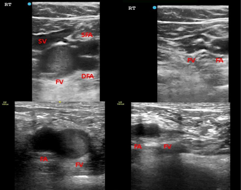
In this study, POCUS was performed on 38 consecutive patients with COVID-19 admitted to our ICU (Fig. 1). We first excluded 7 patients; 4 with missing data and 3 with repeat scans. Of the remaining 31 patients, SEC was observed among 22 patients; however, the average time of scan was 3.93 ± 3.71 days with a significant difference in the SEC group (N = 22, 2.75 days ± 2.3 days) compared to normal venous group (N = 9, 6.56 days ± 4.6 days). To adjust for time variability, an additional 13 patients, who were scanned beyond 72 h, were excluded. Among the 18 patients analyzed in the study, 83% demonstrated SEC in more than 2 sites (Image 1, Video 1). Notably, 87.5% in the SEC group showed bilateral evidence in all sites. This finding was independently verified and confirmed by 2 physicians (S.D; A.M). All 18 patients were receiving anticoagulation (N = 18 on thromboprophylaxis, N = 4 on heparin infusions due to renal replacement therapy [RRT]). A concomitant echocardiogram was performed in 72% of patients among which 83% did not have abnormalities. Elevated inflammatory markers, a higher need for RRT (67% vs 33%) and an increased incidence of deep vein thrombosis (DVT; 40% vs 33%), were detected in the SEC group as compared to those with normal venous flow (ESM Table 1).
Fig. 1.
The Top image shows SEC in right femoral vein with no echogenicity observed in adjacent femoral artery. The vein is fully compressible ruling out DVT at the site. The bottom image again shows SEC in fully compressible right femoral vein, with no echogenicity in adjacent femoral artery. SV saphenous vein, FV femoral vein, FA femoral artery, SFA superficial femoral artery, DFA deep femoral artery
Jensen et al. [5] have described amorphous echogenicity occupying the entire lumen of a fully compressible venous lumen, greater than the adjacent artery as a marker of venous stasis in oncology patients. They also correlated a qualitative SEC with lower peak venous velocities with a doubling in the incidence of DVT. In light of this, the SEC observed among severe COVID-19 patients could likely indicate a precursor for VTE. As a result, higher intensity thromboprophylaxis may be appropriate for this group of SEC patients and future studies are investigating clinical outcomes with anticoagulation strategies.
Electronic supplementary material
Below is the link to the electronic supplementary material.
Acknowledgements
We also acknowledge Dr. Nirupama Mulherkar, Phd for proofreading of this manuscript.
Funding
The authors have no financial support to report for this manuscript. This research did not receive any specific grant from funding agencies in the public, commercial, or not-for-profit sectors.
Institutional IRB approval obtained. IRB# 20-398
Compliance with ethical standards
Conflicts of Interest
On behalf of all authors, the corresponding author states that there is no conflict of interest.
Footnotes
Publisher's Note
Springer Nature remains neutral with regard to jurisdictional claims in published maps and institutional affiliations.
References
- 1.Helms J, Tacquard C, Severac F, et al. High risk of thrombosis in patients in severe SARS-CoV-2 infection: a multicenter prospective cohort study. Intensive Care Med. 2020 doi: 10.1007/s00134-020-06062-x. [DOI] [PMC free article] [PubMed] [Google Scholar]
- 2.Klok FA, Kruip MJHA, van der Meer NJM, et al. Incidence of thrombotic complications in critically ill ICU patients with COVID-19. Thromb Res. 2020 doi: 10.1016/j.thromres.2020.04.013. [DOI] [PMC free article] [PubMed] [Google Scholar]
- 3.Llitjos JF, Leclerc M, Chochois C, et al. High incidence of venous thromboembolic events in anticoagulated severe COVID-19 patients. J Thromb Haemost. 2020 doi: 10.1111/jth.14869. [DOI] [PMC free article] [PubMed] [Google Scholar]
- 4.Kory PD, Pellecchia CM, Shiloh AL, et al. Accuracy of ultrasonography performed by critical care physicians for the diagnosis of DVT. Chest. 2011 doi: 10.1378/chest.10-1479. [DOI] [PubMed] [Google Scholar]
- 5.Jensen CT, Chahin A, Amin VD, et al. Qualitative slow blood flow in lower extremity deep veins on doppler sonography: quantitative assessment and preliminary evaluation of correlation with subsequent deep venous thrombosis development in a tertiary care oncology center. J Ultrasound Med Off J Am Inst Ultrasound Med. 2017;36:1867–1874. doi: 10.1002/jum.14220. [DOI] [PMC free article] [PubMed] [Google Scholar]
Associated Data
This section collects any data citations, data availability statements, or supplementary materials included in this article.


