Figure 1:

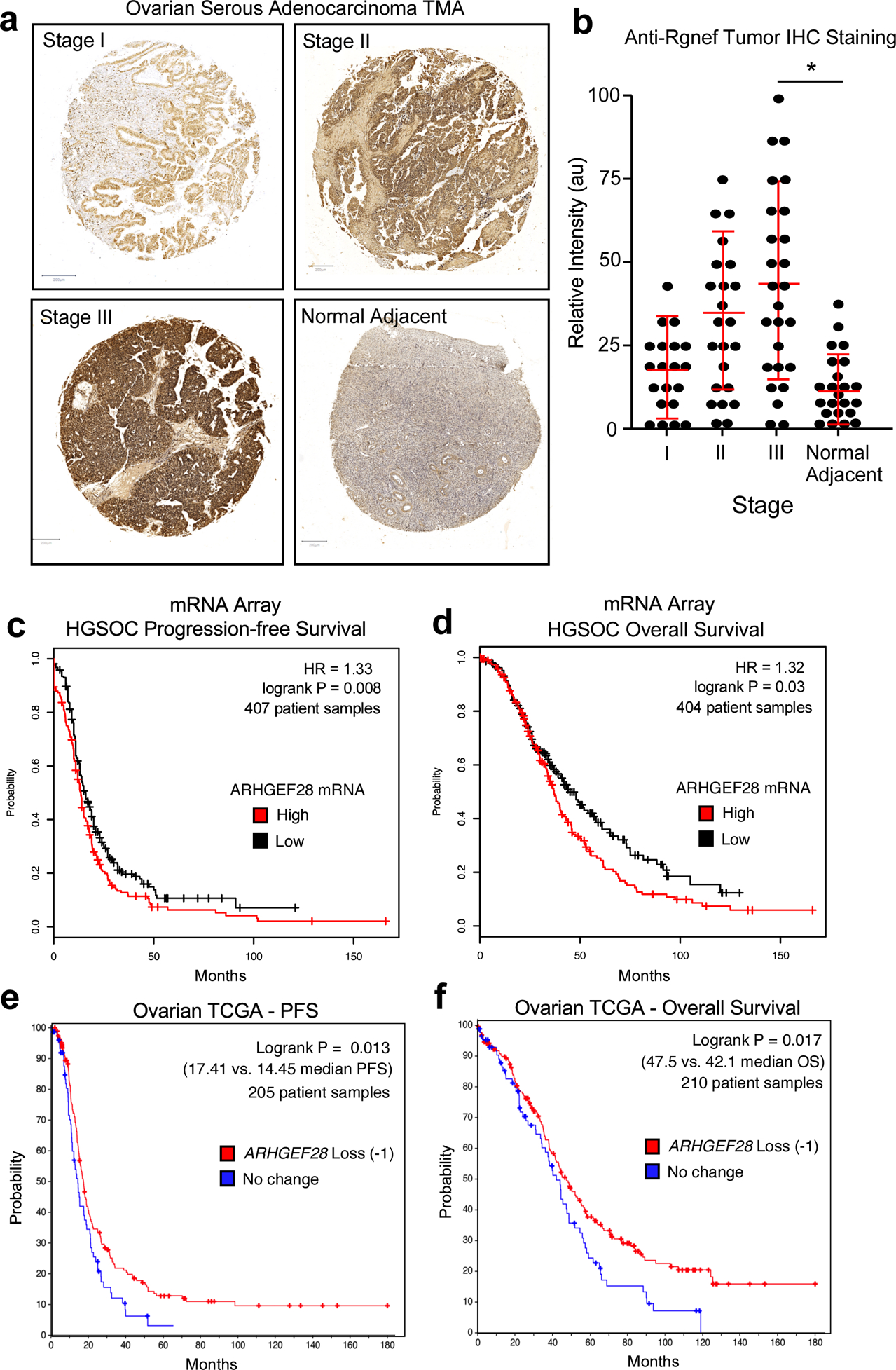
Analysis of Rgnef or ARHGEF28 levels in serous ovarian cancer. (a) Representative high grade serous ovarian cancer tumor micro-array (TMA) cores stained with polyclonal anti-Rgnef antibodies (brown) and nuclei counter-stained with hematoxylin. Stage information was from the manufacturer and normal adjacent is ovarian tissue from tumor-bearing patients. (b) Aperio Image Analysis quantification using the positive pixel count algorithm and relative intensity values (arbitrary units, au) are shown per TMA core. Values are means +/− SD (n=92, P< 0.05, T-Test). (c and d) Kaplan-Meier plotter (http://kmplot.com/ovar) was used to evaluate ARHGEF28 mRNA levels in stage III-IV serous ovarian cancer samples. High ARHGEF28 expression [auto-select cutoff] was significantly associated with (c) progression-free survival (*P = 0.008, n = 407) or (d) overall survival (*P = 0.03, n = 404). Loss of ARHGEF28 at the genomic level (heterozygous loss or homologous deletion vs. maintenance or gain) was similarly evaluated for (e) progression free or overall (f) survival using data available in The Cancer Genome Atlas (TCGA) (*P < 0.02).
