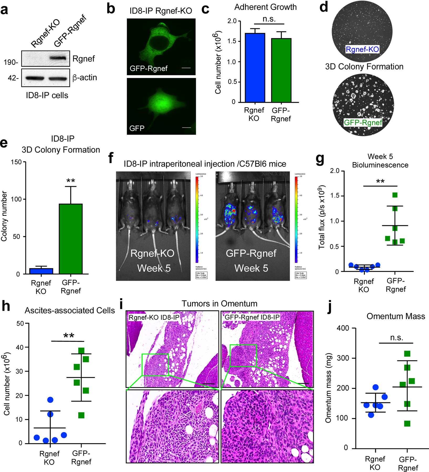
Figure 5:

Rgnef promotes anchorage-independent growth in vitro and in vivo. (a) Anti-Rgnef immunoblotting showing GFP-Rgnef re-expression in ID8-IP Rgnef-KO cells. Actin is a control. (b) GFP-Rgnef or GFP distribution in ID8-IP Rgnef-KO cells visualized by confocal microscopy. Scale is 2 μm. (c) ID8-IP Rgnef-KO and GFP-Rgnef re-expressing cells exhibit no adherent growth difference (n=3 technical replicates, n.s.= no significance, +/− SD). (d) Colony growth is enhanced by GFP-Rgnef re-expression. Representative images (d) and quantification (e) are shown (**P ≤ 0.01, n=3 biological replicates, +/− SD). (f) Representative bioluminescent imaging of ID8-IP Rgnef-KO or GFP-Rgnef re-expressing cells after 5 weeks. (g) Bioluminescent flux quantified on experimental Day 34 (**P ≤ 0.01, n = 6, +/− SD). (h) Quantitation of ascites-associated cells recovered at Day 39 (**P ≤ 0.01, n = 6, +/− SD). (i) Representative H&E stained images of ID8-IP Rgnef-KO or GFP-Rgnef tumor implants within the omentum. Scale is 50 μm. Inset, high magnification. (j) Final omentum mass from ID8-IP Rgnef-KO or GFP-Rgnef tumor-bearing mice. Differences are not significant (n.s.)
