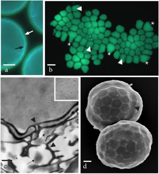
Fig. 4.
Developing tetrads still within the spore mother cell wall 90-100 μm in diameter begin tetrahedral lobing as callose disappears and spore wall ornamentation develops. a Round tetrads showing light blue fluorescence of the sporopollenin in exine next to the cytoplasm (black arrow), yellow fluorescence of callose layer due to aniline blue (white arrow) and dark outer mother cell wall. b Aniline blue fluorescence of callosic special wall that was pushed out of the spore mother cell wall and flattened making the entire wall visible. Areolate exine pattern is evident in thin areas of callose as is the central papilla in each areole. Weak areas where callosic layer separated are areas where the exine muri are most developed. Callose is thicker (brighter fluorescence) at the juncture of spores and the distal most surface. The trilete line of contact between three spores identified by asterisk (*) and juncture of two spores by arrowheads. c TEM at the distal junction between spores showing strong labeling of callose (inset of upper right corner at 4X higher magnification) outside the exine and isolation of label to patches of electron dense material (arrows) within the layers of TPL. Callose degrades progressively from inside the spores to the outside of the tetrad as sporopollenin assembles on TPL. Arrowheads denote irregular seam in the exine between adjacent spores. d SEM of slightly older tetrads still in the mother cell wall that have assumed tetrahedral lobing due to individual spore enlargement. Arrowhead identifies boundary between two spores. Scale bars = 20 μm (a), 30 μm (b), 0.5μm (c), and 10 μm (d)

