Drs. Wilde and Postema discuss
The Brugada syndrome (BrS) case described by Drs. Sichrovsky and Mittal1 is a unique case that may help us to understand the reason for the male preponderance of BrS. Before going into the details, it is important to emphasize that this case is another example of the difficulties that apparently still exist in recognizing a BrS electrocardiogram (ECG) pattern.2 An ECG like this one should not be missed anymore!
The patient described is a genetically female patient who had been using testosterone for about 20 years “to live the life of a transgender male.”1 He presented with an out-of-hospital cardiac arrest and a BrS type 1 pattern ECG (note, the ECG in Sichrovsky et al.’s Figure 2 is from five months before the arrest). Unfortunately, we do not have an ECG from before the start of the intramuscular testosterone injections. Importantly, the claim that testosterone converted this female into a symptomatic BrS male, the essence of this case report and the basis for the nice subtitle “Gender trumps sex as a risk factor,” can of course only with confidence be made with the demonstration of a normal ECG prior to the testosterone therapy. Yet, the odds are in favor of the interpretation presented by the authors. Indeed, testosterone serum levels have been shown to impact the degree of right precordial ST-segment amplitude.3,4 Interestingly, BrS patients who have undergone orchidectomy lose their type 1 pattern after the procedure,3 and androgen deprivation therapy does reduce the ST-segment level in non-BrS patients.4 Furthermore, in BrS patients, testosterone levels have been found to be higher as compared with in age-matched controls.5
The authors clearly adhere to the “repolarization theory” as the pathophysiological mechanism of the right precordial ST-segment elevation. At this point, we can say that all interventions that increase the early potassium currents, and testosterone may be one of them, also impact in a negative way the safety of conduction.6 Hence, the effects of testosterone may also be explained by further deterioration of conduction in the right ventricular outflow tract (RVOT) area.
Finally, we assume that the ectopy shown in this patient is not related to the BrS substrate. Although the origin is in the RVOT area, the coupling interval of ventricular fibrillation (VF)–triggering episodes in BrS patients is shorter as compared with the ectopy in this patient. Earlier studies report a coupling interval of less than 400 ms7 and, here, it is 440 ms to 560 ms (Figure 2 by Sichrovsky et al.). Also, the fact that quinidine was not effective in suppressing the ectopy (while it is very effective in suppressing more serious arrhythmias, as has been described previously in BrS8) is in favor of there being a different mechanism for the patient’s ectopy. This potentially explains why the ablation procedure from the endocardial side was successful, whereas the substrate for BrS-related arrhythmias is expected in the epicardial layer.9 It is also possible that ablation from the endocardial side does affect the epicardial layer of the RVOT, which, after all, is relatively thin.
In summary, the use of testosterone in this patient most likely contributed to the BrS phenotype and underscores the fact that gender indeed impact the phenotype. The RVOT ectopy is presumably unrelated but may serve as a trigger in the setting of a vulnerable substrate in the epicardial layer of the RVOT region.
Arthur A. M. Wilde, md, phd (a.a.wilde@amc.uva.nl)1,2 and Pieter G. Postema, md, phd1
1Department of Clinical and Experimental Cardiology, Amsterdam University Medical Center, Academic Medical Centre, University of Amsterdam, Amsterdam, the Netherlands
2Department of Medicine, Columbia University Irving Medical Center, New York, NY, USA
The authors report no conflicts of interest for the published content.
References
- 1.Sichrovsky TC, Mittal S. Brugada syndrome unmasked by use of testosterone in a transgender male: gender trumps sex as a risk factor. J Innov Cardiac Rhythm Manage. 2019;10(2):3526–3529. doi: 10.19102/icrm.2019.100202. [DOI] [PMC free article] [PubMed] [Google Scholar]
- 2.Gottschalk BH, Anselm DD, Brugada J, et al. Expert cardiologists cannot distinguish between Brugada phenocopy and Brugada syndrome electrocardiogram patterns. Europace. 2016;18(7):1095–1100. doi: 10.1093/europace/euv278. [CrossRef] [PubMed] [DOI] [PubMed] [Google Scholar]
- 3.Matsuo K, Akahoshi M, Seto S, Yano K. Disappearance of the Brugada-type electrocardiogram after surgical castration: a role for testosterone and an explanation for the male preponderance. Pacing Clin Electrophysiol. 2003;26(7 Pt 1):1551–1553. doi: 10.1046/j.1460-9592.2003.t01-1-00227.x. [PubMed] [DOI] [PubMed] [Google Scholar]
- 4.Ezaki K, Nakagawa M, Taniguchi Y, et al. Gender differences in the ST segment: effect of androgen-deprivation therapy and possible role of testosterone. Circulation J. 2010;74(11):2448–2454. doi: 10.1253/circj.cj-10-0221. [PubMed] [DOI] [PubMed] [Google Scholar]
- 5.Shimizu W, Matsuo K, Kokubo Y, et al. Sex hormone and gender difference–-role of testosterone on male predominance in Brugada syndrome. J Cardiovasc Electrophysiol. 2007;18:415–421. doi: 10.1111/j.1540-8167.2006.00743.x. [CrossRef] [PubMed] [DOI] [PubMed] [Google Scholar]
- 6.Wilde AAM, Postema PG, Diego JM, et al. The pathophysiological mechanism underlying Brugada syndrome: depolarization versus repolarization. J Mol Cell Cardiol. 2010;49(4):543–553. doi: 10.1016/j.yjmcc.2010.07.012. [CrossRef] [PubMed] [DOI] [PMC free article] [PubMed] [Google Scholar]
- 7.Kakishita M, Kurita T, Matsuo K, et al. Mode of onset of ventricular fibrillation in patients with Brugada syndrome detected by implantable cardioverter defibrillator therapy. J Am Coll Cardiol. 2000;36(5):1646–1653. doi: 10.1016/s0735-1097(00)00932-3. [CrossRef] [PubMed] [DOI] [PubMed] [Google Scholar]
- 8.Viskin S, Wilde AA, Tan HL, Antzelevitch C, Shimizu W, Belhassen B. Empiric quinidine therapy for asymptomatic Brugada syndrome: time for a prospective registry. Heart Rhythm. 2009;6(3):401–404. doi: 10.1016/j.hrthm.2008.11.030. [CrossRef] [PubMed] [DOI] [PMC free article] [PubMed] [Google Scholar]
- 9.Nademanee K, Veerakul G, Chandanamattha P, et al. Prevention of ventricular fibrillation episodes in Brugada syndrome by catheter ablation over the anterior right ventricular outflow tract epicardium. Circulation. 2011;123(12):1270–1279. doi: 10.1161/CIRCULATIONAHA.110.972612. [CrossRef] [PubMed] [DOI] [PubMed] [Google Scholar]
Dr. Brugada remarks
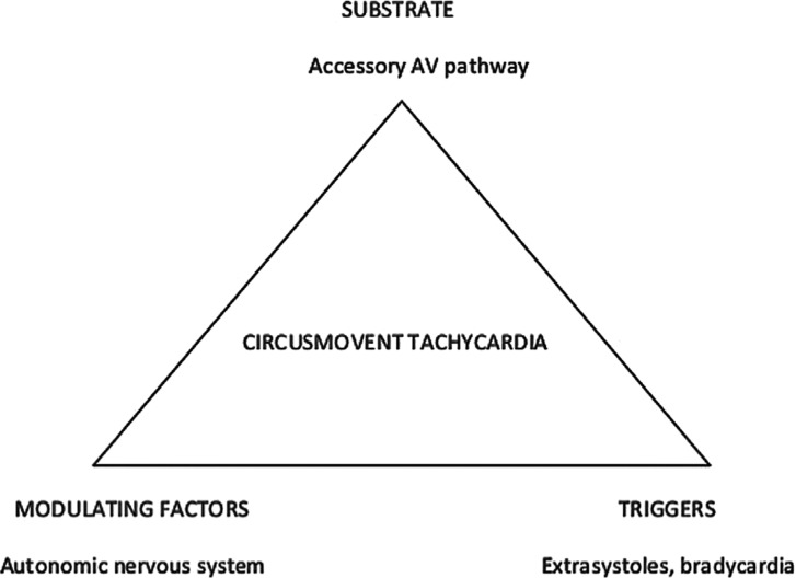
To honor the magnificent contributions of the late Philippe Coumel in the understanding of the mechanisms of cardiac arrhythmias, I coined the term “Coumel’s triangle.”1 Coumel’s philosophy on cardiac arrhythmias was based on the concept that a complex situation (a cardiac arrhythmia) is likely the result of multiple causation: the interaction between the substrate of the arrhythmia with the modulating factors facilitates the start of the arrhythmia by way of the appropriate triggers. A presented example can be used to clarify these mechanisms, as follows: a patient with Wolff–Parkinson–White (WPW) syndrome is born with an accessory atrioventricular (AV) pathway (the substrate). The arrhythmia, circusmovement tachycardia, can be triggered by extrasystoles or junctional rhythm during bradycardia. However, the arrhythmia will only initiate and perpetuate if the conduction properties of the different pathways involved in reentry are appropriate. The appropriateness of the conduction properties is modulated, among other ways, by the autonomic nervous system. An application of the Coumel’s triangle in the case of WPW syndrome is shown in Figure 1.
Figure 1:
A succinct application of Coumel’s triangle to understand why circusmovement tachycardia in a patient with WPW occurs. See text.
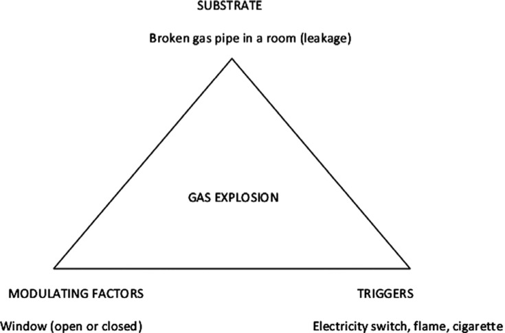
Coumel’s triangle can be used to understand cardiac arrhythmias and, in fact, it can be applied to understand any phenomenon or event in any domain of knowledge. Figure 2 shows another succinct application of Coumel’s triangle to understand a gas explosion in a room.
Figure 2:
Understanding the factors that play a role in the occurrence of a gas explosion in a room. The substrate is the leakage in the gas pipe. The explosion can only occur if the window is closed, allowing for the accumulation of the gas inside the room. The explosion will occur when triggered by turning on an electricity switch, a flame, or a lighted cigarette in the room.
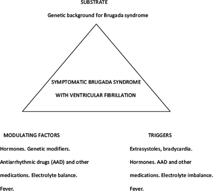
In the present issue, Drs. Sichrovsky and Mittal present2 a phenomenal case of a female-born patient with BrS who has been living as a transgender male via the chronic use of testosterone. According to their description, the patient developed a BrS ECG pattern under this medication. Subsequently, his BrS pattern turned into a full-blown symptomatic syndrome with repeated cardiac arrest. Appropriate protection with an implantable cardioverter-defibrillator was provided to the patient, together with variable quinidine therapy. Symptomatic frequent ventricular extrasystoles prompted endocardial ablation at the RVOT, with a subsequent event-free follow-up. Based on these observations, the authors concluded that “gender trumps sex when it comes to arrhythmia risk in … Brugada syndrome.” Looking at this case with the glasses of Coumel’s triangle, however, I think it is important to state that I disagree with this statement. Biological sex (sex—the substrate) and social sex (gender—a modulating factor) do not compete against each other in importance to trigger VF in BrS. Rather, both are part of the same triangle and are equally relevant (Figure 3). In terms of the triggers, I do believe that the extrasystoles (and the bradycardia) were examples in this patient. Elimination of the ventricular extrasystoles with ablation probably eliminated one of the triggers; however, it is also possible that the authors inadvertently also eliminated the BrS substrate. Successful endocardial (instead of epicardial) ablation of BrS substrate has been reported.3
Figure 3:
Analysis of the case by Drs. Sichrovsky and Mittal2 based on Coumel’s triangle; in this, gender and sex do not compete in importance but rather are both equally relevant.
This observation is not surprising, given the very small thickness of the RVOT. Endocardial or epicardial ablation in that area most likely always results in a transmural lesion.3 Drs. Sichrovsky and Mittal should be congratulated by their extraordinary observation. This case makes it also clear that testosterone has to be added to the list of drugs to be avoided in confirmed or suspected BrS (www.brugadadrugs.org).
Pedro Brugada, md, phd (pedro@brugada.org)1
1Cardiovascular Division, University Hospital Brussels (UZ Brussel), Brussels, Belgium
Dr. Brugada reports that he is a consultant for Biotronik and the director of the Biotronik Midwest Bio Alliance for Scientific Growth program.
References
- 1.Brugada P, Wellens H. Abstracts: 20 years of programmed electrical stimulation of the heart. June 1-5, 1987, Maastricht, The Netherlands. Pacing Clin Electrophysiol. 1987;10(3 Pt 1):591–622. [PubMed] [PubMed] [Google Scholar]
- 2.Sichrovsky TC, Mittal S. Brugada syndrome unmasked by use of testosterone in a transgender male: gender trumps sex as a risk factor. J Innov Cardiac Rhythm Manage. 2019;10(2):3526–3529. doi: 10.19102/icrm.2019.100202. [DOI] [PMC free article] [PubMed] [Google Scholar]
- 3.Tauber PE, Mansilla V, Brugada P, et al. Endocardial approach for substrate ablation in Brugada syndrome: epicardial, endocardial or transmural substrate? J Clin Exp Res Cardiol. 2018;4(1):101–113. [Google Scholar]
Drs. Belhassen and Milman comment
With respect to the present case, we must acknowledge that we have not previously encountered a patient similar to that reported by Drs. Sichrovsky and Mittal.1 A thorough Medline search also confirmed the exceptional nature of this case report.
Based on our latest experience with the Survey on Arrhythmic Events (AEs) in BrS (SABRUS),2,3 we would like to highlight a few points. The SABRUS gathered 678 patients with BrS and AEs from 23 large centers with prior experience in treating BrS. Fifty-nine (8.7%) of the SABRUS patients were female. Although the patient described in the present case is male in all his being, his genetics are of female origin and so we would like to highlight a few critical points in this regard.
Arrhythmic events in “females” with Brugada syndrome
As emphasized by the authors, BrS occurs eight to 10 times more frequently in males despite the disease being equally inherited by both genders. The male/female ratio observed in SABRUS was 10.43; however, this ratio varied according to patients’ age and ethnic origin. Considering the 63-year-old Caucasian, genetically female patient at hand yields a male/female ratio of 2.3 It is noteworthy that this ratio is much higher in Asian patients of the same age group (ratio of 14).3
Age at onset of initial arrhythmic event
Adult female patients (identified as those aged older than 16 years) included in SABRUS suffered their first AE at a mean age of 49.5 years ± 14.4 years, which was significantly older than that in males (43.0 ± 12.7 years; p = 0.001).2 We hypothesized that AE onset in females might correlate with circulating estrogen levels because of a predominance for AEs to occur at ages at which estrogen levels are at their lowest.2,3 In the present case, the occurrence of the AE after the age of 60 years could relate more to the marked decrease in estradiol activity rather than to the 20-year use of testosterone.
Mode of arrhythmic event documentation
When considering the patient’s female genetics, it is not surprising that his primary presentation was of an aborted cardiac arrest, as a majority of females in SABRUS (36/59; 61%) developed their initial AE as a first manifestation of the disease.3
Electrocardiogram at the time of arrhythmic event
Similar to our SABRUS female patients (35/59; 59%),3 the ECG at the time of AE did not show a typical type 1 BrS ECG pattern. Interestingly, the latter manifested five months before the event, when the patient was evaluated for an episode of atrial fibrillation.
Quinidine treatment
The authors used a daily low dose of quinidine (quinidine gluconate ER 324 mg), which was similar to the doses of quinidine (≤ 600 mg daily) reported by Marquez et al.4 as effective in preventing AEs in 11 of 14 patients with BrS. It is interesting that such a low dose was apparently effective in both suppressing arrhythmic storms and preventing further AEs during follow-up. This dose is lower than those found to be effective by Anguera et al.5 in 19 (66%) of their 29 patients with multiple AEs [quinidine bisulfate (mean dose: 591 ± 239 mg/day) and hydroquinidine (mean dose: 697 ± 318 mg/day)]. In addition, the small dose used in the present paper by Sichrovsky and Mittal1 was much lower than the doses necessary to achieve drug efficacy based on serial electrophysiologic testing6 (ie, mean dose of 1,406 ± 242 mg of quinidine bisulfate and mean dose of 900 mg of hydroquinidine).
Bearing in mind the present outstanding case report, it seems advisable to closely follow with serial ECGs those subjects using chronic exogenous testosterone supplementation. Although anabolic steroids are known to cause sudden death through ischemic coronary disease,7,8 the present case report by Sichrovsky and Mittal highlights the need to consider other possible etiologies such as BrS.
Bernard Belhassen, md (bblhass@gmail.com)1,2 and Anat Milman, md, phd2,3
1Department of Cardiology, Tel-Aviv Sourasky Medical Center, Tel-Aviv, Israel
2Sackler School of Medicine, Tel-Aviv University, Tel-Aviv, Israel
3The Heart Center, Chaim Sheba Medical Center, Tel Hashomer, Israel
The authors report no conflicts of interest for the published content.
References
- 1.Sichrovsky TC, Mittal S. Brugada syndrome unmasked by use of testosterone in a transgender male: gender trumps sex as a risk factor. J Innov Cardiac Rhythm Manage. 2019;10(2):3526–3529. doi: 10.19102/icrm.2019.100202. [DOI] [PMC free article] [PubMed] [Google Scholar]
- 2.Milman A, Andorin A, Sacher F, et al. Age of first arrhythmic event in Brugada syndrome: data from the SABRUS (Survey on Arrhythmic Events in Brugada Syndrome) in 678 Patients. Circ Arrhythm Electrophysiol. 2017;10(12) doi: 10.1161/CIRCEP.117.005222. pii: e005222. [CrossRef] [PubMed] [DOI] [PubMed] [Google Scholar]
- 3.Milman A, Gourraud JB, Andorin A, et al. Gender differences in patients with Brugada syndrome and arrhythmic Events: data from a survey on arrhythmic events in 678 patients. Heart Rhythm. 2018;15(10):1457–1465. doi: 10.1016/j.hrthm.2018.06.019. [CrossRef] [PubMed] [DOI] [PubMed] [Google Scholar]
- 4.Márquez MF, Bonny A, Hernández-Castillo E, et al. Long-term efficacy of low doses of quinidine on malignant arrhythmias in Brugada syndrome with an implantable cardioverter-defibrillator: a case series and literature review. Heart Rhythm. 2012;9(12):1995–2000. doi: 10.1016/j.hrthm.2012.08.027. [CrossRef] [PubMed] [DOI] [PubMed] [Google Scholar]
- 5.Anguera I, García-Alberola A, Dallaglio P, et al. Shock reduction with long-term quinidine in patients with Brugada syndrome and malignant ventricular arrhythmia episodes. J Am Coll Cardiol. 2016;67(13):1653–1654. doi: 10.1016/j.jacc.2016.01.042. [CrossRef] [PubMed] [DOI] [PubMed] [Google Scholar]
- 6.Belhassen B, Rahkovich M, Michowitz Y, Glick A, Viskin S. Management of Brugada syndrome: thirty-three-year experience using electrophysiologically guided therapy with class 1A antiarrhythmic drugs. Circ Arrhythm Electrophysiol. 2015;8(6):1393–1402. doi: 10.1161/CIRCEP.115.003109. [CrossRef] [PubMed] [DOI] [PubMed] [Google Scholar]
- 7.Inoue H, Nishida N, Ikeda N, et al. The sudden and unexpected death of a female-to-male transsexual patient. J Forensic Leg Med. 2007;14(6):382–386. doi: 10.1016/j.jcfm.2006.07.012. [CrossRef] [PubMed] [DOI] [PubMed] [Google Scholar]
- 8.Lusetti M, Licata M, Silingardi E, Reggiani Bonetti L, Palmiere C. Pathological changes in anabolic androgenic steroid users. J Forensic Leg Med. 2015;33:101–104. doi: 10.1016/j.jflm.2015.04.014. [CrossRef] [PubMed] [DOI] [PubMed] [Google Scholar]
Dr. Antzelevitch considers
The male predominance in BrS has long been recognized.1,2 The recent SABRUS study served to highlight the marked male predominance of the syndrome (91.3%) with minor involvement of pediatric (4.3%) and elderly populations (1.5%). The peak for arrhythmic events occurred at an average age of 42 years.3 Women with BrS generally present more benign clinical characteristics, less spontaneous type 1 ECG pattern, and are more likely to be asymptomatic.1
The increased risk of males and the protection of females has been the subject of considerable study over the past many years.2,4–7 Gender differences in the degree of ST-segment elevation are observed even in individuals with normal hearts. Ezaki et al.8 reported on the degree of ST-segment elevation measured in leads V2 and V5, representative of the right and left ventricles, in 640 healthy males and females ranging in age from five years to 89 years. In both sexes, ST-segment elevation was higher in V2 than in V5. At younger ages (ie, 5–12 years), ST-segment elevation was similar and very low in both males and females. It remained at a low level throughout life in females, but increased dramatically in males soon after puberty and subsequently steadily declined with advancing age, suggesting an influence of changing androgen levels. To test this hypothesis, Ezaki et al. evaluated the effects of androgen-deprivation therapy on ST-segment levels in 21 patients with prostate cancer. Notably, androgen-deprivation therapy significantly decreased ST-segment levels in both leads, in support of the hypothesis that testosterone modulates the early phase of ventricular repolarization. Hayashi et al.9 reported similar gender- and age-dependent differences in early repolarization in a large cohort of healthy Japanese subjects.
In 2003, Matsuo et al.10 reported ECG normalization following orchiectomy in two cases of prostate cancer patients who had displayed a type 1 ST-segment elevation typical of BrS for many years. The disappearance of ST-segment elevation after surgical castration once again suggests an association between the manifestation of the Brugada ECG pattern and testosterone. It is noteworthy that male patients with BrS have been reported to have higher testosterone levels as compared with age-matched controls.5 Interestingly, Yamaki et al.11 shared that J-wave manifestation in BrS patients peaks in the early morning hours (2:00 am), coincidently in conjunction with peak levels of serum testosterone. These findings may explain why sudden cardiac death in BrS often occurs during sleep in the early morning hours.
Thus, both clinical and basic science studies point to testosterone as the basis for the gender differences in J-wave manifestation underlying the elevation of the ST segment and consequently the expression of the BrS phenotype. However, what is responsible for the testosterone-mediated ST-segment elevation? Our group has previously tested the hypothesis that testosterone influences the expression of Ito. In preliminary studies, Barajas-Martinez et al. reported that chronic exposure to testosterone increases the expression of the transient outward current (Ito) in human-induced, pluripotent-stem-cell-derived cardiomyocytes.12 Ito is responsible for phase I of the action potential and thus for early repolarization of the ventricular myocardium, and its augmentation underlies the accentuation of the J-wave and ST-segment elevation that contributes to the arrhythmogenic substrate in BrS.13,14
Inhibition of Ito has proven to be ameliorative regardless of the cause of BrS. Unfortunately, ion-channel-specific and cardioselective Ito blockers are not accessible. The best drug currently available in the clinic capable of blocking Ito is quinidine. Clinical evidence for the effectiveness of quinidine has been reported in numerous studies and case reports.15–30 Belhassen et al., who pioneered the use of quinidine in VF,31 reported a 90% efficacy in the prevention of VF induction following treatment with quinidine, despite the use of very aggressive protocols of extrastimulation.31
In the fascinating case here reported by Sichrovsky and Mittal,32 a genetically female patient who presumably was carrying a BrS susceptibility gene variant but who was totally asymptomatic and protected because of her birth gender lost that protection when chronically receiving 200 mg testosterone intramuscularly twice weekly in order to maintain her transgender identity as a male. After experiencing two aborted cardiac arrests at night, the patient was implanted with a dual-chamber implantable cardioverter-defibrillator capable of atrial pacing to avoid the nocturnal bradycardia, which is known to precipitate episodes of ventricular tachycardia/VF. After receiving four appropriate shocks for VF, the patient was placed on quinidine sulfate, which was subsequently changed to quinidine gluconate ER. After a few days, the patient developed angioedema with every dose of quinidine. The addition of the antihistamine ocetrizine subsequently allowed him to tolerate once-daily dosing of quinidine. Although quinidine was effective in suppressing all sustained arrhythmias, a high burden of premature ventricular contractions (PVCs) persisted. The PVCs were successfully eliminated via ablation of the endocardial aspect of the RVOT.
Quinidine’s action to suppress ventricular tachycardia/VF in this case is likely attributable to its inhibition of Ito. The persistence of the PVCs may be due to the relatively low dose of the quinidine prescribed, owing to a desire to minimize angioedema. In addition to its action to block Ito, quinidine also exerts an anticholinergic effect and is an effective inhibitor of the delayed rectifier potassium current (IKr), which prolongs the QT interval and is the basis for its use in the treatment of short QT syndrome. Both of these actions may have contributed to its ameliorative action in this patient, particularly because, prior to the administration of quinidine, his QT interval was relatively short (QT interval = 300 ms; corrected QT interval = 335 ms) (see Sichrovsky et al.’s Figure 2). Genetic screening was not performed in this case, but the relatively short QT interval suggests the possibility that the case involved a mutation in a calcium channel gene (eg, CACNA1C, CACNB2, or CACNA2D1).33
Charles Antzelevitch, phd (cantzelevitch@gmail.com)1–3
1Lankenau Institute for Medical Research, Wynnewood, PA, USA
2Lankenau Heart Institute, Wynnewood, PA, USA
3Sidney Kimmel Medical College of Thomas Jefferson University, Philadelphia, PA, USA
Dr. Antzelevitch acknowledges support from the National Heart, Lung, and Blood Institute (HL138103) and from the Martha and Wistar Morris Fund.
References
- 1.Benito B, Sarkozy A, Mont L, et al. Gender differences in clinical manifestations of Brugada syndrome. J Am Coll Cardiol. 2008;52(19):1567–1573. doi: 10.1016/j.jacc.2008.07.052. [CrossRef] [PubMed] [DOI] [PubMed] [Google Scholar]
- 2.Antzelevitch C. Androgens and male predominance of the Brugada syndrome phenotype. Pacing Clin Electrophysiol. 2003;26(7 Pt 1):1429–1431. doi: 10.1046/j.1460-9592.2003.t01-1-00206.x. [CrossRef] [PubMed] [DOI] [PubMed] [Google Scholar]
- 3.Milman A, Andorin A, Gourraud JB, et al. Profile of Brugada syndrome patients presenting with their first documented arrhythmic event: data from the Survey on Arrhythmic Events in Brugada Syndrome (SABRUS) Heart Rhythm. 2018;15(5):716–724. doi: 10.1016/j.hrthm.2018.01.014. [CrossRef] [PubMed] [DOI] [PubMed] [Google Scholar]
- 4.Di Diego JM, Cordeiro JM, Goodrow RJ, et al. Ionic and cellular basis for the predominance of the Brugada syndrome phenotype in males. Circulation. 2002;106(15):2004–2011. doi: 10.1161/01.cir.0000032002.22105.7a. [PubMed] [DOI] [PubMed] [Google Scholar]
- 5.Shimizu W, Matsuo K, Kokubo Y, et al. Sex hormone and gender difference--role of testosterone on male predominance in Brugada syndrome. J Cardiovasc Electrophysiol. 2007;18(4):415–421. doi: 10.1111/j.1540-8167.2006.00743.x. [CrossRef] [PubMed] [DOI] [PubMed] [Google Scholar]
- 6.Fish JM, Antzelevitch C. Cellular and ionic basis for the sex-related difference in the manifestation of the Brugada syndrome and progressive conduction disease phenotypes. J Electrocardiol. 2003;36:173–179. doi: 10.1016/j.jelectrocard.2003.09.054. [CrossRef] [PubMed] [DOI] [PubMed] [Google Scholar]
- 7.Verkerk AO, Wilders R, de Geringel W, Tan HL. Cellular basis of sex disparities in human cardiac electrophysiology. Acta Physiol (Oxf) 2006;187(4):459–477. doi: 10.1111/j.1748-1716.2006.01586.x. [CrossRef] [PubMed] [DOI] [PubMed] [Google Scholar]
- 8.Ezaki K, Nakagawa M, Taniguchi Y, et al. Gender differences in the ST segment: effect of androgen-deprivation therapy and possible role of testosterone. Circ J. 2010;74(11):2448–2454. doi: 10.1253/circj.cj-10-0221. [CrossRef] [PubMed] [DOI] [PubMed] [Google Scholar]
- 9.Hayashi H, Miyamoto A, Ishida K, et al. Prevalence and QT interval of early repolarization in a hospital-based population. J Arrhythmia. 2010;26(2):127–133. doi: 10.1016/S1880-4276(10)80017-1. [DOI] [Google Scholar]
- 10.Matsuo K, Akahoshi M, Seto S, Yano K. Disappearance of the Brugada-type electrocardiogram after surgical castration: a role for testosterone and an explanation for the male preponderance. Pacing Clin Electrophysiol. 2003;26(7 Pt 1):1151–1153. doi: 10.1046/j.1460-9592.2003.t01-1-00227.x. [CrossRef] [PubMed] [DOI] [PubMed] [Google Scholar]
- 11.Yamaki M, Sato N, Okada M, et al. A case of Brugada syndrome in which diurnal ECG changes were associated with circadian rhythms of sex hormones. Int Heart J. 2009;50(5):669–676. doi: 10.1536/ihj.50.669. [CrossRef] [PubMed] [DOI] [PubMed] [Google Scholar]
- 12.Barajas-Martinez H, Hu D, Urrutia J, et al. Chronic exposure to testosterone increases expression of transient outward current in human induced pluripotent stem cell (hiPSC)-derived cardiomyocytes (CM) Heart Rhythm. 2013;10(11):1741. doi: 10.1016/j.hrthm.2013.09.012. [DOI] [Google Scholar]
- 13.Antzelevitch C YG. J wave syndrome: Brugada and early repolarization syndromes. Heart Rhythm. 2015;12(8):1852–1866. doi: 10.1016/j.hrthm.2015.04.014. [CrossRef] [PubMed] [DOI] [PMC free article] [PubMed] [Google Scholar]
- 14.Antzelevitch C, Yan GX, Ackerman MJ, et al. J-Wave syndromes expert consensus conference report: Emerging concepts and gaps in knowledge. Heart Rhythm. 2016;13(10):e295–e324. doi: 10.1016/j.hrthm.2016.05.024. [CrossRef] [PubMed] [DOI] [PMC free article] [PubMed] [Google Scholar]
- 15.Belhassen B, Glick A, Viskin S. Efficacy of quinidine in high-risk patients with Brugada syndrome. Circulation. 2004;110(13):1731–1737. doi: 10.1161/01.CIR.0000143159.30585.90. [CrossRef] [PubMed] [DOI] [PubMed] [Google Scholar]
- 16.Belhassen B, Shapira I, Shoshani D, Paredes A, Miller H, Laniado S. Idiopathic ventricular fibrillation: inducibility and beneficial effects of class I antiarrhythmic agents. Circulation. 1987;75:809–816. doi: 10.1161/01.cir.75.4.809. [PubMed] [DOI] [PubMed] [Google Scholar]
- 17.Belhassen B, Viskin S, Antzelevitch C. The Brugada syndrome: is an implantable cardioverter defibrillator the only therapeutic option? Pacing Clin Electrophysiol. 2002;25(11):1634–1640. doi: 10.1046/j.1460-9592.2002.01634.x. [CrossRef] [PubMed] [DOI] [PubMed] [Google Scholar]
- 18.Alings M, Dekker L, Sadee A, Wilde A. Quinidine induced electrocardiographic normalization in two patients with Brugada syndrome. Pacing ClinElectrophysiol. 2001;24(9 Pt 1):1420–1422. doi: 10.1046/j.1460-9592.2001.01420.x. [CrossRef] [PubMed] [DOI] [PubMed] [Google Scholar]
- 19.Belhassen B, Viskin S. Pharmacologic approach to therapy of Brugada syndrome: quinidine as an alternative to ICD therapy? In: Antzelevitch C, Brugada P, Brugada J, Brugada R, editors. The Brugada Syndrome: From Bench to Bedside. Oxford, UK: Blackwell Futura; 2004. pp. 202–211. [Google Scholar]
- 20.Viskin S, Wilde AA, Tan HL, Antzelevitch C, Shimizu W, Belhassen B. Empiric quinidine therapy for asymptomatic Brugada syndrome: time for a prospective registry. Heart Rhythm. 2009;6(3):401–404. doi: 10.1016/j.hrthm.2008.11.030. [CrossRef] [PubMed] [DOI] [PMC free article] [PubMed] [Google Scholar]
- 21.Belhassen B, Glick A, Viskin S. Excellent long-term reproducibility of the electrophysiologic efficacy of quinidine in patients with idiopathic ventricular fibrillation and Brugada syndrome. Pacing Clin Electrophysiol. 2009;32(3):294–301. doi: 10.1111/j.1540-8159.2008.02235.x. [CrossRef] [PubMed] [DOI] [PubMed] [Google Scholar]
- 22.Marquez MF, Bonny A, Hernandez-Castillo E, et al. Long-term efficacy of low doses of quinidine on malignant arrhythmias in Brugada syndrome with an implantable cardioverter-defibrillator: a case series and literature review. Heart Rhythm. 2013;9(12):1995–2000. doi: 10.1016/j.hrthm.2012.08.027. [DOI] [PubMed] [Google Scholar]
- 23.Pellegrino PL, Di BM, Brunetti ND. Quinidine for the management of electrical storm in an old patient with Brugada syndrome and syncope. Acta Cardiol. 2013;68(2):201–203. doi: 10.1080/ac.68.2.2967280. [CrossRef] [PubMed] [DOI] [PubMed] [Google Scholar]
- 24.Probst V, Evain S, Gournay V, et al. Monomorphic ventricular tachycardia due to brugada syndrome successfully treated by hydroquinidine therapy in a 3-year-old child. J Cardiovasc Electrophysiol. 2006;17(1):97–100. doi: 10.1111/j.1540-8167.2005.00329.x. [CrossRef] [PubMed] [DOI] [PubMed] [Google Scholar]
- 25.Schweizer PA, Becker R, Katus HA, Thomas D. Successful acute and long-term management of electrical storm in Brugada syndrome using orciprenaline and quinine/quinidine. Clin Res Cardiol. 2010;99(7):467–470. doi: 10.1007/s00392-010-0145-7. [CrossRef] [PubMed] [DOI] [PubMed] [Google Scholar]
- 26.Hasegawa K, Ashihara T, Kimura H, et al. Long-term pharmacological therapy of Brugada syndrome: is J-wave attenuation a marker of drug efficacy? InternMed. 2014;53(14):1523–1526. doi: 10.2169/internalmedicine.53.1829. [CrossRef] [PubMed] [DOI] [PubMed] [Google Scholar]
- 27.Marquez MF, Rivera J, Hermosillo AG, et al. Arrhythmic storm responsive to quinidine in a patient with Brugada syndrome and vasovagal syncope. Pacing Clin Electrophysiol. 2005;28(8):870–873. doi: 10.1111/j.1540-8159.2005.00183.x. [CrossRef] [PubMed] [DOI] [PubMed] [Google Scholar]
- 28.Viskin S, Antzelevitch C, Marquez MF, Belhassen B. Quinidine: a valuable medication joins the list of ’endangered species’. Europace. 2007;9(12):1105–1106. doi: 10.1093/europace/eum181. [CrossRef] [PubMed] [DOI] [PubMed] [Google Scholar]
- 29.Ohgo T, Okamura H, Noda T, et al. Acute and chronic management in patients with Brugada syndrome associated with electrical storm of ventricular fibrillation. Heart Rhythm. 2007;4(6):695–700. doi: 10.1016/j.hrthm.2007.02.014. [CrossRef] [PubMed] [DOI] [PubMed] [Google Scholar]
- 30.Rosso R, Glick A, Glikson M, et al. Outcome after implantation of cardioverter defribrillator in patients with Brugada syndrome: a multicenter Israeli study (ISRABRU) Isr Med Assoc J. 2008;10(6):435–439. [PubMed] [PubMed] [Google Scholar]
- 31.Belhassen B, Rahkovich M, Michowitz Y, Glick A, Viskin S. Management of Brugada syndrome: a 33-year experience using electrophysiologically-guided therapy with class 1A antiarrhythmic drugs. Circ Arrhythm Electrophysiol. 2015;8(6):1393–1402. doi: 10.1161/CIRCEP.115.003109. [DOI] [PubMed] [Google Scholar]
- 32.Sichrovsky TC, Mittal S. Brugada syndrome unmasked by use of testosterone in a transgender male: gender trumps sex as a risk factor. J Innov Cardiac Rhythm Manage. 2019;10(2):3526–3529. doi: 10.19102/icrm.2019.100202. [DOI] [PMC free article] [PubMed] [Google Scholar]
- 33.Burashnikov E, Pfeiffer R, Barajas-Martinez H, et al. Mutations in the cardiac L-type calcium channel associated J wave syndrome and sudden cardiac death. Heart Rhythm. 2010;7(12):1872–1882. doi: 10.1016/j.hrthm.2010.08.026. [CrossRef] [PubMed] [DOI] [PMC free article] [PubMed] [Google Scholar]


