European Journal of Translational Myology logoLink to European Journal of Translational Myology
. 2020 Apr 1;30(1):8826. doi: 10.4081/ejtm.2019.8826

Thirty years of translational research in Mobility Medicine: Collection of abstracts of the 2020 Padua Muscle Days

Ugo Carraro (1),(2),
PMCID: PMC7254447  PMID: 32499887

Abstract

More than half a century of skeletal muscle research is continuing at Padua University (Italy) under the auspices of the Interdepartmental Research Centre of Myology (CIR-Myo), the European Journal of Translational Myology (EJTM) and recently also with the support of the A&CM-C Foundation for Translational Myology, Padova, Italy. The Volume 30(1), 2020 of the EJTM opens with the collection of abstracts for the conference “2020 Padua Muscle Days: Mobility Medicine 30 years of Translational Research”. This is an international conference that will be held between March 18-21, 2020 in Euganei Hills and Padova in Italy. The abstracts are excellent examples of translational research and of the multidimensional approaches that are needed to classify and manage (in both the acute and chronic phases) diseases of Mobility that span from neurologic, metabolic and traumatic syndromes to the biological process of aging. One of the typical aim of Physical Medicine and Rehabilitation is indeed to reduce pain and increase mobility enough to enable impaired persons to walk freely, garden, and drive again. The excellent contents of this Collection of Abstracts reflect the high scientific caliber of researchers and clinicians who are eager to present their results at the PaduaMuscleDays. A series of EJTM Communications will also add to this preliminary evidence.

Key Words: 2020 Padua Muscle Days, translational research, Mobility Medicine

Ethical Publication Statement

Author confirms that he have read the Journal’s position on issues involved in ethical publication and affirms that this report is consistent with those guidelines.

Myologists of Padua University (Italy) were able to begin and continue a tradition of skeletal muscle studies that started half a century ago with a research project whose aim was to investigate whether skeletal muscle was responsible for fever by burning bacterial toxins.1 While this concept sounds strange today, recent results on the effects of myokines may shape new research interests and clinical outcomes.2 In 1991 the first issue of Basic and Applied Myology (BAM) was published by Unipress, a private company independent from the University of Padova.3 The journal was retitled ten years ago to the European Journal of Translational Myology (EJTM) to stress the approaches of the scientific community publishing in the journal, though original basic papers continue to be published by the new publisher PAGEpress, Pavia (Italy).4 The retitled journal was only e-published under the new Open Source rules. Thus all papers are now retrievable for free from the websites of EJTM (Link to: https://pagepressjournals. org/index. php/am/issue/archive) or of BAM On-line (Link to: http://www.bio.unipd.it/bam/). Furthermore, from 2010 all papers were indexed in PUBMED and free available in PMC. Recently, EJTM was also indexed in SCOPUS of Elsevier, presenting a decent Citescore Index of 0.91 from October 2019. The BAM/EJTM story started earlier than 1991. Indeed, the journal is the long term result of a series of muscle rehabilitation-related conferences that have been occasionally organized from 1985 onwards, in Abano Terme (Padova), Italy. These were presented as conferences of scientific associations and then, regularly from 1991, were run under the auspices of Padua University with the shortened name of Padua Muscle Days (PMD). The organization and institution of the Interdepartmental Research Center of Myology of the University of Padova (CIR-Myo, Myology Center) in 2006, strengthened the collaboration on Muscle/Mobility topics among researchers and clinicians belonging to several departments of the University of Padova. This allowed for broadening of the core interests of the Padua Muscle Community from strictly myology issues to wider clinical interests. Soon after, interactions of pain and mobility and of cellular and genetic approaches emerged to complement the traditional fields of Anatomy, Physiology, Physiopathology, Neurology (Human and Veterinary Sciences), allowing to organize studies on etiology, pathogenesis, prevention, managements and rehabilitation of mobility related diseases and syndromes.

Fig 1.
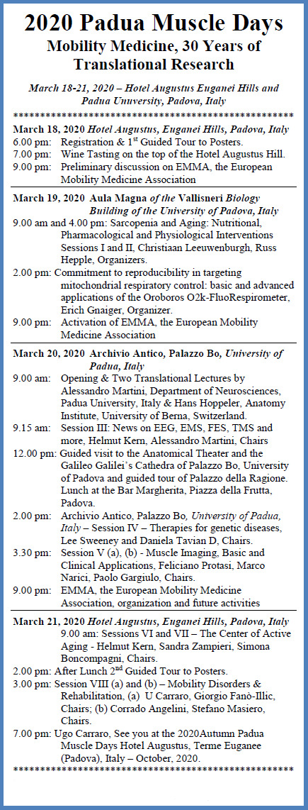
Fig 1.

Program of the 2020 Padua Muscle Days, Euganei Hills and Padova University - March 18-21, 2020.

The 2020 Padua Muscle Days (PMD) conference will be held in Euganei Hills and Padova, from March 18 to 21, 2020, under the caption “Mobility Medicine, 30 years of Translational Research”. This is to stress both the 30 years of publication of BAM/EJTM and the wider scientific, clinical and engineering interests that have emerged during the last 30 years of national and international collaborations. The programmed events of the 2020PMD are attracting not only the core group of researchers that have gathered year after year in Padova, but new speakers that are filling the sessions of a three day program. The collection of scientific sessions listed in Figure 1 provides a good summary of the interests and proposals of researchers, clinicians and bioengineers who will join together on 18 March, 2020 at the Hotel Augustus, Euganei Hills, Padova, Italy. The locations of Padova and the Venetian Euganei Hills are easy venues to sell to organize Scientific Meetings. Junior and senior researchers gather to learn from each other and to find opportunities for new collaborations. They have time to participate in cultural events and, during times outside sessions, have opportunities to meet senior researchers. This year there are many reasons to join the PaduaMuscleDays. One key reason is to discuss about the organization and institution of EMMA, the European Mobility Medicine Association. EMMA will be a European organization, but with international partners. So we are proud that, beside speakers from European countries (Austria, Germany, Iceland (that is half European and half American), Italy, Portugal, Slovakia, Slovenia and Switzerland), attendees from China, Japan, and the USA will also join the 2020PMD. Participants of the 2020PMD will have three after-dinner opportunities to follow discussions on EMMA and to eventually join. Complementing these opportunities is the organization of a new section of EJTM, entitled: “History and Future of Mobility Medicine”. Giorgio Fanò-Illic was the first to accept being one of the Editors. Then Marina Bouché and Patrizia Mecocci joined as Editors and Carmelinda Ruggiero as an Advisor for the new EJTM Section.

As to the concept of Mobility Medicine, it is worthy to stress that increased mobility levels are recognized management methods, not only for immobilization-related impairments of skeletal muscle structure and functions, but also for many diseases where impaired mobility has a heavy influence on quality of life of persons. Understanding etiology/physiopathogenesis and finding prevention/treatment approaches for symptoms and signs, including pain, are common needs to manage these mobility-related diseases. A the typical aim of Physical Medicine and Rehabilitation is indeed to reduce pain and increase mobility enough to enable impaired persons to walk freely, garden, and drive again, but the same goal is shared by surgeons, sports specialists, nutritionists and diverse basic and biomedical scientists and engineers.

Very different specialists and sub-specialists will join the 2020PMD to learn from each other. These delegates will range from geneticists, molecular and cellular experts to clinicians and engineers. Our decision to attract attendees with very different backgrounds and expertise is a moral obligation in an era of expanding needs and shrinking resources, i.e., to try to contrast the fragmentation of knowledge and expertise.

The following Collection of Abstracts of the 2020 PMD cover translational research involving physical, pharmacological, cellular and genetic strategies to maintain or rehabilitate the structure and function of skeletal muscles, and mobility of patients, in aging or premature aging due to diverse diseases. Many of the results, patented or not, in the following abstracts of the 2020 PMD are indeed mature enough to be translated into applications. This has happened in the past,5-38 and seems likely to also happen in the future.

Acknowledgments and Funding

This typescript is sponsored by the A&CM-C Foundation for Translational Myology, Padova, Italy.

References

  • 1.Carraro U. From BAM to BEM, a personal journey through EJTM and PaduaMuscleDays. Biol Eng Med 2017; 2(2): 1-2 doi: 10.15761/BEM.1000117. [Google Scholar]
  • 2.Gabellini D, Musarò A. Report and Abstracts of the 14th Meeting of IIM, the Interuniversity Institute of Myology, - Assisi (Italy), October 12-15, 2017. Eur J Transl Myol 2017;27:185-224. [DOI] [PMC free article] [PubMed] [Google Scholar]
  • 3.Carraro U. BAM: Is a New Professional Activity Arising? 1991. Basic and Applied Myology 1991:1:4. Editorial. [Google Scholar]
  • 4.Carraro U. European Journal of Translational Myology and 2010 Spring Padua Muscle Days. 2010;1:4. Editorial. [DOI] [PMC free article] [PubMed] [Google Scholar]
  • 5.Edmunds K, Gíslason M, Sigurðsson S, et al. Advanced quantitative methods in correlating sarcopenic muscle degeneration with lower extremity function biometrics and comorbidities. PLoS One 2018;13:e0193241. doi: 10.1371/journal.pone.0193241.eCollection2018. [DOI] [PMC free article] [PubMed] [Google Scholar]
  • 6.Vahed LK, Arianpur A, Gharedaghi M, Rezaei H. Ultrasound as a diagnostic tool in the investigation of patients with carpal tunnel syndrome. Eur J Transl Myol 2018 Apr 24;28(2):7380. doi: 10.4081/ejtm.2018.7406. eCollection 2018 Apr 24. [DOI] [PMC free article] [PubMed] [Google Scholar]
  • 7.Masiero S, Carraro U. (eds) Rehabilitation Medicine for Elderly Patients. Practical Issues in Geriatrics. Springer International Publishing AG, part of Springer Nature. doi: 10.1007/978-3-319-57406-6_40. [Google Scholar]
  • 8.Taylor MJ, Fornusek C, Ruys AJ. Reporting for Duty: The duty cycle in Functional Electrical Stimulation research. Part I: Critical commentaries of the literature. Eur J Transl Myol 2018 Nov 7;28(4):7732. doi: 10.4081/ejtm.2018.7732. eCollection 2018 Nov 2. [DOI] [PMC free article] [PubMed] [Google Scholar]
  • 9.Laursen CB, Nielsen JF, Andersen OK, Spaich EG. Feasibility of Using Lokomat Combined with Functional Electrical Stimulation for the Rehabilitation of Foot Drop. Eur J Transl Myol 2016;26(3):6221. eCollection 2016 Jun 13. [DOI] [PMC free article] [PubMed] [Google Scholar]
  • 10.Angeli CA, Boakye M, Morton RA, et al. Recovery of Over-Ground Walking after Chronic Motor Complete Spinal Cord Injury. N Engl J Med 2018; 379:1244-1250. doi: 10.1056/NEJMoa1803588. [DOI] [PubMed] [Google Scholar]
  • 11.Power GA, Dalton BH, Gilmore KJ, et al. Maintaining Motor Units into Old Age: Running the Final Common Pathway. Eur J Transl Myol 2017;27:6597. doi: 10.4081/ejtm.2017.6597. eCollection 2017 Feb 24. [DOI] [PMC free article] [PubMed] [Google Scholar]
  • 12.Coletti D, Adamo S, Moresi V. Of Faeces and Sweat. How Much a Mouse is Willing to Run: Having a Hard Time Measuring Spontaneous Physical Activity in Different Mouse Sub-Strains. Eur J Transl Myol 2017;27:6483. doi: 10.4081/ejtm.2017.6483. eCollection 2017 Feb 24. [DOI] [PMC free article] [PubMed] [Google Scholar]
  • 13.Scicchitano BM, Sica G, Musarò A. Stem Cells and Tissue Niche: Two Faces of the Same Coin of Muscle Regeneration. Eur J Transl Myol 2016;26:6125. doi: 10.4081/ejtm.2016.6125. eCollection 2016 Sep 15. [DOI] [PMC free article] [PubMed] [Google Scholar]
  • 14.Lavorato M, Gupta PK, Hopkins PM, Franzini-Armstrong C. Skeletal Muscle Microalterations in Patients Carrying Malignant Hyperthermia-Related Mutations of the e-c Coupling Machinery. Eur J Transl Myol 2016;26:6105. doi: 10.4081/ejtm.2016.6105. eCollection 2016 Sep 15. [DOI] [PMC free article] [PubMed] [Google Scholar]
  • 15.Carotenuto F, Coletti D, Di Nardo P, Teodori α-Linolenic Acid Reduces TNF-Induced Apoptosis in C2C12 Myoblasts by Regulating Expression of Apoptotic Proteins. Eur J Transl Myol 2016;26:6033. doi: 10.4081/ejtm.2016.6033. eCollection 2016 Sep 15. [DOI] [PMC free article] [PubMed] [Google Scholar]
  • 16.Edmunds KJ, Gíslason MK, Arnadottir ID, et al. Quantitative Computed Tomography and Image Analysis for Advanced Muscle Assessment. Eur J Transl Myol 2016;26:6015. doi: 10.4081/ejtm.2016.6015. eCollection 2016 Jun 13. [DOI] [PMC free article] [PubMed] [Google Scholar]
  • 17.Coletti D, Daou N, Hassani M, et al. Serum Response Factor in Muscle Tissues: From Development to Ageing. Eur J Transl Myol 2016;26:6008. doi: 10.4081/ejtm.2016. 6008. eCollection 2016 Jun 13. [DOI] [PMC free article] [PubMed] [Google Scholar]
  • 18.Tramonti C, Rossi B, Chisari C. Extensive Functional Evaluations to Monitor Aerobic Training in Becker Muscular Dystrophy: A Case Report. Eur J Transl Myol 2016;26:5873. doi: 10.4081/ejtm.2016.5873. eCollection 2016 Jun 13. [DOI] [PMC free article] [PubMed] [Google Scholar]
  • 19.Riuzzi F, Beccafico S, Sorci G, Donato R. S100B protein in skeletal muscle regeneration: regulation of myoblast and macrophage functions. Eur J Transl Myol 2016;26:5830. doi: 10.4081/ejtm.2016.5830. eCollection 2016 Feb 23. [DOI] [PMC free article] [PubMed] [Google Scholar]
  • 20.Barberi L, Scicchitano BM, Musaro A. Molecular and Cellular Mechanisms of Muscle Aging and Sarcopenia and Effects of Electrical Stimulation in Seniors. Eur J Transl Myol 2015;25:231-6. doi: 10.4081/ejtm.2015.5227. eCollection 2015 Aug 24. Review. [DOI] [PMC free article] [PubMed] [Google Scholar]
  • 21.Gabrielli E, Fulle S, Fanò-Illic G, Pietrangelo T. Analysis of Training Load and Competition During the PhD Course of a 3000-m Steeplechase Female Master Athlete: An Autobiography. Eur J Transl Myol 2015;25:5184. doi: 10.4081/ejtm.2015.5184. eCollection 2015 Sep 11. [DOI] [PMC free article] [PubMed] [Google Scholar]
  • 22.Gargiulo P, Helgason T, Ramon C, et al. CT and MRI Assessment and Characterization Using Segmentation and 3D Modeling Techniques: Applications to Muscle, Bone and Brain. Eur J Transl Myol 2014;24:3298. doi: 10.4081/ejtm. 2014 3298. eCollection 2014 Mar 31. [DOI] [PMC free article] [PubMed] [Google Scholar]
  • 23.Ortolan P, Zanato R, Coran A, et al. Role of Radiologic Imaging in Genetic and Acquired Neuromuscular Disorders. Eur J Transl Myol 2015;25:5014. doi: 10.4081/ejtm.2015.5014. eCollection 2015 Mar 11. Review. [DOI] [PMC free article] [PubMed] [Google Scholar]
  • 24.Ravara B, Gobbo V, Carraro U, et al. Functional Electrical Stimulation as a Safe and Effective Treatment for Equine Epaxial Muscle Spasms: Clinical Evaluations and Histochemical Morphometry of Mitochondria in Muscle Biopsies. Eur J Transl Myol 201525:4910. doi: 10.4081/ejtm.2015.4910. eCollection 2015 Mar 11. [DOI] [PMC free article] [PubMed] [Google Scholar]
  • 25.Franzini-Armstrong C. Electron Microscopy: From 2D to 3D Images with Special Reference to Muscle. Eur J Transl Myol 2015;25:4836. doi: 10.4081/ejtm.2015.4836. eCollection 2015 Jan 7. Review. [DOI] [PMC free article] [PubMed] [Google Scholar]
  • 26.Costa A, Rossi E, Scicchitano BM, et al. Neurohypophyseal Hormones: Novel Actors of Striated Muscle Development and Homeostasis. Eur J Transl Myol 2014;24:3790. doi: 10.4081/ejtm.2014.3790. eCollection 2014 Sep 23. Review. [DOI] [PMC free article] [PubMed] [Google Scholar]
  • 27.Veneziani S, Doria C, Falciati L, Castelli CC, Illic GF. Return to Competition in a Chronic Low Back Pain Runner: Beyond a Therapeutic Exercise Approach, a Case Report. Eur J Transl Myol 2014;24:2221. doi: 10.4081/ejtm.2014. 2221. eCollection 2014 Sep 23. [DOI] [PMC free article] [PubMed] [Google Scholar]
  • 28.Kern H, Hofer C, Löfler S, et al. Atrophy, ultra-structural disorders, severe atrophy and degeneration of denervated human muscle in SCI and Aging. Implications for their recovery by Functional Electrical Stimulation, updated 2017. Neurol Res 2017;39:660-666. doi: 10.1080/01616412.2017.1314906. Epub 2017 Apr 13. [DOI] [PubMed] [Google Scholar]
  • 29.Kern H, Carraro U, Adami N, et al. Home-based functional electrical stimulation rescues permanently denervated muscles in paraplegic patients with complete lower motor neuron lesion. Neurorehabil Neural Repair 2010; 24:709-21. [DOI] [PubMed] [Google Scholar]
  • 30.Albertin G, Ravara B, Kern H, et al. Two-years of home based functional electrical stimulation recovers epidermis from atrophy and flattening after years of complete Conus-Cauda Syndrome. Medicine (Baltimore) 2019;98:e18509. doi: 10.1097/MD.0000 000000018509. [DOI] [PMC free article] [PubMed] [Google Scholar]
Eur J Transl Myol. 2020 Apr 1;30(1):8826.

Abstracts of the 2020 Padua Muscle Days

March 19, 2020 - Aula Magna of the Vallisneri Biology Building of the University of Padova, Italy

SARCOPENIA AND AGING, Nutritional, Pharmacological and Physiological Interventions - Session I

1. Effects of epicatechins on endothelial, mitochondrial and physical function: Clinical trial results

Christiaan Leeuwenburgh 1,

Abstract

Multiple biological pathways contribute to aging. Some prominent pathways and causes are genome instability, immunosenescence, inflammation, the NAD+ salvage pathways, proteostasis, and mitochondrial dysfunction. Altered gene regulation influenced by epigenetics, circadian rhythms, diet and physical activity levels also influences the rate of aging. Our Institute on Aging and collaborators are involved with multiple clinical trials with the goal to target these pathways and to improve physical and cognitive function or slow the rate of decline typically seen during aging. The ultimate goal is to develop interventions that can maintain health and independence of older adults. Trials ongoing and/or completed have utilized compounds shown to extend health-span and/or life span in invertebrates (c. elegans) or vertebrates (mice/rats). The compounds used range from nutritional (nicotinamide riboside, epicatechin, resveratrol, omega 3, fermented papaya) to pharmacological (metformin, aspirin, rapamycin, telmisartin/losartan, testosterone), lifestyle-physiological interventions (calorie restriction, exercise, fasting regimes). These biological compounds and lifestyle interventions target single or multiple pathways to potentially improve mitochondrial function, mitobiogenesis, autophagy, angiogenesis, nitric oxide production, and levels of NAD+ and/or reduce pathways or biomarkers of inflammation or senescence. In most of these trials we collect skeletal muscle, fat, white blood cells and plasma to determine if the compound alters the proposed biological pathway. We will briefly discuss some clinical trials and highlight findings on a recently completed clinical trial using epicatechins a flavonoid, which improved endothelial and mitochondrial function as well as walking speed. We will also highlight the challenges and limitations in conducting clinical trials, such as dose, duration of study, targeting single vs. multiple pathways, combining an intervention with exercise and/or intermittent fasting.1-5

Keywords: biology of aging, ”anti-aging”, strategies, epicatechins, clinical outcomes

References

  • 1.Cocoa to Improve Walking Performance in Older People with Peripheral Artery Disease: The COCOA-PAD Pilot Randomized Clinical Trial. Circulation Research 2020. [DOI] [PMC free article] [PubMed] [Google Scholar]
  • 2.Picca A, Mankowski RT, Kamenov G, et al. Advanced Age Is Associated with Iron Dyshomeostasis and Mitochondrial DNA Damage in Human Skeletal Muscle. Cells 2019;8(12). pii: E1525. doi: 10.3390/cells8121525. [DOI] [PMC free article] [PubMed] [Google Scholar]
  • 3.Anton SD, Lee SA, Donahoo WT, et al. The Effects of Time Restricted Feeding on Overweight, Older Adults: A Pilot Study. Nutrients 2019;11(7).pii: E1500. doi: 10.3390/nu11071500. [DOI] [PMC free article] [PubMed] [Google Scholar]
  • 4.Miller RA, Harrison DE, Astle CM, et al. Glycine supplementation extends lifespan of male and female mice. Aging Cell 2019;18:e12953. doi: 10.1111/acel.12953. [DOI] [PMC free article] [PubMed] [Google Scholar]
  • 5.Pahor M, Anton SD, Beavers DP, et al. Effect of Losartan and Fish Oil on Plasma IL-6 and Mobility in Older Persons. The ENRGISE Pilot Randomized Clinical Trial. J Gerontol A Biol Sci Med Sci. 2019;74:1612-19. doi: 10.1093/gerona/gly277. [DOI] [PMC free article] [PubMed] [Google Scholar]
Eur J Transl Myol. 2020 Apr 1;30(1):8826.

2. The impact of Fermented Papaya Product (FPP) on cognitive and brain function in older adults: a pilot clinical trial

Adam J Woods (1),(2),*, Stephen Anton (3), Christiaan Leeuwenburgh (3)

Abstract

Age-related cognitive decline has become a major public health concern. Even in the absence of frank neurodegenerative disorders, both cognitive and brain function decline as a function of advancing age. At present, there are few effective intervention options for remediating age-related cognitive and neural decline. Fermented Papaya Product has previously shown promise in human and animal models as a potential nutraceutical compound for impacting mitochondrial function, inflammation, and other age-related processes that contribute to cognitive and neural decline.1-3 The current study presents data from a pilot crossover clinical trials in 28 healthy older adults undergoing 8 weeks of treatment on either 9mg/day FPP vs. 9mg/day placebo (table sugar) with a 6-week washout period. Participants underwent multimodal magnetic resonance imaging and spectroscopy before and after each arm, as well as comprehensive neurocognitive assessment. Results demonstrated promising effect sizes on default mode resting state connectivity (Cohen’s d = .44), measures of verbal fluency (d = 1.16) and attention (d = .61), as well as two out of three novel markers of mitochondrial function from 31P magnetic resonance spectroscopy and neuroinflammation from free-water quantification in diffusion weighted imaging (d = .38). While results failed to survive significance after multiple comparison correction, the pattern of effect size improvement in FPP vs. placebo demonstrate significant promise for further investigation in larger Phase II randomized clinical trials. Potential implications for both healthy older adults and adults with neurodegenerative disease will be discussed.

Keywords: Fermented papaya product, cognitive aging, brain aging

References

  • 1.Barbagallo M, Marotta F, Dominguez LJ. Oxidative stress in patients with Alzheimer's disease: effect of extracts of fermented papaya powder. Mediators of Inflammation 2015;2015:624801. doi: 10.1155/2015/624801. [DOI] [PMC free article] [PubMed] [Google Scholar]
  • 2.Logozzi M, Mizzoni D, Di Raimo R, et al. Oral Administration of Fermented Papaya (FPP®) Controls the Growth of a Murine Melanoma through the In Vivo Induction of a Natural Antioxidant Response. Cancers (Basel). 2019;11pii:E1449. doi: 10.3390/cancers11101449. [DOI] [PMC free article] [PubMed] [Google Scholar]
  • 3.Mankowski R, Leeuwenbergh C, Manini T, et al. Effects of fermented papaya preparation (FPP) on safety outcomes in older adults - A short report of a placebo-controlled clinical trial. Journal of Frailty & Aging 2018;7(2):142-6. doi: 10.14283/jfa.2018.6. [DOI] [PubMed] [Google Scholar]
Eur J Transl Myol. 2020 Apr 1;30(1):8826.

3. IMMUNAGE for cancer and aging prevention

Stefano Fais 1,*, Davide Mizzoni 1, Rossella Di Raimo 1, Mariantonia Logozzi 1

Abstract

Prolonged oxidative stress may play a key role in tumor development. Antioxidants molecules are contained in many foods and seem to have potential role in future anti-tumor strategies. Among the natural antioxidants, the beneficial effect of Fermented Papaya (FPP®) is known. The aim of this study was to investigate the effects of orally administered FPP® in either prevention or treatment of a murine model of melanoma. The tumor growth was analyzed together with the blood levels of both oxidants (ROS) and anti-oxidants (SOD-1 and GSH). The results showed that FPP® controlled tumor growth, reducing the tumor mass of about 3 to 7 times vs untreated mice. The most significant effect was obtained with sublingual administration of FPP® close to the inoculation of melanoma. At the time of the sacrifice, none of mice treated with FPP® had metastases, the subcutaneous tumors were significantly smaller, and amelanotic compared to untreated mice. Moreover, the FPP® anti-tumor effect was consistent with the decrease of total ROS levels and the increase in the blood levels of GSH and SOD-1. This study shows that a potent anti-oxidant treatment through FPP® may contribute to both preventing and inhibiting tumors growth. The results of the above study suggested that FPP® while showing a clear anti-tumor effect it occurred through the in vivo induction of a potent anti-oxidant reaction. In a new set of experiments, we wanted to verify whether FPP had a clear and scientifically solid in vivo anti-aging effect together with the induction of the anti-oxidant reaction. To this purpose we used a mouse model suitable for aging studies (C576J) treating daily each mouse from 4 weeks of life to 10 months with the same dose of IMMUNEAGE dissolved into the daily water as compared to mice receiving only tap water. At the end of the treatment period (10 months) we measured some biological parameters related to aging cell processes: i) the total anti-oxydant capacity in the plasma of mice treated or untreated with FPP®; ii) the telomerase activity in the plasma of mice treated or untreated with FPP®; iii) the telomeres length in the bone marrow and ovaries of mice treated or untreated with FPP®. The results showed that the blood of treated mice, at the end of the treatment period (10 months) had 2-3 folds more anti-oxidant power and telomerase activities than the untreated mice. In the same mice at the sacrifice, we collected both the bone marrow (from the tibias) and the ovaries. We measured the telomere lengths in both cellular preparations. The results showed that daily FPP® assumption induced 3 folds increase in telomeres length in bone marrow and ovary of treated mice as compared to the untreated mice. This suggests that FPP® induce a clear improvement of the aging biomarkers and that the treated mice were for those variables younger than the untreated ones.

Keywords: Fermented papaya product, FPP®, IMMUNAGE, cancer prevention, rejuvenation

References

  • 1.Logozzi M, Mizzoni D, Di Raimo R, et al. Oral Administration of Fermented Papaya (FPP®) Controls the Growth of a Murine Melanoma through the In Vivo Induction of a Natural Antioxidant Response. Cancers (Basel) 2019, 11, pii: E118. doi: 10.3390/cancers11010118. [DOI] [PMC free article] [PubMed] [Google Scholar]
  • 2.Spugnini E, Fais S. Proton pump inhibition and cancer therapeutics: A specific tumor targeting or it is a phenomenon secondary to a systemic buffering? Seminars in Cancer Biology 2017;43:111–8. doi:10.1016/j.semcancer.2017.01.003 [DOI] [PubMed] [Google Scholar]
  • 3.Pillai SR, Damaghi M, Marunaka Y, et al. Causes, consequences, and therapy of tumors acidosis. Cancer and Metastasis Reviews 2019;38:205-22. doi:10.1007/s10555-019-09792-7. [DOI] [PMC free article] [PubMed] [Google Scholar]
  • 4.Gillies RJ, Pilot C, Marunaka Y, Fais S. Targeting acidity in cancer and diabetes. Biochimica et Biophysica Acta (BBA) - Reviews on Cancer 2019;1871:273-80. doi:10.1016/j.bbcan.2019.01. 003 [DOI] [PMC free article] [PubMed] [Google Scholar]
  • 5.Astigiano S, Puglisi A, Mastracci L, et al. Systemic alkalinisation delays prostate cancer cell progression in TRAMP mice. Journal of Enzyme Inhibition and Medicinal Chemistry 2017;32:363-8. doi:10.1080/14756366.2016.1252760. [DOI] [PMC free article] [PubMed] [Google Scholar]
Eur J Transl Myol. 2020 Apr 1;30(1):8826.

4. Dietary phosphorus overload aggravates the phenotype of the Dystrophin-deficient mdx mouse. Therapeutic effect of calcium and Vitamin C on muscle degeneration in elevated Pi or at low temperature cultures

Eiji Wada (1), Namiko Kikkawa-Nagata (1), Mizuko Yoshida (1), Yosuke Nagata (1), Ikuya Nonaka (2), Kazuya Ohashi (2), Masataka Shiozuka (1), Munehiro Date (3), Tatsuo Higashi (4), Ichizo Nishino (2), Ryoichi Matsuda (1),(5),*

Abstract

Increased intake of a diet high in phosphorus (P) or phosphate (Pi) is associated with an increased risk of mortality in patients with chronic kidney disease or cardiovascular disease. Muscle fibers with ectopic calcification are observed in young patients with severe muscle degeneration. Ectopic calcification was also observed in the skeletal and cardiac muscles of mdx mice. In this study, we determined the effects of dietary phosphorus intake on ectopic calcification in skeletal muscle, muscle performance (specific maximal forces and daily running activity), and pathological features during exercise-induced stress in mdx mice. Increased level of dietary P or Pi intake also increased ectopic calcification and decreased muscle performance in mdx mice. On the other hand, decreased level of dietary P or Pi intake decreased ectopic calcification and muscle degeneration and improved muscle performance. When we added calcium absorber in high phosphorus diet, the ectopic calcification decreased significantly. In C2C12 muscle cell cultures, the increased concentration of Pi in culture medium inhibited myogenesis in a dose-dependent manner. Therefore, we propose that phosphorus (P) or phosphate (Pi) is one of the modifiers in regulating muscle degradation/inflammation in vivo and in vitro. In addition, we also found that the long-lasting Vitamin C, ascorbic acid-2 phosphate added into culture medium supported myogenesis even under high Pi- or low temperature-culture conditions. These results suggest that vitamin C may have therapeutic effect on muscle degeneration caused by increased level of Pi or lowered culture temperature.

Keywords: dietary phosphorus overload, mdx mouse, calcium, Vitamin C

References

  • 1.Wada E, Yoshida M, Kojima Y, et al. Dietary Phosphorus Overload Aggravates the Phenotype of the Dystrophin-Deficient mdx Mouse. Am J Pathol 2014;184:3094-104. [DOI] [PubMed] [Google Scholar]
  • 2.Kikkawa M, Ohno T, Nagata Y, et al. Ectopic calcification is caused by elevated levels of serum inorganic phosphate in mdx mice. Cell Struct Funct 2009;34:77-88 [DOI] [PubMed] [Google Scholar]
  • 3.Shima A, Pham J, Blanco E, et al. IGF-I and vitamin C promote myogenic differentiation of mouse and human skeletal muscle cells at low temperatures. Exp Cell Res 2011;317:356-66. [DOI] [PubMed] [Google Scholar]
Eur J Transl Myol. 2020 Apr 1;30(1):8826.

5. Natural products regulate agrin-induced acetylcholine receptor clustering

Masayuki Arakawa (1),*, Akira Wagatsuma (2)

Abstract

The neuromuscular junctions (NMJ) are specialized synapses between motor neuron and muscle fibers. They are targets for a variety of neuromuscular diseases. Maintenance and remodeling of NMJ are critical to treat these diseases. Different therapeutic strategies for neuromuscular diseases have been developed ranging from gene therapy to antisense-mediated exon skipping and nonsense mutation suppression by small molecules. We are attempting to search for therapeutic potential molecules from natural sources such as several microorganisms and chemical libraries. Among others, natural bioactive molecules, glucocorticoids and vitamin D have been proposed as potential treatments to delay progression in neuromuscular diseases and aging. However, the direct mechanisms for skeletal muscle have not been fully characterized. Recently, using cultured C2C12 myotubes, we showed that glucocorticoids and vitamin D enhance agrin-induced acetylcholine receptors (AChR) clustering compared to agrin alone. However, sex hormones such as estradiol and testosterone are less effective. To elucidate the physiological role of these compounds in agrin-induced AChR clustering, we investigated and will discuss their effects on expression of key molecules involved in AChR cluster formation and maintenance. This work was supported in part by Japan Society for the Promotion of Science, JSPS Grants-in-Aid for Scientific Research, Grant number:17K10089.

Keywords: Neuromuscular diseases, natural products, neuromuscular junction, acetylcholine receptors

References

  • 1.Mendell JR, Al-Zaidy S, Shell R, et al. Single-dose gene-replacement therapy for spinal muscular atrophy. N Eng J Med 2017;377:1713-22. doi: 10.1056/NEJMoa1706198. [DOI] [PubMed] [Google Scholar]
  • 2.Mercuri E, Darras BT, Chriboga CA, et al. ENDEAR Study Group. Nusinersen versus sham control in infatile-onset spinal muscular atrophy. N Eng J Med 2018;378:625-35. doi: 10.1056/NEJMoa1710504. [DOI] [PubMed] [Google Scholar]
  • 3.MacDonald CM, Campbell C, Torricelli RE, et al. Ataluren in patients with nonsense mutation Duchenne muscular dystrophy (ACT DMD): a multicenter, randomized, double-blind, placebo-controled, phase 3 trial. Lancet 2017;390(10101): 1489-98. doi: 10.1016/S0140-6736(17)31611-2. [DOI] [PubMed] [Google Scholar]
  • 4.Arakawa M, Shiozuka M, Nakayama Y, et al. Negamycin-therapy in skeletal and cardiac muscles of mdx mice. Basic Appl Myol 2003;13:313-20. [DOI] [PubMed] [Google Scholar]
  • 5.Wagatsuma A, Sakuma K. Vitamin D signaling in myogenesis: potential for treatment of sarcopenia. Biomed Res Int 2014;2014:121254. doi: 10.1155/2014/121254. [DOI] [PMC free article] [PubMed] [Google Scholar]
Eur J Transl Myol. 2020 Apr 1;30(1):8826.

6. Challenge of Osato Research Institute for Preventive Medicine with the aim to reduce medical costs in super-aging societies

Yuki Hayashi 1,

Abstract

Japan is facing a “super-aging” society. Seniors over 65 is 28.4% of the population and the life expectancy is the highest in the world, however, the nation’s annual medical cost is now over 42.6 trillion yen. If this trend continues, the Japanese government will not be able to sustain the social services. Therefore, Osato Research Institute has been conducting researches, aimed at reducing medical costs through Preventive Medicine. Our three core activities for achieving this goal are Education, Researches on Fermented Foods, and Improving QOL of senior generation. Firstly, we think the best preventive medicine is education. For example, AIDS is a typical disease that can be prevented by education. We have been working on young people’s education for AIDS prevention with Prof. Montagnier, president of World Foundation AIDS Research and Prevention at UNESCO. Secondly, we have been focusing our attention on “Fermented Food” since traditional fermented foods are considered to be a key to the world's highest longevity of Japanese people. Among our numerous studies, I will present 5 researches on Fermented Papaya Preparation,1-5 which may be a desired candidate for preventive medicine. Finally, we would like to introduce our activities of “Project ORI wine”. We established vineyards to make a place for social activities especially for local retired people. It would give them fun and challenge to live better and keep them productive and healthy. In this way, we hope we could have happy world not only for young but also for senior people.

Keywords: Preventive Medicine, super-aging society, young people’s education, fermented papaya preparation, QOL for seniors

References

  • 1.Yoshino F, Lee M.-C, Kobayashi K, et al. Assessment of the effect of fermented papaya preparation on oxidative damage in spontaneously hypertensive rat brain using electron spin resonance (ESR) imaging and L-band ESR spectroscopy. J Funct Foods 1 2009:375-80. [Google Scholar]
  • 2.Marotta F, Marcellino M, Soilmene U, et al. A 2-year double-blind RCT follow-up study with fermented papaya preparation (FPP) modulating key markers in middle-age subjects with clustered neurodegenerative disease-risk factors. Clin Pharmacol Biopharm 2017;6pii:170. doi: 10.4172/2167-065X.1000170. [DOI] [PMC free article] [PubMed] [Google Scholar]
  • 3.Dickerson R, Banerjee J, Rauckhorst A, et al. Does oral supplementation of a fermented papaya preparation correct respiratory burst function of innate immune cells in type 2 diabetes mellitus patients? Antioxid Redox Signal 2015;22:339-45. doi: 10.1089/ars.2014.6138. [DOI] [PMC free article] [PubMed] [Google Scholar]
  • 4.Barbagallo M, Marotta F, Dominguez LJ. Oxidative stress in patients with Alzheimer’s disease: Effect of extracts of fermented papaya powder. Mediators Inflamm 2015;2015:624801. doi: 10.1155/2015/ 624801. [DOI] [PMC free article] [PubMed] [Google Scholar]
  • 5.Irigaray P, Garrel C, Houssay C, et al. Beneficial effects of a Fermented Papaya Preparation for the treatment of electrohypersensitivity self-reporting patients: results of a phase I-II clinical trial with special reference to cerebral pulsation measurement and oxidative stress analysis. Functional Foods in Health and Disease 2018;8(2). [Google Scholar]
Eur J Transl Myol. 2020 Apr 1;30(1):8826.

March 19, 2020 Aula Magna of the Vallisneri Biology Building of the University of Padova, Italy

Commitment to reproducibility in targeting mitochondrial respiratory control: basic and advanced applications of the O2k-FluoRespirometer

7. Commitment to reproducibility in targeting mitochondrial respiratory control: basic and advanced applications of the O2k-FluoRespirometer

Carolina Doerrier (1), Marco Di Marcello (1), Erich Gnaiger (1),(2),

Abstract

Mitochondria play a crucial role in health and disease, imposing a growing demand for evaluation of mitochondrial function in inherited and preventable degenerative diseases. These are associated with lack of physical exercise and obesity, causing decline of mitochondrial fitness and early aging. High-resolution respirometry (HRR) is based on the state-of-the-art instrument (Oroboros O2k, Innsbruck, Austria) for mitochondrial and cell respiration.1 The O2k combines real-time respirometry with simultaneous measurement of H2O2 production, mt-membrane potential, ADP-ATP exchange, pH, Ca2+, or nitric oxide. HRR can be applied with a wide spectrum of sample preparations (e.g. isolated mitochondria, tissue homogenate, permeabilized fibers, permeabilized cells, and living cells). Optimized substrate-uncoupler-inhibitor titration (SUIT) protocols offer a powerful tool for comprehensive mitochondrial physiology analysis by HRR.2 Peripheral blood mononuclear cells, platelets and permeabilized fibers are relevant models for diagnostic analysis of oxidative phosphorylation (OXPHOS) in clinical applications (e.g. aging, sarcopenia, obesity). Science and particularly mitochondrial respiratory research face a reproducibility crisis.3 Consequently, Oroboros is strongly committed towards reproducibility by promoting: (1) O2k quality control (i.e. calibrations, instrumental background, DatLab software); (2) proficiency tests with reference samples for inter-laboratory harmonization4,5; and (3) training by scientific experts. Supported by the COST Action MitoEAGLE network, we generate reference values for an Open-Access mitochondrial respirometry database, which requires harmonization of nomenclature.6 With a large O2k-Network, the Oroboros Ecosystem operates in the frame of collaborative Open-Innovation (NextGen-O2k with Q-redox sensor, NAD(P)H autofluorescence and PhotoBiology module) to promote quality and reproducibility in mitochondrial physiology. Support. Contribution to European Union Framework Programme Horizon 2020 COST Action CA15203 MitoEAGLE. Supported by project NextGen-O2k which has received funding from the European Union’s Horizon 2020 research and innovation programme under grant agreement No. 859770.

References

  • 1.Doerrier C, Garcia-Souza LF, Krumschnabel G, et al. High-Resolution FluoRespirometry and OXPHOS protocols for human cells, permeabilized fibers from small biopsies of muscle, and isolated mitochondria. Methods Mol Biol 2018;1782:31-70. [DOI] [PubMed] [Google Scholar]
  • 2.Gnaiger E. Mitochondrial pathways and respiratory control. An introduction to OXPHOS analysis. 5th ed. Mitochondr Physiol Network 25.05. Oroboros MiPNet Publications, Innsbruck: 2020. [Google Scholar]
  • 3.Baker M. 1,500 scientists lift the lid on reproducibility. Survey sheds light on the ‘crisis’ rocking research. Nature 2016;533:452–4. [DOI] [PubMed] [Google Scholar]
  • 4.Doerrier C, Gama-Perez P, Distefano G, et al. Inter-laboratory harmonization of respiratory protocols in permeabilized human muscle fibers. MitoFit Preprint Arch 2019. doi:10.26124/mitofit:ea19. MiPSchool .0009. [Google Scholar]
  • 5.Chabi B, Ost M, Gama-Perez P, et al. Generating reference values on mitochondrial respiration in permeabilized muscle fibers. MitoFit Preprint Arch 2019. doi:10.26124/mitofit:ea19.MitoEAGLE.0001. [Google Scholar]
  • 6.Gnaiger E, Aasander Frostner E, Abdul Karim N, et al. Mitochondrial respiratory states and rates. MitoFit Preprint Arch 2019. doi:10.26124/ mitofit:190001.v6. [Google Scholar]
Eur J Transl Myol. 2020 Apr 1;30(1):8826.

8. Methods to monitor mitochondrial activity in skeletal muscle

Gaia Gherardi 1,, Rosario Rizzuto 1, Cristina Mammucari 1

Abstract

Skeletal muscle plays a significant role in the regulation of whole-body metabolism, and mitochondria are essential organelles for ATP production, thus the studies focused on skeletal muscle mitochondrial activity are necessary to understand the molecular mechanisms controlling the skeletal muscle homeostasis.1 One of the key regulators of mitochondrial metabolism is the second messenger Ca2+. Upon physiological stimuli, skeletal muscle mitochondria rapidly and efficiently accumulate Ca2+ into their matrix via an electrogenic pathway, which relies on the driving force of a steep electrochemical gradient. A large [Ca2+]mt peak occurs dynamically in parallel to agonist-induced [Ca2+]cyt increases, thanks to the activity of the Mitochondrial Calcium Uniporter (MCU), the highly selective channel responsible for mitochondrial Ca2+ accumulation.2 Mitochondrial Ca2+ stimulates aerobic metabolism, tightly regulating three dehydrogenases of the TCA cycle, thus the alteration of mitochondrial Ca2+ homeostasis leads to a dysregulation of metabolism.3 In this talk, we will show an overview of techniques to monitor mitochondrial activity in skeletal muscle, starting with the ex vivo measure of Ca2+ signaling, in terms of cytosolic Ca2+ concentration and mitochondrial Ca2+ uptake, in single isolated fibers thanks to the use of genetically encoded Ca2+ probes.4 Then, to investigate skeletal muscle mitochondrial metabolism we take advantage of genetically encoded indicators for ATP and NADH (Perceval and Peredox probes), and O2 consumption rate measurements.5 Finally, mitophagy, the removal of damaged mitochondria, is a necessary cellular process to maintain healthy muscle homeostasis. Mt-Keima probe is an easy tool to monitor this phenomenon in muscle fibers. Together, all these techniques allow studying many processes that regulate mitochondrial metabolism, a key aspect in the pathophysiology of skeletal muscle.

Key words: skeletal muscle, mitochondrial metabolism, mitochondrial Ca2+ signalling

References

  • 1.Mammucari C, Raffaello A, Vecellio Reane D, et al. Mitochondrial calcium uptake in organ physiology: from molecular mechanism to animal models, Pflugers Arch Eur J Physiol 2018. doi:10.1007/s00424-018-2123-2. [DOI] [PMC free article] [PubMed] [Google Scholar]
  • 2.De Stefani D, Raffaello V, Teardo E, et al. A forty-kilodalton protein of the inner membrane is the mitochondrial calcium uniporter. Nature 476 (2011) 336–40. doi:10.1038/nature10230. [DOI] [PMC free article] [PubMed] [Google Scholar]
  • 3.Denton RM. Regulation of mitochondrial dehydrogenases by calcium ions, Biochim Biophys Acta - Bioenerg 2009;1787:1309-16. doi:10.1016/J.BBABIO.2009.01.005. [DOI] [PubMed] [Google Scholar]
  • 4.Gherardi G, Mammucari C. Ex Vivo Measurements of Ca2+ Transients in Intracellular Compartments of Skeletal Muscle Fibers by Means of Genetically Encoded Probes, in: Methods Mol Biol 2019: pp. 103-9. doi:10.1007/978-1-4939-9018-4_9. [DOI] [PubMed] [Google Scholar]
  • 5.Gherardi G, Nogara L, Ciciliot S, et al. Loss of mitochondrial calcium uniporter rewires skeletal muscle metabolism and substrate preference. Cell Death Differ 2018;1. doi:10.1038/s41418-018-0191-7. [DOI] [PMC free article] [PubMed] [Google Scholar]
Eur J Transl Myol. 2020 Apr 1;30(1):8826.

SARCOPENIA AND AGING, Session II - Nutritional, Pharmacological and Physiological Interventions

9. Mitochondrial Permeability Transition causes muscle atrophy in advanced age

Yana Konokhova (1), Trace Thome (2), Terence Ryan (2), Sarah K Burke (1),(3), Sally Spendiff (4), Russell T Hepple (1),(4),*

Abstract

Mitochondria are implicated in atrophy of aging skeletal muscle, yet the link between specific changes in mitochondrial function and aging muscle atrophy has not been established. Amongst many changes occurring with aging, there is a sensitization to mitochondrial permeability transition (MPT) in aged skeletal muscle.1 Building on this observation, we took advantage of heterogeneity in atrophy with aging between hindlimb muscles of the rat, in combination with a muscle cell culture system, to evaluate the hypothesis that MPT is a mechanism of aging muscle atrophy. Our results showed that, exclusive to muscles that atrophy with aging, mitochondria are sensitized to MPT and there is a marked increase in nuclei positive for the mitochondrial-derived protein, apoptosis inducing factor (AIF). Within a given muscle, aged muscle fibers harboring AIF-positive nuclei are severely atrophied relative to normal muscle fibers. In a muscle cell culture system, induction of MPT using doxorubicin is coincident with AIF translocation to myotube nuclei and causes atrophy that can be blocked using inhibitors of MPT (cyclosporin A, bonkrekic acid), inhibiting mitochondrial reactive oxygen species (ROS; using MitoTempo), or knocking down caspase 3 (using shRNA). Together, these results show that: (1) MPT is linked to aging muscle atrophy from the whole muscle to single fiber level; and (2) MPT causes muscle cell atrophy in cell culture in a manner that depends upon both mitochondrial ROS and caspase 3. Our findings thus support the hypothesis that MPT is a mechanism of aging muscle atrophy

Keywords: Aging, sarcopenia, mitochondria, mitochondrial permeability transition pore, reactive oxygen species

References

  • 1.Gouspillou G, Sgarioto N, Kapchinsky S, et al. Increased sensitivity to mitochondrial permeability transition and myonuclear translocation of endonuclease G in atrophied muscle of physically active older humans. FASEB J. 28 (2014) 1621-33. Doi: 10.1096/fj.13-2427505. [DOI] [PubMed] [Google Scholar]
Eur J Transl Myol. 2020 Apr 1;30(1):8826.

10. Neuromuscular activity of C. elegans and zebrafish models of FBXL4-based mitochondrial respiratory chain disease: translational platforms for drug screening

Manuela Lavorato (1),*, Neal D Mathew (1), Eiko Nakamaru-Ogiso (1), Marni J Falk (1),(2)

Abstract

FBXL4-related encephalomyopathic mitochondrial DNA depletion syndrome is an autosomal recessive severe, multi-systemic mitochondrial disease with 48 known pathogenic variants. To better understand FBXL4 function and therapies, we characterized novel C. elegans and D. rerio models of FBXL4 disease. This study particularly focuses on analyzing the neuromuscular function of these genetic models using manual and automated assays. We investigated fbxl-1 worms’ neuromuscular function analyzing pharyngeal pump rate, body bends rate and overall motor activity using manual and automated assays. A novel automated video-tracking approach was used to evaluate overall worms’ activity in liquid media. In the latter case, we created a protocol based on the use of the Zebrabox (Viewpoint), automated software created and used for analyzing zebrafish larvae activity and only in this study, applied to worms. Worms’ neuromuscular function was impaired with significantly increased and uncoordinated pharyngeal pumping rate in young adult and decreased rate during aging. Motility was reduced by ~70% compared to N2 control worms. Automated Video-tracking analysis was also used to analyze swim activity in fbxl4sa12470 zebrafish larvae carrying a missense mutation in the FBXL4 allele. Data shows decreased (~50 %) swimming activity in basic condition and after applying stimuli of light on/off cycles. Overall, these data demonstrate that FBXL4 deficiency can be effectively modeled in simple translational animals; new protocols were created for using automated software applications offering robust translational platforms in which therapies can now be modeled to reverse organ-level and behavioral dysfunction directly relevant to the human disease.

Keywords: Mitochondrial disease, neuromuscular activity, C. elegans, zebrafish, FBXL4

References

  • 1.Gai X, Ghezzi D, Johnson MA, et al. Mutations in FBXL4, Encoding a Mitochondrial Protein, Cause Early-Onset Mitochondrial Encephalomyopathy. Am J Hum Genet 2013;93:482–95. doi: 10.1016/j.ajhg.2013.07.016. [DOI] [PMC free article] [PubMed] [Google Scholar]
  • 2.Bonnen PE, Yarham JW, Besse A, et al. Mutations in FBXL4 cause mitochondrial encephalopathy and a disorder of mitochondrial DNA maintenance. Am J Hum Genet 2013;93:471–81. [DOI] [PMC free article] [PubMed] [Google Scholar]
  • 3.Antoun G, McBride S, Vanstone J R, et al. Detailed biochemical and bioenergetic characterization of FBXL4-related encephalomyopathic mitochondrial DNA depletion. JIMD Rep 2016;27,1–9. doi: 10.1007/8904_2015_4914. [DOI] [PMC free article] [PubMed] [Google Scholar]
Eur J Transl Myol. 2020 Apr 1;30(1):8826.

11. Impact of ageing and exercise on the neuromuscular junction

Marco Narici (1),(2), Martina Pizzichemi (1), Giuseppe Marcolin (1), Elena Monti (1), Benedetta Rizzi (1), Martino V Franchi (1), Sandra Zampieri (1),(3)

Abstract

The neuromuscular junction (NMJ) plays an essential role in enabling the cross-talk between the motoneuron (MN) and skeletal muscle. With progressive age, oxidative damage due to mitochondrial dysfunction, seems to cause a ‘dying-back’ phenomenon, where damage at the axon terminal, causes degeneration of the entire cell.1 The ensuing death of MNs leads to muscle fibre denervation and loss of the entire motor units, a main cause of sarcopenia.2 NMJ degeneration may also arise from changes at the muscle level. In ageing muscle, several phenomena could also cause NMJ damage: mitochondrial dysfunction, increased ROS production, decreased PGC-1alpha expression, known to trigger NMJ changes with ageing.3 Inactivity also promotes muscle fibre denervation as increased neural cell adhesion molecule (N-CAM), marker of denervation/reinnervation, has been found after short periods of bed rest (10-14 days) in young and older men.4,5 In this study, we tested the hypothesis that regular physical activity, such as recreational dancing, would protect against NMJ degeneration while improving functional performance of older individuals. Thirty-one participants aged 60>years (70.4 ± 5.1 years) were recruited for this study after Ethical approval and participants’ informed consent. Among these, 15 (9 female, 6 male) were sedentary (S), while 16 (10 female, 6 male) practiced regular recreational dancing (D) at least 1.5 h/week for a minimum of 1 year. NMJ degeneration was assessed from serum c-terminal agrin fragment (CAF) levels using a commercially available Elisa kit assay. Gait performance was tested with the timed 10m walk test (10WT), dynamic balance (WD) was tested with a stabilometric tablet measuring ankle oscillations, while timed get-up-and go (TUG) was tested as the time taken to get up from a chair, walk 3m and sit back. Muscle size and architecture of the knee extensors were assessed using ultrasonography. Significance was set at p<0.05. CAF levels were 17% lower in D than in S (P<0.02). Notably, 10WT, WD and TUG were significantly better (p<0.02) in the D population. Also NMJ degeneration (CAF levels) negatively correlated with WD. It was also noteworthy that NMJ damage was present in S despite no difference in muscle size and architecture compared to the D group. In conclusion, these findings show that an active lifestyle affords protection against the neurodegenerative phenomena associated with ageing and inactivity, positively impacting also on functional performance, such as dynamic balance. The presence of NMJ degeneration in the sedentary individuals despite no differences in muscle size and architecture compared to the dancers, seems to confirm the hypothesis that NMJ changes precede sarcopenia.6

Keywords: ageing, exercise, neuromuscular junction

References

  • 1.Rygiel KA, Picard M, Turnbull DM. The ageing neuromuscular system and sarcopenia: a mitochondrial perspective. J Physiol 2016. 15;594:4499-512. [DOI] [PMC free article] [PubMed] [Google Scholar]
  • 2.Gilmore KJ, Morat T, Doherty TJ, Rice CL. Motor unit number estimation and neuromuscular fidelity in 3 stages of sarcopenia. Muscle Nerve 2017;55:676-84. [DOI] [PubMed] [Google Scholar]
  • 3.Kang C, Chung E, Diffee G, Ji LL. Exercise training attenuates aging-associated mitochondrial dysfunction in rat skeletal muscle: role of PGC-1α. Exp Gerontol 2013;48:1343-50. [DOI] [PubMed] [Google Scholar]
  • 4.Demangel R, Treffel L, Py G, et al. Early structural and functional signature of 3-day human skeletal muscle disuse using the dry immersion model. J Physiol. 2017;595:4301-15. [DOI] [PMC free article] [PubMed] [Google Scholar]
  • 5.Arentson-Lantz EJ, English KL, Paddon-Jones D, Fry CS. Fourteen days of bed rest induces a decline in satellite cell content and robust atrophy of skeletal muscle fibers in middle-aged adults. J Appl Physiol (1985). 2016;120:965-75. [DOI] [PMC free article] [PubMed] [Google Scholar]
  • 6.Deschenes MR, Roby MA, Eason MK, Harris MB. Remodeling of the neuromuscular junction precedes sarcopenia related alterations in myofibers. Exp Gerontol 2010;45:389–93 [DOI] [PMC free article] [PubMed] [Google Scholar]
Eur J Transl Myol. 2020 Apr 1;30(1):8826.

12. Neurohypophyseal hormones and skeletal muscle: a tale of two faces

Alexandra Benoni 1, Sergio Adamo 1,*

Abstract

Stimulation of myogenic cells with vasopressin (AVP), oxytocin (OT), their analogs and antagonists revealed a previously unknown role of the neurohypophyseal hormones (NH) in modulating myogenic differentiation and trophism1 through the activation of complex intracellular signaling, the expression of the MRFs and the stimulation of the hypertrophy pathways2,3. Other studies demonstrated the expression of functional OT receptors (OTR) in human and murine satellite cells (reviewed in2). Extending these studies to in vivo models, we could show a highly stimulated expression of the V1a AVP receptor (V1aR) in post-injury muscle regeneration and assess that the overexpression of V1aR suffices, without exogenous hormone administration, to sustain muscle regeneration2. Furthermore, the administration of AVP rescued the inhibitory effect of TNF in a mouse model of cancer cachexia (reviewed in2). Works conducted in bovine and ovine livestock showed that animals treated with anabolizing steroids displayed dramatically increased OT expression in skeletal muscle, leading to a significant increase of plasma OT level and that the hypertrophying effect of anabolic steroids is mediated by increased OTR signaling (reviewed in 4). Furthermore, in a mouse model of aging, it was shown that reduced circulating OT levels accompany sarcopenia, and that satellite cells obtained from aged mice express reduced levels of OTR5. Efficient regeneration could be restored in aged mice by administering exogenous OT. Taken together, these studies not only confirm that NH target skeletal muscle increasing differentiation and trophism, but also point to muscle as a physiologic source of OT.

Keywords: vasopressin, oxytocin, muscle trophism, cancer cachexia, sarcopenia

References

  • 1.Nervi C, Benedetti L, Minasi A, et al. Arginine-vasopressin induces differentiation of skeletal myogenic cells and upregulates myogenin and myf-5. Cell Growth & Differentiation 1995;6:81-9 PMID: 7718487. [PubMed] [Google Scholar]
  • 2.Costa A, Rossi E, Scicchitano BM, et al. Neurohypophyseal hormones: novel actors of striated muscle development and homeostasis, Eur J Trasl Myol 2014;24:217-25 doi: 10.4081/ejtm.2014.3790 [DOI] [PMC free article] [PubMed] [Google Scholar]
  • 3.Sorrentino S, Barbiera A, Proietti G, et al. Inhibition of PI3K/PKB signaling hampers the Vasopressin-dependent stimulation of myogenic differentiation. Intl J Mol Sci 2019. 20: 4188 doi:10.3390/ijms20174188 [DOI] [PMC free article] [PubMed] [Google Scholar]
  • 4.Adamo S, Pigna E, Lugarà R, et al. Skeletal Muscle: A Significant Novel Neurohypophyseal Hormone-Secreting Organ. Front Physiol 2019;9:1885. doi:10.3389/fphys.2018.01885. [DOI] [PMC free article] [PubMed] [Google Scholar]
  • 5.Elabd C, Cousin W, Upadhyayula P, et al. Oxytocin is an age-specific circulating hormone that is necessary for muscle maintenance and regeneration. Nat Commun 2014;5:4082. doi:10.1038/ncomms 5082. [DOI] [PMC free article] [PubMed] [Google Scholar]
Eur J Transl Myol. 2020 Apr 1;30(1):8826.

13. Single-cell analysis revealed the importance of non-coding RNAs for muscle plasticity

Enrico Alessio (1), Lisa Buson (1), Francesco Chemello (1), Caterina Peggion (2), Maria Lina Massimino (3), Paolo Martini (1), Beniamina Pacchioni (1),(4), Caterina Millino (1),(4), Alessandro Bertoli (2), Chiara Romualdi (1), Luca Scorrano (1),(5), Gerolamo Lanfranchi (1),(4),(6), Stefano Cagnin (1),(4),(6),*

Abstract

Non-coding RNAs are emerging as important players in the regulation of several aspects of cellular biology. The expression and therefore the activity of non-coding RNAs is more tissue and cell specific than that of coding RNAs. Therefore, for a better comprehension of their function, it is fundamental to determine their tissue or cell specificity and to identify their subcellular localization. Myofibers are the smallest complete contractile system of skeletal muscle influencing its contraction velocity and metabolism. We compiled a comprehensive catalog of ncRNAs expressed in skeletal muscle, associating the fiber-type specificity and subcellular location to each of them. We demonstrated that many ncRNAs can be involved in the biological processes de-regulated during muscle atrophy. Focusing our attention on a specific long non-coding RNA (Pvt1), activated early during muscle atrophy, we revealed that it impacts on mitochondrial respiration and morphology and affects mito/autophagy, apoptosis and myofiber size in vivo. This work corroborates the importance of long non-coding RNAs in the regulation of metabolism and neuromuscular pathologies and offers a valuable resource to study the metabolism in single cells characterized by pronounced plasticity.

Keywords: non-coding RNAs, single myofiber, skeletal muscle, atrophy, Amyotrophic Lateral Sclerosis

References

  • 1.Mikovic J, Sadler K, Butchart L, et al. MicroRNA and Long Non-coding RNA Regulation in Skeletal Muscle From Growth to Old Age Shows Striking Dysregulation of the Callipyge Locus. Front. Genet. 2018; 9: 548-60 PMID: 30505320. [DOI] [PMC free article] [PubMed] [Google Scholar]
  • 2.Cabili MN, Trapnell C, Goff L, et al. Integrative annotation of human large intergenic noncoding RNAs reveals global properties and specific subclasses. Genes & Development 2011: 25(18): 1915–1927 PMID 21890647. [DOI] [PMC free article] [PubMed] [Google Scholar]
  • 3.Liu SJ, Nowakowski TJ, Pollen AA, et al. Single-cell analysis of long non-coding RNAs in the developing human neocortex. Genome Biology 2016: 17: 67-84 PMID 27081004. [DOI] [PMC free article] [PubMed] [Google Scholar]
Eur J Transl Myol. 2020 Apr 1;30(1):8826.

14. Prospects for exercise training in the absence of muscle IGF-I

Ray Spradlin 1, Kaitlyn Gregorio 1, Elisabeth Barton 1,*

Abstract

One of the consequences of aging is the reduction of the growth hormone/insulin-like growth factor I (GH/IGF-I) axis. For skeletal muscle, this leads to a loss of anabolic and regenerative capacity, which, in part, underlies the onset of sarcopenia. Maintenance of activity in aging can help offset functional deficits, and these benefits are both systemic and specific to skeletal muscle. It is an open question as to the contribution of muscle IGF-I activity to these beneficial adaptations. To address this, we imposed an endurance exercise regimen on a recently developed mouse model with muscle specific inducible deletion of IGF-I (MID mouse), and strain matched controls (CON), comparing their performance to sedentary mice of the same genotypes. After 4 weeks of daily endurance training at 15 m/min for 60 minutes, both MID and CON mice displayed similar and significant improvements in a run-to-exhaustion test. Body composition also displayed a reduction in proportional fat with training. However, in the subsequent 4 weeks of daily endurance training at 18 m/min for 60 minutes, only the CON mice continued to improve in the run-to-exhaustion test, even though there were no differences in the body composition changes in the exercised groups. Over the course of 8 weeks, the sedentary mice progressively gained fat, and by the end of the study, the sedentary MID mice ran for less time in the run-to-exhaustion test compared to sedentary CON mice. Taken together, this suggests that benefits to moderate intensity training can occur in the absence of muscle IGF-I. However, the capacity of muscle to adapt to higher intensity regimens may be limited when actions of local IGF-I signaling are impaired.

Keywords: endurance exercise, sarcopenia, mouse models

References

  • 1.Giannoulis MG, Martin FC, Nair KS, et al. Hormone replacement therapy and physical function in healthy older men. Time to talk hormones? Endocr Rev 2012;33:314-77. [DOI] [PMC free article] [PubMed] [Google Scholar]
  • 2.Vassilakos G, Lei H, Yang Y, et al. Deletion of muscle IGF-I transiently impairs growth and progressively disrupts glucose homeostasis in male mice. FASEB J 2019;33:181-19. [DOI] [PMC free article] [PubMed] [Google Scholar]
  • 3.Ferreira JC, Rolim NP, Bartholomeu JB, et al. Maximal lactate steady state in running mice: effect of exercise training. Clin Exp Pharmacol Physiol 2007;34:760-5. [DOI] [PubMed] [Google Scholar]
  • 4.Lawler JM, Hindle A. Living in a box or call of the wild? Revisiting lifetime inactivity and sarcopenia. Antioxid Redox Signal 2011;15:2529-41. [DOI] [PMC free article] [PubMed] [Google Scholar]
Eur J Transl Myol. 2020 Apr 1;30(1):8826.

15. A possible strategy to prevent skeletal muscle atrophy induced by immobilization

Paula Tavares (1),(2),, Eurico Ribeiro (1),(2), Victor Pinheiro (3), João Martins (2), Sandra Simões (2), Francisco Ambrósio (2), Carlos A Fontes Ribeiro (1),(2)

Abstract

Skeletal muscle atrophy induced by immobilization is one of the greatest issues in athletes as well in general population. The mechanisms underlying skeletal muscle atrophy, are reasonable known, however, there is no evidence of works that was able to avoid it. A strategy that could preclude the large recovery needed after immobilization would be of great importance. Our group made a research protocol that aims to study the effect of Branched-Chain Amino Acids (BCAA’s) administration to avoid skeletal muscle atrophy. In order to achieve our goals, we use an animal model divided in several groups that includes control, cast-immobilization, exercise animals and animals which have taken BCAA’s. We observed the structure of skeletal muscle, fibre type changes and the satellite cells turnover. To evaluate satellite cells, we used antibodies against Pax-7, Myf-5 and c-met, identification by fluorescence which allows to count the immunoreactive cells and study its evolution and differentiation. As expected, cast immobilization caused atrophy and muscle damage. The satellite cells were activated to regenerate the skeletal muscle, as seen by the increase in Pax-7 and Myf-5 expression with a decrease of c-met, suggesting mobilization of those cells. Visible cell niches in the muscle reinforced these data. The supplement had a partial protective effect when it was taken during immobilization, but it seems to hamper the muscle regeneration, when exercise is performed after immobilization. Thus, our results suggested that BCAA prevent skeletal muscle atrophy by acting over its satellite cells. Partially Supported: FCT, Portugal (Strategic Project UID/NEU/04539/2013), FEDER-COMPETE.

Keywords: prevention, atrophy, skeletal muscle, immobilization

References

  • 1.Foschini RMSA, Ramalho F S, Bicas HEA. Myogenic satellite cells. Arq Bras Oftalmol 2004;4:681-7 [Google Scholar]
  • 2.Chen X, Xiang L, Jia G, et al. Leucine regulates slow-twitch muscle fibers expression and mitochondrial function by Sirt1/AMPK signaling in porcine skeletal muscle satellite cells. Anim Sci J 2019; 90:255-63. [DOI] [PubMed] [Google Scholar]
  • 3.She Y, Deng H, Cai H, Liu H. Regulation of the expression of key signalling molecules in mTOR pathway of skeletal muscle satellite cells in neonatal chicks: Effects of leucine and glycine–leucine peptide. J Anim Physiol Anim Nutr 2019;103:786-90. [DOI] [PubMed] [Google Scholar]
  • 4.Kokabu S, Nakatomi C, Matsubara T, et al. The transcriptional co-repressor TLE3 regulates myogenic differentiation by repressing the activity of the MyoD transcription factor. J Biol Chem 2017;292:12885-94. [DOI] [PMC free article] [PubMed] [Google Scholar]
Eur J Transl Myol. 2020 Apr 1;30(1):8826.

March 20, 2020 Archivio Antico of the University of Padova, Italy

OPENINGS & TRANSLATIONAL LECTURES

16. Translational Lecture 1. Ear Stimulation, from Padova Anatomic School 1600 to cochlear implants

Alessandro Martini 1,

Abstract

According to Bartholomaei Eustachii, Aristotle was the first to describe the complicate interlacement of canaliculi which are in the petrosal bone, identifyng the “coclea”. Aristotle, to explain his idea from an anatomical point of view, compared the inner ear to the strovmboi, i.e. spiral shell (cochlea). Regarding the term labirinto, probably Falloppio was the first to use the term labyrinthus – of egyptian and not greek origin - to describe the inner ear: quum haec cavitas tot habeat meatus et cuniculos, merito labyrinthus dicetur, in quem prospicit fenestra ovalis, clausa a stapede, ecc.». The Anatomic school of Padova gave during the XVI century an enormous contribution to the knowledge of the ear:

• Andreas Vesalius 1514-1564

• Andrea Falloppio 1523-1562

• Jeronimus Fabricius ab Acquapendente 1537-1619

• Bartolomeus Eustachi 1514-1574

• Matteo Colombo 1515-1559

• Giovanni Filippo Ingrassia 1510-1580

• Iulius Casserius 1552-1616

One of the major contributions of Vesalius was his suggestion that the organ of hearing should be removed from the skull for investigation. We remember here 3 important contributions: Bartolomeus Eustachi in his work entitled “Epistola de auditus organis” (Eustachi, 1564); Girolamo Fabrici ab Acquapendente De Visione Voce Auditu (1600) and Iulius Casserius De Vocis Auditusque Organis Historia 1601. In 1740, Antonio Valsalva published his anatomical observations on the human auditory system in which he pointed out the importance of the ossicular chain and the oval window for hearing and also observed that the innervation target for the auditory nerve was not the osseous spiral lamina, but was instead the membranous portions of the cochlea and that these areas of sensory epithelium represented, in the opinion of Valsalva, the true receptors of sound. Later, Antonio Scarpa, pupil of Giovanni Battista Morgagni and Marco Antonio Caldani, reported a liquid present within the cochlea's inner membranous compartment, that is, scala media. The Count Alessandro Volta is generally qualified as the first to stimulate the ear with the electricity. Volta, carried out on himself in the late 1790s the first experiment on electrical stimulation of the auditory nerve. Because of the unpleasant sensation experienced by the scientist, any other experiment was carried out over the next half century to study this effect.

From the first half of the 18th century onwards, tremendous curiosity about electrical phenomena spread throughout Europe. Machines producing electrostatic electricity were produced, and lectures on electricity attracted members of academia as well as the ruling elite. A field known at the time as “medical electricity” emerged following the electrical researches and the discovery of the effects of electricity on the human body.

In 1950, Lundberg performed one of the first recorded attempts to stimulate the auditory nerve with a sinusoidal current during a neurosurgical operation. His patient could only hear noise. In 1957 by Djourno and Eyries provided the first detailed description of the effects of directly stimulating the auditory nerve in deafness. They placed a wire on the auditory nerves that were exposed during an operation for cholesteatoma. When the current was applied to the wire, the patient described generally high-frequency sounds that resembled a “roulette wheel”. The signal generator provided up to 1,000 Hz and the patient gradually developed limited recognition of common words and improved lip-reading capabilities.

At this point the clinical application of a cochlear implant started. A cochlear implant is a surgically implanted electronic device that provides a sense of sound to a person who is profoundly deaf or hard of hearing; at now approximately 500,000 people worldwide had received cochlear implants. Padova Audio/Otological group is strongly engaged in the diagnosis and treatment of deafness particularly in childhood and in basic research (Padua Bioacustic lab). Recently an international center was established in Venice: I APPROVE, the International Auditory Processing PROject in Venice

Keywords: cochlea, electric stimulation, cochlear implant, Padova Anatomy School

References

  • 1.Martini A., Girasoli L., Bovo R. Appunti per una storia della stimolazione elettrica dell’udito (dal ’700 all’impianto cocleare) Accademia Galileiana, Memorie 2019 in press [Google Scholar]
  • 2.Martini A., Mylonakis I. Aproino, Galileo e il primo amplificatore artificiale del suono Accademia Galileiana, Memorie 2019 in press [Google Scholar]
  • 3.Ghiselli S, Gheller F, Trevisi P, et al. Restoration of auditory network after cochlear implant in prelingual deafness: a P300 study using LORETA. Acta Otorhinolaryngol Ital 2019. Sep 30. [DOI] [PMC free article] [PubMed] [Google Scholar]
  • 4.Castiglione A, Martini A. Correspondence Between Cognitive and Audiological Evaluations Among the Elderly: A Preliminary Report of an Audiological Screening Model of Subjects at Risk of Cognitive Decline With Slight to Moderate Hearing Loss. Front Neurosci. 2019;13:1279. [DOI] [PMC free article] [PubMed] [Google Scholar]
  • 5.Sorrentino F, Gheller F, Lunardi G, et al. Cochlear implantation in adults with auditory deprivation: What do we know about it? Am J Otolaryngol. 2019. Dec 2. [DOI] [PubMed] [Google Scholar]
  • 6.Simoni E, Gentilin E, Candito M, Martini A, Astolfi L. Polydimethylsiloxanes biocompatibility in PC12 neuronal cell line. Colloids Surf B Biointerfaces 2019 Jan 1 [DOI] [PubMed] [Google Scholar]
  • 7.Bettini S, Franceschini V, Astolfi L, et al. Human mesenchymal stromal cell therapy for damaged cochlea repair in nod-scid mice deafened with kanamycin. Cytotherapy 2018;20:189-203. [DOI] [PubMed] [Google Scholar]
Eur J Transl Myol. 2020 Apr 1;30(1):8826.

17. Translational Lecture 2. The genetic underpinning of VO2max and trainability

Hans Hoppeler 1,

Abstract

Maximal oxygen consumption (O2max) denotes the reproducible upper limit of oxygen (energy) flux through the respiratory system into skeletal muscle mitochondria that can be reached during intense exercise with a large muscle mass. A high O2max is a key requisite for success in all endurance sports such as cycling, cross-country skiing or running over longer distances. However, O2max has also been strongly and negatively associated with cardiovascular diseases and all-cause mortality. O2max can vary by more than twofold between untrained, sedentary subjects with a heritability value greater than 50%.1 Trainability for an individual's O2max also varies massively between subjects. Trainability is independent of sedentary O2max with a similarly high heritability as sedentary O2max.2 The high heritability of sedentary O2max and trainability and its importance for athletic performance as well as health has prompted a massive search for its genetic underpinning. Candidate-gene studies, gene-expression studies and genome-wide-association studies (GWAS) have failed to identify a genetic signature of the high O2max phenotype.3 This may be due to the fact that there are vast multigenetic regulatory networks in skeletal muscle and in other organs that are responsible both for the set-point and the malleability of O2max. Multigenetic phenotypes such as O2max appear to be emergent properties of multiple underlying transcriptomic networks modified by epistasis, the epigenome and the epitranscriptome. It is unclear currently whether an artificial intelligence approach on sufficiently large datasets can make reliable predictions on multigenetic phenotypes such as O2max.

Keywords: endurance, exercise, health, epigenetics

References

  • 1.Schutte NM, Nederend I, Hudziak JJ, et al. Twin-sibling study and meta-analysis on the heritability of maximal oxygen consumption. Physiol Genomics 2016;48, 210-9. doi:10.1152/physiolgenomics.00117.2015. [DOI] [PMC free article] [PubMed] [Google Scholar]
  • 2.Bouchard C, An P, Rice T, et al. Familial aggregation of VO(2max) response to exercise training: results from the HERITAGE Family Study. J Appl Physiol 1999;87,1003-8. doi:10.1152/jappl.1999.87.3.1003. [DOI] [PubMed] [Google Scholar]
  • 3.Sarzynski MA, Ghosh S, Bouchard C. Genomic and transcriptomic predictors of response levels to endurance exercise training. J Physiol 2017;595:2931-9. doi:10.1113/JP272559. [DOI] [PMC free article] [PubMed] [Google Scholar]
Eur J Transl Myol. 2020 Apr 1;30(1):8826.

SESSION III: News on EEG, EMS, FES, TMS and more

18. Surface Electro Stimulation for the prevention of atrophy in denervated facial muscles

Gerd Fabian Volk (1),*, Dirk Arnold (2), Jovanna Thielker (1), Anne Ruck (1), Oliver Mothes (1),(3), Mastryukova Valeria (1), Winfried Mayr (4), Orlando Guntinas-Lichius (1)

Abstract

In the last decades sparse evidence of the therapeutic potentialities of surface electrical stimulation for the treatment of facial palsy has been published.1,2,3 Our study represents one of the few systematic evaluations of this approach within a homogeneous population suffering from complete unilateral facial paralysis. For this study the stimulation was delivered on the region closest to the upper mouth corner and denervation recovery was monitored for the entire period. Five patients were recruited, undergoing surface electrostimulation for a maximum of 1y, twice a day for 15min (5min pause every 5min stimulation). The parameters set during the first visit were confirmed/adapted every month thereafter. At each visit the patients underwent 3D photos, electromyography, ultrasound,4 magnetic resonance controls,5 and Sunnybrook evaluation, answered to the FaCE and FDI questionnaire. For all the patients effective parameters leading to an utterly Zygomaticus m.-specific stimulation could be found below the discomfort threshold. The required stimulation pulse width decreased with time without requiring a significant amplitude increase to remain effective and specific. Sunnybrook results significantly (p=0.04) improved after 6m stimulation. FaCE (total score) showed a significant (p=0.04) improvement already after 2m, while the social component of the FDI significantly (p=0.04) improved after 7m. Short- (hours) and mid-term (weeks) improvements of muscle tone could also be shown by 3D imaging. Our results showed that surface electrostimulation can be used to deliver a specific response of the Zygomaticus m. without adverse events and without non-specific activation of other facial muscles. Patients were extremely compliant with the treatment protocol and received a significant improvement in their quality of life.

Keywords: Electrostimulation, unilateral facial paralysis, electromyography, M. Zygomaticus

References

  • 1.Gittins J, Martin K, Sheldrick J, et al. Electrical stimulation as a therapeutic option to improve eyelid function in chronic facial nerve disorders. Investig Ophthalmol Vis Sci 1999;40: 547–54. [PubMed] [Google Scholar]
  • 2.Fargher KA, Coulson SE. Effectiveness of electrical stimulation for rehabilitation of facial nerve paralysis. Physical Therapy Reviews 2017;22:169-76 DOI: 10.1080/10833196.2017.1368967. [Google Scholar]
  • 3.Mayr W, Hofer C, Bijak M, et al. Functional Electrical Stimulation (FES ) of Denervated Muscles : Existing and Prospective Technological Solutions. Eur J Transl Myol 2002;12:1–4. [Google Scholar]
  • 4.Volk GF, Sauer M, Pohlmann M, Guntinas-Lichius O. Reference values for dynamic facial muscle ultrasonography in adults. Muscle Nerve. 2014;50:348-57. doi: 10.1002/mus.24204. [DOI] [PubMed] [Google Scholar]
  • 5.Volk GF, Karamyan I, Klingner CM, et al. Quantitative magnetic resonance imaging volumetry of facial muscles in healthy patients with facial palsy. doi: 10.1097/GOX.0000000000000128. [DOI] [PMC free article] [PubMed] [Google Scholar]
Eur J Transl Myol. 2020 Apr 1;30(1):8826.

19. Remediating age-related cognitive decline with transcranial direct current stimulation (tDCS)

Adam J Woods (1),(2),*, Nicole Nissim (1),(2), Andrew O’Shea (1), Eric Porges (1), Ronald Cohen (1), Jessica Kraft (1),(2), Alejandro Albizu (1),(2), Aprinda Indahlastari (1)

Abstract

Age-related cognitive decline has become a major public health concern. There is currently a paucity of effective interventions to prevent or treat cognitive decline and prevent dementia in the elderly. Decline in working memory is a central facet of the cognitive aging process. Prior research has attempted to use transcranial direct current stimulation (tDCS) to enhance working memory performance in older adults with and without neurodegenerative disease, often in the context of cognitive training paradigms(1, 2). Alone, these methods have shown a degree of promise in remediating age-related working memory decline. In combination, these interventions may prove more effective by pairing two interventions targeting neuroplasticity. However, the neural mechanisms and efficacy of combined cognitive training and tDCS improvements in working memory is not well understood. This talk will discuss data from 3 studies investigating the benefits neural mechanisms of cognitive training paired with tDCS to remediate age-related cognitive decline.1-3 Study 1 was a mechanistic investigation of the impact of tDCS with CT during BOLD fMRI in 14 healthy older adults. Study 2 was a pilot randomized clinical trial of 28 older adults randomized to active tDCS or sham tDCS with cognitive training. Study 3 is an ongoing Phase III RCT investigating the benefits of tDCS with cognitive training in 360 older adults. Neural mechanisms of working memory improvement, efficacy of near transfer from tDCS vs. sham with cognitive training in working memory measures, and future directions of investigation will be discussed.

Keywords: Transcranial direct current stimulation (tDCS), cognitive aging, brain aging

References

  • 1.Nissim N, O’Shea A, Indahlastari A, et al. Effects of transcranial direct current stimulation paired with cognitive training on functional connectivity of the working memory network in older adults. Frontiers in Aging Neuroscience. 2019;11:340. doi: 10.3389/fnagi.2019.00051. [DOI] [PMC free article] [PubMed] [Google Scholar]
  • 2.Nissim N, O’Shea A, Indahlastari A, et al. Effects of in-scanner bilateral frontal tDCS on functional connectivity of the working memory network in older adults. Frontiers in Aging Neuroscience 2019;11:51. doi.org/10.3389/fnagi.2019.00051 [DOI] [PMC free article] [PubMed] [Google Scholar]
  • 3.Woods AJ, Cohen R, Marsiske M, et al. Augmenting cognitive training in older adults (The ACT Study): Design and methods of a Phase III tDCS and cognitive training trial. Contemporary Clinical Trials 2018;65:19-32. doi: 10.1016/j.cct.2017.11.017 [DOI] [PMC free article] [PubMed] [Google Scholar]
Eur J Transl Myol. 2020 Apr 1;30(1):8826.

20. Using high density EEG to assess TMS treatment in patients with schizophrenia

Sara Marcu 1, Elena Pegolo 1, Eysteinn Ívarsson 1, Aron D Jónasson 1, Viktor D Jónasson 1, Paolo Gargiulo 1, Ovidiu C Banea 1,*

Abstract

We present preliminary results from the ongoing study entitled “Icelandic AVH TMS” which aim is to study the effectiveness of repetitive transcranial magnetic stimulation (rTMS) treatment for patients with schizophrenia and with persistent auditory verbal hallucinations (AVH) using high-density EEG system (256 channels). The main objectives of this work were to describe P50 and P300 cortical topography pre and post treatment, and to define a robust methodology of signal quantification using high density EEG.1-4 Our results show differences in sensory gating and a stronger response to rare audio stimulus 1 week post treatments.

Moreover we show the value of assessing brain electrical activity from high-density EEG (256 channels) analyzing the results in different regions of interest.

Keywords: transcranial magnetic stimulation, schizophrenia, high density EEG, P50, P300

References

  • 1.Sur S, Sinha VK. Event-related potential: an overview. Ind Psychiatry J 2009;18:70-3. [DOI] [PMC free article] [PubMed] [Google Scholar]
  • 2.Bramon E, Rabe-Hesketh S, Sham P, et al. Meta-analysis of the P300 and P50 waveforms in schizophrenia. Schizophr Res 2004;70:315-29. [DOI] [PubMed] [Google Scholar]
  • 3.Strik WK, Dierks T, Franzek E, et al. P300 in schizophrenia: interactions between amplitudes and topography. Biol Psychiatry 1994;35:850-6. [DOI] [PubMed] [Google Scholar]
  • 4.Toyomaki A, Hashimoto N, Kako Y, et al. Different P50 sensory gating measures reflect different cognitive dysfunctions in schizophrenia. Schizophr Res Cogn 2015;2:166-9. [DOI] [PMC free article] [PubMed] [Google Scholar]
  • 4.Toyomaki A, Hashimoto N, Kako Y, et al. Different P50 sensory gating measures reflect different cognitive dysfunctions in schizophrenia. Schizophr Res Cogn 2015;2:166-9. [DOI] [PMC free article] [PubMed] [Google Scholar]
Eur J Transl Myol. 2020 Apr 1;30(1):8826.

21. Characterisation of diabetic myophathy by high density EMG

Dean Minnock (1), Rui Wu (1), Giuseppe De Vito (1),(2),*

Abstract

Reduction in muscle mass and muscle strength together with alterations in the metabolic and cellular machinery of the skeletal muscle are common in type 1 diabetes (T1D)1 Collectively these negative changes have been termed diabetic myopathy.2 Moreover, whole-body fatigue is a frequent complaint in individuals living with T1D but it is unclear if this would depend on muscle alterations per se or on a concomitant impairment in motor units (MU) recruitment capacity. MU recruitment can be now investigated non-invasively using high-density surface electromyography (HD-EMGs) using multi-channels electrodes.3 Sixteen participants, 8 T1D (4M; 33.7 ± 5.2 yrs.; HbA1c 8.1 ± 3.1 %) and 8 healthy (4M; 31.5 ± 5.6 yrs.) volunteered for the study. In one 2-hour visit to the lab participants were tested for maximal isometric strength (MVIC) of knee extensors (KE), sustained KE isometric contractions at 10, 20, 40 and 60 (%MVIC), plus a sustained contraction at 40% MVIC until task failure. During these measures we recorded HD-EMGs from vastus lateralis (VL) muscle with a grid of 64 electrodes. At baseline no differences, between the 2 groups, were observed in MVIC, rate of torque development, and HD-EMGs parameters. In addition, and contrary to our expectations, no differences were observed concerning the fatiguing test either in terms of time to fatigue or of changes in muscle fiber conduction velocity (a marker of MU recruitment). In conclusion, no major differences were detected between the two groups, which could justify the presence of diabetic myopathy (whether neurogenic or myogenic) in our T1D participants.

Keywords: type 1 diabetes, muscle fiber conduction velocity, multi arrays electrodes, muscle and neural fatigue

References

  • 1.Krause MP, Riddell MC, Hawke TJ. Review. Effects of type 1 diabetes mellitus on skeletal muscle: clinical observations and physiological mechanisms. Pediatric Diabetes 2011;12:345-64. [DOI] [PubMed] [Google Scholar]
  • 2.Monaco CMF, Perry CGR, Hawke TJ. Diabetic Myopathy: current molecular understanding of this novel neuromuscular disorder. Curr Opin Neurol 2017;30:545-52. [DOI] [PubMed] [Google Scholar]
  • 3.Merletti R, Holobar A, Farina D. Analysis of motor units with high-density surface electromyography. J Electromyogr Kinesiol 2008;18:879-90. [DOI] [PubMed] [Google Scholar]
Eur J Transl Myol. 2020 Apr 1;30(1):8826.

22. Body mass excess, muscle mass, obesity and mitochondrial fitness

Erich Gnaiger (1),(2),

Abstract

Skeletal muscle is the driver of whole-body aerobic capacity measured by spiroergometry as VO2max/M [mL O2∙min-1∙kg-1]. Obesity is defined as accumulation of excess fat tissue mass, MFE=MF-MF° [kg/x]. MF° is the fat mass [kg] per individual [x] in the healthy reference population at a given height and total body mass M° without overweight.1 Body fat excess, BFE=MFE/M°, is directly related to total body mass excess, BME=ME/M°, where ME=M-M°. In model 1, BFE does not reduce VO2max [mL O2∙min-1∙x-1], but BFE lowers VO2max/M by increasing M. Experimentally, however, VO2max/M declines with BME much steeper than predicted by model 1. The more pronounced loss of ergometric fitness is due to the decline of mitochondrial respiratory capacity per muscle mass, mM,2,3 as a function of BME. Yet this model 2 predicts an even lower VO2max/M at overweight. Finally, model 3 includes the well-known ‘weight-lifting’ effect of obesity on increasing muscle mass with low mitochondrial density, providing a quantitatively complete link between low mitochondrial and whole body aerobic fitness in obesity before onset of sarcopenia. The decline of muscular mitochondrial fitness in overweight states is a biomarker of the systemic mitObesity syndrome: Compromised mitochondrial fitness across metabolically active organs provides the mechanistic link between obesity and comorbidities such as diabetes, cardiovascular and neurodegenerative diseases and various types of cancer bound to redox imbalance, inflammation, oxidative stress and insulin resistance. Today mitObesity is the world-wide leading cause of deaths and early aging, which can be prevented by an active lifestyle and improvement of the quality of life by exercise and caloric balance.

Supported by project NextGen-O2k which has received funding from the European Union’s Horizon 2020 research and innovation programme under grant agreement No. 859770. Contribution to COST Action CA15203 MitoEAGLE funded by the Horizon 2020 Framework Programme of the European Union.

Keywords: mitochondrial respiratory capacity, BME, VO2max, mitObesity, comorbidities, early aging

References

  • 1.Gnaiger E. OXPHOS capacity in human muscle tissue and body mass excess – the MitoEAGLE mission towards an integrative database. 2019 Mitochondrial Physiology Conference, Belgrade: https://www.mito physiology.org/index.php/Gnaiger_2019_MiP2019 (Version 6; 2020-01-12). [Google Scholar]
  • 2.Gnaiger E. Capacity of oxidative phosphorylation in human skeletal muscle. New perspectives of mitochondrial physiology. Int J Biochem Cell Biol 2009;41:1837-45. [DOI] [PubMed] [Google Scholar]
  • 3.Gnaiger E, Boushel R, Søndergaard H, et al. Mitochondrial coupling and capacity of oxidative phosphorylation in skeletal muscle of Inuit and caucasians in the arctic winter. Scand J Med Sci Sports 2015;25(Suppl 4):126–34. [DOI] [PubMed] [Google Scholar]
Eur J Transl Myol. 2020 Apr 1;30(1):8826.

23. Label-free live imaging of mitochondrial dynamics and transplantation using Nanolive instruments

Luca Clario 1,

Abstract

By combining the digital holography with the tomography, Nanolive technology is an example of emerging intelligent nanoscopy techniques1. The 3D Cell Explorer microscope guarantees a high spatial-temporal resolution imaging of transparent unlabeled specimens, with the advantage over fluorescence techniques of not requiring sample labeling, thus reduce potential damages to living cells2. This intuitive and affordable technology enables scientists to investigate macro cellular dynamics like cell health, proliferation, movement and function as well as micro organelle dynamics and interactions in a e.g. mitochondrial network label-free characterization.3

Key words: label-free microscopy, mitochondrial fusion and fission, mitochondrial transplantation, energy metabolism, stem cells

References

  • 1.Cotte Y., Toy F., Jourdain, P., et al. Marker-free phase nanoscopy. Nature Photon 2013;7:113–7. doi:10.1038/nphoton.2012.329. [Google Scholar]
  • 2.Sandoz PA, Tremblay C, van der Goot FG, Frechin M. Image-based analysis of living mammalian cells using label-free 3D refractive index maps reveals new organelle dynamics and dry mass flux. PLOS Biology 2019;17(12):e3000553. doi 10.1371/journal.pbio.3000553 [DOI] [PMC free article] [PubMed] [Google Scholar]
  • 3.Sun C, Liu X, Wang B, et al. Endocytosis-mediated mitochondrial transplantation: Transferring normal human astrocytic mitochondria into glioma cells rescues aerobic respiration and enhances radiosensitivity. Theranostics 2019;9:3595-607. doi:10.7150/thno.33100. [DOI] [PMC free article] [PubMed] [Google Scholar]
Eur J Transl Myol. 2020 Apr 1;30(1):8826.

SESSION IV – Therapies for genetic diseases

24. Gene Therapies for Duchenne Muscular Dystrophy

H Lee Sweeney 1,
Eur J Transl Myol. 2020 Apr 1;30(1):8826.

25. PABPN1 nuclear aggregates in oculopharyngeal muscular dystrophy

Fanny Roth 1, Jamila Dhiab 1, Alexis Boulinguiez 1, Laura Muraine 1, Elisa Negroni 1, Jean Lacau St Guily 1, Vincent Mouly 1, Capucine Trollet 1, Gillian Butler-Browne 1,

Abstract

Intracellular aggregates of RNA binding proteins and perturbed RNA metabolism are typical features of many human neurological and neuromuscular diseases, including oculopharyngeal muscular dystrophy (OPMD), a rare genetic repeat expansion disease (1). In OPMD, a short abnormal polyalanine expansion in PABPN1 (poly(A)-binding protein nuclear 1) leads to the accumulation of intranuclear tubulofilament aggregates in muscles of patients and ultimately to muscle dysfunction. Symptoms usually appear in the fifth decade of life and we have shown a good genotype phenotype correlation (2). The disease is characterized by weakness of the eyelid and pharyngeal muscles, leading to eyelid drooping and dysphagia. A strong collaboration with clinicians gave us access to a large collection of muscle biopsies from OPMD patients. We characterized PABPN1 nuclear aggregates to evaluate if age and genotype influence their features (submitted). In addition, using mammalian models of OPMD, we assessed the efficacy of the anti-aggregates molecule guanabenz (GA). We showed that aging and genotype of OPMD patients influence the amount, the size, the percentage and the composition of nuclear aggregates. Treatment of cellular and mouse models of OPMD with GA allowed a reduction in the percentage and the size of nuclear aggregates as well as an improvement of the mice muscle phenotype. GA acts through the unfolded protein response to endoplasmic reticulum stress (ER) showing for the first time that ER stress is activated in OPMD. (3) This study suggests that the use of pharmacological molecules modulating ER stress is a promising strategy to treat OPMD.

Key words: oculopharyngeal muscular dystrophy, skeletal muscle, nucelar aggregates, PABPN1, endoplasmic reticulum stress

References

  • 1.Trollet C, Gidaro T, Klein P, et al. Oculopharyngeal Muscular Dystrophy. Adam MP, Ardinger HH, Pagon RA, Wallace SE, Bean LJH, Stephens K, Amemiya A, editors. GeneReviews® [Internet]. Seattle (WA): University of Washington, Seattle; 1993-2020.2001 Mar 8 [updated 2014 Feb 20]. [Google Scholar]
  • 2.Richard P, Trollet C, Stojkovic T, et al. Neurologists of French Neuromuscular Reference Centers CORNEMUS and FILNEMUS. Correlation between PABPN1 geneotype and disease severity in Oculopharyngeal Muscular Dystrophy. Neurology 2017;88:359-65. doi: 10.1212/WNL.000000000000 3554. Epub 2016 Dec 23. [DOI] [PMC free article] [PubMed] [Google Scholar]
  • 3.Malerba A, Roth F, Harish P, et al. Pharmacological modulation of the ER stress response ameiliorates Oculopharyngeal Muscular Dystrophy. Hum Mol Genet 2019;28:1694-1708. doi: 10.1093/hmg/ddz 007. [DOI] [PubMed] [Google Scholar]
Eur J Transl Myol. 2020 Apr 1;30(1):8826.

26. A mitochondrial therapy for muscular dystrophies

Marco Schiavone (1), Alessandra Zulian (1), Anna Stocco (1), Natalia Smolina (1), Valeria Petronilli (1), Justina Šileikytė (2), Michael Forte (2), Michael Cohen (3), Jordan Devereaux (4), Francesco Argenton (5), Luciano Merlini (6), Patrizia Sabatelli (7), Paolo Bernardi (1),*
Eur J Transl Myol. 2020 Apr 1;30(1):8826.

27. Relationship between ETFDH mutations, expression levels of serum myomiRNAs and response to treatment in MADD patients

Corrado Angelini (1), Valentina Pegoraro (1), Roberta Marozzo (1), Daniela Tavian (2),(3), Sara Missaglia (2),(3),*

Abstract

Multiple acyl-CoA dehydrogenase deficiency (MADD) is a rare autosomal recessive lipid storage myopathy, often associated with ETFDH gene mutations.1 This gene encodes electron transfer flavoprotein dehydrogenase, a mitochondrial protein involved in the electron-transfer system.2 In this study we investigated the correlation between ETFDH mutations, expression profile of serum muscle‐specific miRNAs (myomiRs)3 and response to treatment in 2 Italian MADD patients. Patient 1 (53 years) presented late onset of MADD, at the age of 38 years. She carried two different ETFDH missense mutations.4 In patient 2 (54 years) the symptoms appeared at 2 years of age and molecular analysis of ETFDH showed a missense and a splice site variation.5 Muscle biopsies of both patients displayed type I fibers vacuolated and muscle atrophy. Moreover, MRI of patient 2 revealed slight alteration of posterior thigh muscles. Bioinformatic analysis demonstrated that the missense mutations can determine conformational modification of protein structure, decreasing enzyme stability and activity. Serum myomiRs analysis displayed an up-regulation of miR-1, miR-133a, miR-133b and miR-206 expression in the two patients. Treatment led to an improvement of clinical condition in both patients, in particular of muscle vacuolization. In this preliminary study the results seem to indicate that the presence of two ETFDH missense mutations correlate not only with a minor severity of MADD phenotype but also with a partial dysregulation of circulating myomiRs, mainly miR-1 and miR-206. In these cases, various treatments can improve ETFDH stability, resulting in skeletal muscle recovery.

Keywords: Multiple acyl-CoA dehydrogenase deficiency, fatty acid oxidation, ETFDH, myomiRs, riboflavin

References

  • 1.Angelini C, Pennisi E, Missaglia S, et al. Metabolic lipid muscle disorders: biomarkers and treatment. Ther Adv Neurol Disord 2019;12:175628641984-3359. doi: 10.1177/1756286419843359. [DOI] [PMC free article] [PubMed] [Google Scholar]
  • 2.Olsen RK, Andresen BS, Christensen E, et al. Clear relationship between ETF/ETFDH genotype and phenotype in patients with multiple acyl-CoA dehydrogenation deficiency. Hum Mutat 2003;22:12–23. doi:10.1002/humu.10226. [DOI] [PubMed] [Google Scholar]
  • 3.Siracusa J, Koulmann N, Banzet S. Circulating myomiRs: a new class of biomarkers to monitor skeletal muscle in physiology and medicine. J Cachexia Sarcopenia Muscle 2018;9:20-7. doi:10.1002/jcsm.12227. [DOI] [PMC free article] [PubMed] [Google Scholar]
  • 4.Angelini C, Tavian D, Missaglia S. Heterogeneous phenotypes in lipid storage myopathy due to ETFDH gene mutations. JIMD Rep 2018;38:33–40. doi: 10.1007/8904_2017_27. [DOI] [PMC free article] [PubMed] [Google Scholar]
  • 5.Missaglia S, Tavian D, Moro L, Angelini C. Characterization of two ETFDH mutations in a novel case of riboflavin-responsive multiple acyl-CoA dehydrogenase deficiency. Lipids Health Dis 2018;17:254. doi: 10.1186/s12944-018-0903-5. [DOI] [PMC free article] [PubMed] [Google Scholar]
Eur J Transl Myol. 2020 Apr 1;30(1):8826.

SESSION V (a) - Muscle Imaging

28. Discovery of Calcium Entry Units: when electron microscopy still counts

Feliciano Protasi (1),(2),*, Simona Boncompagni (1),(3)

Abstract

Store-operated Ca2+ entry (SOCE) is a ubiquitous cellular Ca2+ influx mechanism, first described in non-excitable cells, that is triggered by depletion of intracellular Ca2+ stores (endoplasmic reticulum, ER). A major breakthrough in the field was the identification of the two essential molecular players in SOCE: STIM1, the Ca2+ sensor in the ER, and Orai1, a Ca2+ permeable channel in the plasma membrane.1,2 SOCE is also well-documented in skeletal muscle4 where it limits muscle fatigue during repetitive stimulation.3 Also in muscle SOCE is mediated by interactions between STIM1 in the SR and Orai1 channels in the PM.4 However, the precise subcellular location of STIM1-Orai1 SOCE complexes in skeletal muscle is still debated. We discovered that exercise in mice drives formation of new junctions between stacks of sarcoplasmic reticulum (SR) cisternae and transverse-tubules (TTs) containing STIM1 and Orai1, two proteins that mediate store-operated Ca2+ entry (SOCE). We proposed that these previously unidentified SR-TT junctions function as Ca2+ Entry Units (CEUs), providing a preferential pathway for rapid reuptake of Ca2+ into the SR during repetitive muscle activity.5 Using electron microscopy and two different functional assays we also studied muscles from mice subjected to an incremental treadmill running and sacrificed within 1hr or after 6-24hrs.6 Data collected indicates that: a) while the number of SR-stacks increased up to at least 6hrs to return to control values only after 24hrs of recovery, the extension of TTs (significantly increased at 1 hr), returned to control values already after 6hrs; b) fatigue resistance of EDL muscles during high-frequency stimulation and Mn2+ quench of Fura-2 fluorescence in FDB fibers were both increased after 1hr, but were not different from control after 6 and 24hrs of recovery. Our work represents a pioneer study that identified exercise-driven dynamic formation of new intracellular structures, a mechanism potentially quite important for the delay of muscle fatigue. As altered SOCE activity contributes to muscle dysfunction in ageing and various myopathies, our findings may also have implications for the understanding of mechanisms involved in muscular dysfunction.

Keywords: Ca2+ entry unit (CEU), sarcoplasmic reticulum (SR), store-operated Ca2+ entry (SOCE)

References

  • 1.Roos J, Di Gregorio PJ, Yeromin AV, et al. STIM1, an essential and conserved component of store-operated Ca2+ channel function. J Cell Biol 2005;169:435-45. [DOI] [PMC free article] [PubMed] [Google Scholar]
  • 2.Vig M, Peinelt C, Beck A, et al. CRACM1 is a plasma membrane protein essential for store-operated Ca2+ entry. Science 2006;312(5777):1220-3. [DOI] [PMC free article] [PubMed] [Google Scholar]
  • 3.Lyfenko AD, Dirksen RT. Differential dependence of store-operated and excitation-coupled Ca2+ entry in skeletal muscle on STIM1 and Orai1. J Physiol 2008;586:4815-24. [DOI] [PMC free article] [PubMed] [Google Scholar]
  • 4.Wei-Lapierre L, Carrell EM, Boncompagni S, et al. Orai1-dependent calcium entry promotes skeletal muscle growth and limits fatigue. Nat Commun 2013;4:2805. [DOI] [PMC free article] [PubMed] [Google Scholar]
  • 5.Boncompagni S, Michelucci A, Pietrangelo L, et al. Exercise-dependent formation of new junctions that promote STIM1-Orai1 assembly in skeletal muscle. 2017; Sci Rep 7(1):14286. [DOI] [PMC free article] [PubMed] [Google Scholar]
  • 6.Michelucci A, Boncompagni S, Pietrangelo L, et al. Transverse tubule remodeling enhances Orai1-dependent Ca2+ entry in skeletal muscle. Elife. 2019;28(8):e47576. [DOI] [PMC free article] [PubMed] [Google Scholar]
Eur J Transl Myol. 2020 Apr 1;30(1):8826.

29. HERG Expression in C2C12 Myotubes leads to upregulation of genes related to Interferon gamma

Amber L Pond (1),, Clayton Whitmore (1), Judy K Davie (2), Sulbha Choudhari (3), Jyothi Thimmapuram (3), Gregory H Hockerman (4)

Abstract

The HERG potassium channel is detected as a heteromultimer of 2 alternative splice variants (1A and 1B) in heart and has been shown to be partially responsible for repolarization of the cardiac action potential.1 Both alternative splice variants have been reported in certain cancer cells, but their role in these cells is not clear.2 The HERG1A variant has been detected at low abundance in normal skeletal muscle, but is up-regulated in atrophying skeletal muscle, where it has been shown to increase protein degradation by modulation of both intracellular calcium levels and ubiquitin proteasome proteolysis (UPP).3,4,5 The pathways by which this modulation occurs is not clear. Therefore, we virally transduced C2C12 myotubes with either an adenovirus encoding HERG or an appropriate control virus (n=6). After 48 hours, we extracted total RNA from these cells and reverse transcribed them into cDNA, selecting for coding sequences (i.e., mRNA) by using poly(T) oligomers; the cDNA libraries were sequenced on Illumina’s NovaSeq platform. Sequence quality was assessed using FastQC (v 0.11.7; https://www.bioinformatics.babraham.ac.uk/projects/fastqc) for all samples and quality trimming was done using FASTX-Toolkit (v 0.0.14; http://hannonlab.cshl.edu. fastx_toolkit/) to remove bases with Phred33 score of less than 30. Resulting reads of at least 50 bases were mapped against the reference genome using STAR.6 STAR derived mapping results and annotation file for reference genome were used as input for HTSeq7 (v 0.7.0) to obtain read counts. Counts from all replicates were merged together to produce a read count matrix for all samples and this count matrix was used for downstream differential gene expression analysis (DGEA). DGEA between treatment and control was carried out using ‘R’ (v 3.5.1; http://www.r-project.org/). The results show that HERG does result in numerous changes in gene expression. Limiting results to those with a p<0.1 that we find most interesting, we find that HERG potentially modulates expression of numerous genes connected with the UPP and with the cytokine interferon, which has been connected with muscle atrophy.8 Indeed, these results suggest HERG plays a role in protein degradation in skeletal muscle.

Keywords: HERG Expression, C2C12 Myotubes, gene upregulation, Interferon gamma, UPP

References

  • 1.Curran ME, Splawski I, Timothy KW, et al. A molecular basis for cardiac arrhythmia: herg mutations cause long QT syndrome. Cell 1995;80:795-803. [DOI] [PubMed] [Google Scholar]
  • 2.Jehle J, Schweizer PA, Katus HA, Thomas D. Novel roles for hERG K+ channels in cell proliferation and apoptosis. Cell Death and Disease 2:e193, 2011. [DOI] [PMC free article] [PubMed] [Google Scholar]
  • 3.Wang X, Hockerman GH, Green HW, 3rd, et al. Merg1a K+ channel induces skeletal muscle atrophy by activating the ubiquitin proteasome pathway. FASEB J 2006;20:1531-3. PMCID: 16723379. [DOI] [PubMed] [Google Scholar]
  • 4.Pond AL, Nedele C, Wang W-H, et al. The MERG1a channel modulates skeletal muscle MuRF1, but not MAFbx, expression. Muscle Nerve. 2013;49:378-88. PMCID: PMC4056345. [DOI] [PMC free article] [PubMed] [Google Scholar]
  • 5.Whitmore C, Pratt EPS, Anderson L, et al. The ERG1a potassium channel increases basal intracellular calcium concentration and calpain activity in skeletal muscle cells. Accepted for publication in Skeletal Muscle, 2019. [DOI] [PMC free article] [PubMed] [Google Scholar]
  • 6.Dobin A, Davis CA, Schlesinger F, et al. STAR: ultrafast universal RNA-seq aligner. Bioinformatics. 2013:2915–21. [DOI] [PMC free article] [PubMed] [Google Scholar]
  • 7.Anders S, Pyl PT, Huber W. HTSeq—a Python framework to work with high-throughput sequencing data. Bioinformatics 2015;31:166–9. Doi 10.1093/ BIOINFORMATICS/BTU638. [DOI] [PMC free article] [PubMed] [Google Scholar]
  • 8.Onesti JK, Guttridge DC. Inflammation based regulation of cancer cachexia. BioMed Research International 2014:1-7, 2014. doi.org/10.1155/2014/168407 [DOI] [PMC free article] [PubMed] [Google Scholar]
Eur J Transl Myol. 2020 Apr 1;30(1):8826.

30. Formation of Tubular Aggregates in muscle: role of STIM1 and Orai1

Simona Boncompagni 1,

Abstract

Store-operated Ca2+ entry (SOCE) is a ubiquitous cellular Ca2+ influx mechanism, first described in non-excitable cells, that is triggered by depletion of intracellular Ca2+ stores (endoplasmic reticulum, ER). A major breakthrough in the field was the identification of the two essential molecular players in SOCE: STIM1, the Ca2+ sensor in the ER, and Orai1, a Ca2+ permeable channel in the plasma membrane.1,2 SOCE is also well-documented in skeletal muscle4 where it limits muscle fatigue during repetitive stimulation.3 Also in muscle SOCE is mediated by interactions between STIM1 in the SR and Orai1 channels in the PM.4 However, the precise subcellular location of STIM1-Orai1 SOCE complexes in skeletal muscle is still debated. We discovered that exercise in mice drives formation of new junctions between stacks of sarcoplasmic reticulum (SR) cisternae and transverse-tubules (TTs) containing STIM1 and Orai1, two proteins that mediate store-operated Ca2+ entry (SOCE). We proposed that these previously unidentified SR-TT junctions function as Ca2+ Entry Units (CEUs), providing a preferential pathway for rapid reuptake of Ca2+ into the SR during repetitive muscle activity.5 Using electron microscopy and two different functional assays we also studied muscles from mice subjected to an incremental treadmill running and sacrificed within 1hr or after 6-24hrs.6 Data collected indicates that: a) while the number of SR-stacks increased up to at least 6hrs to return to control values only after 24hrs of recovery, the extension of TTs (significantly increased at 1 hr), returned to control values already after 6hrs; b) fatigue resistance of EDL muscles during high-frequency stimulation and Mn2+ quench of Fura-2 fluorescence in FDB fibers were both increased after 1hr, but were not different from control after 6 and 24hrs of recovery. Our work represents a pioneer study that identified exercise-driven dynamic formation of new intracellular structures, a mechanism potentially quite important for the delay of muscle fatigue. As altered SOCE activity contributes to muscle dysfunction in ageing and various myopathies, our findings may also have implications for the understanding of mechanisms involved in muscular dysfunction.

Keywords: Ca2+ entry unit (CEU), sarcoplasmic reticulum (SR), store-operated Ca2+ entry (SOCE)

References

  • 1.Roos J, Di Gregorio PJ, Yeromin AV, et al. STIM1, an essential and conserved component of store-operated Ca2+ channel function. J Cell Biol 2005;169:435-45. [DOI] [PMC free article] [PubMed] [Google Scholar]
  • 2.Vig M, Peinelt C, Beck A, et al. CRACM1 is a plasma membrane protein essential for store-operated Ca2+ entry. Science 2006;312(5777):1220-3. [DOI] [PMC free article] [PubMed] [Google Scholar]
  • 3.Lyfenko AD, Dirksen RT. Differential dependence of store-operated and excitation-coupled Ca2+ entry in skeletal muscle on STIM1 and Orai1. J Physiol 2008;586:4815-24. [DOI] [PMC free article] [PubMed] [Google Scholar]
  • 4.Wei-Lapierre L, Carrell EM, Boncompagni S, et al. Orai1-dependent calcium entry promotes skeletal muscle growth and limits fatigue. Nat Commun 2013;4:2805. [DOI] [PMC free article] [PubMed] [Google Scholar]
  • 5.Boncompagni S, Michelucci A, Pietrangelo L, et al. Exercise-dependent formation of new junctions that promote STIM1-Orai1 assembly in skeletal muscle. 2017; Sci Rep 7(1):14286. [DOI] [PMC free article] [PubMed] [Google Scholar]
  • 6.Michelucci A, Boncompagni S, Pietrangelo L, et al. Transverse tubule remodeling enhances Orai1-dependent Ca2+ entry in skeletal muscle. Elife. 2019;28(8):e47576. [DOI] [PMC free article] [PubMed] [Google Scholar]
Eur J Transl Myol. 2020 Apr 1;30(1):8826.

SESSION V (b) - Clinical imaging of human muscles

31. Skeletal muscle mechanics in the aging muscle: Advanced Fast MRI technologies provide insights into skeletal muscle dynamics and physiology

Shantanu Sinha 1,

Abstract

MRI is a powerful non-invasive imaging technique that exploits different contrast mechanisms to provide an unprecedented view into both structure and function. We have used velocity encoded phase contrast technique to map muscle motion in-vivo1 and applied to study age and disuse atrophy related changes. A limitation of this dynamic technique is that it requires of the order of 70 consistent contractions cycles in order to image muscle motion. Thus, it is not easy to extend the MR dynamic studies to senior subjects and to higher % Maximum Voluntary Contractions (%MVC). We have implemented a fairly recent innovation called ‘compressed sensing (CS)’ to dynamic MRI for mapping muscle kinematics during isometric contraction. The implementation of this ‘fast’ technique allowed us to study muscle kinematics in 3D and at three %MVCs in the calf muscle. Eight young and eight senior subjects were imaged using the CS-VEPC sequence and the 3x3 strain and strain rate tensors were calculated in the principle axis and in the muscle fiber basis at 35, 45 and 60 %MVC at much reduced number of contractions cycles. Region of interest measurements are reported for the medial gastrocnemius and in the soleus: principle strains and strain rates were significantly different for 30 and 60% MVC (MG and soleus) and shear strain and shear strain rates were significantly different with age. Surprisingly, the strain and strain rates in the fiber basis were much smaller than in the principal basis and no significant differences were found between %MVCs or in age. The implications of these findings with respect to muscle force loss with age will be discussed.

Keywords: Aging muscle, dynamic MRI, compressed sensing, 3D strain imaging, %MVC

References

  • 1.Sinha S, Hodgson JA, Finni T, et al. Muscle kinematics during isometric contraction: Development of phase contrast and spin tag techniques to study healthy and atrophied muscles. J Magn Reson Imaging 2004;20:1008-19. [DOI] [PubMed] [Google Scholar]
  • 2.Sinha U, Malis V, Csapo R, et al. Age-related differences in strain rate tensor of the medial gastrocnemius muscle during passive plantarflexion and active isometric contraction using velocity encoded MR imaging: potential index of lateral force transmission. Magn Reson Med 2015;73:1852-63. [DOI] [PMC free article] [PubMed] [Google Scholar]
  • 3.Malis V, Sinha U, Csapo R, et al. Relationship of changes in strain rate indices estimated from velocity-encoded MR imaging to loss of muscle force following disuse atrophy. Magn Reson Med 2018;79:912-22. doi: 10.1002/mrm.26759. Epub 2017 May 30. [DOI] [PMC free article] [PubMed] [Google Scholar]
  • 4.Lustig M, Donoho D, Pauly JM. Sparse MRI: The application of compressed sensing for rapid MR imaging. Magn Reson Med 2007;58:1182-95. [DOI] [PubMed] [Google Scholar]
  • 5.Malis V, Sinha U, Sinha S. Compressed sensing velocity encoded phase contrast imaging: Monitoring skeletal muscle kinematics. Magn Reson Med 2019, epub. [DOI] [PMC free article] [PubMed] [Google Scholar]
Eur J Transl Myol. 2020 Apr 1;30(1):8826.

32. Bye, Bye Biopsy: Extracting muscle tissue composition and microstructure from Magnetic Resonance Imaging (MRI), Initial validation to biopsy, andaApplication to the aging muscle

Usha Sinha 1,

Abstract

Soft tissue contrast afforded by MRI makes it an ideal candidate for imaging the musculoskeletal system. We will discuss our results using a combination of advanced MR pulse sequences to explore age related changes in tissue composition and tissue microstructure. Tissue composition includes estimation of fat (adipose) fraction, connective tissue/macromolecular fraction and water fraction in different muscles and compartments1. A combination of MRI sequences, IDEAL, UTEs and Magnetization transfer2 contrast sequences were applied to cohorts of ten young and ten senior subjects to determine the intramuscular and intermuscular tissue composition in calf muscle. Initial results show that fat and connective tissue fraction increased while macromolecular fraction (MTsat) decreased with age. Tissue microstructure was extracted from diffusion tensor imaging3,4 using a STEAM-DTI sequence and Random Permeable Barrier Modeling (RPBM)5. DTI studies were performed after IRB approval on a GE 3T scanner on seven young and six senior subjects. Time dependent diffusion was measured at ten values of the mixing time, TM (20 ms to 600 ms). The RPBM fits were made to the time dependence of the average of the secondary and tertiary diffusion eigenvalues λ2 and λ3 (D ); the values of D were the average over the medial gastrocnemius (MG) muscle segmented from all three slices. Muscle fiber diameters from the fit showed a small increase with age which is surprising as fiber atrophies with age. Fiber membrane permeability increased with age potentially indicating compromised sarcolemma integrity. Initial results of validation with biopsy analysis will also be presented.

Keywords: Aging Muscle, adipose content, connective tissue content, diffusion MRI, diffusion modeling

References

  • 1.Csapo R, Malis V, Sinha U, et al. Age-associated differences in triceps surae muscle composition and strength - an MRI-based cross-sectional comparison of contractile, adipose and connective tissue. BMC Musculoskelet Disord 2014;15:209. [DOI] [PMC free article] [PubMed] [Google Scholar]
  • 2.Li K, Dortch RD, Kroop SF, et al. A rapid approach for quantitative magnetization transfer imaging in thigh muscles using the pulsed saturation method. Magn Reson Imaging 2015;33:709-17. [DOI] [PMC free article] [PubMed] [Google Scholar]
  • 3.Sinha S, Sinha U, Edgerton VR. In vivo diffusion tensor imaging of the human calf muscle. J Magn Reson Imaging 2006;24:182-90. [DOI] [PubMed] [Google Scholar]
  • 4.Malis V, Sinha U, Csapo R, et al. Diffusion tensor imaging and diffusion modeling: Application to monitoring changes in the medial gastrocnemius in disuse atrophy induced by unilateral limb suspension. J Magn Reson Imaging 2018. [DOI] [PMC free article] [PubMed] [Google Scholar]
  • 5.Winters KV, Reynaud O, Novikov DS, et al. Quantifying myofiber integrity using diffusion MRI and random permeable barrier modeling in skeletal muscle growth and Duchenne muscular dystrophy model in mice. Magn Reson Med 2018;80:2094-108. [DOI] [PMC free article] [PubMed] [Google Scholar]
Eur J Transl Myol. 2020 Apr 1;30(1):8826.

33. Biomarkers of muscle atrophy and of neuromuscular maladaptation during 10-day bed rest

Marco Narici (1),(2),(3),, Elena Monti (1), Martino Franchi (1), Fabio Sarto (1), Carlo Reggiani (1), Luana Toniolo (1), Emiliana Giacomello (3), Sandra Zampieri (1),(4), Marta Murgia (1),(5), Leonardo Nogara (1), Bostjan Simunič (5), Rado Pisot (5)

Abstract

Chronic inactivity is a main cause of muscle wasting and weakness.1 However, muscle atrophy may not be solely due to a reduction in mechanical loading since muscle denervation and neuromuscular junction damage seem also triggered by inactivity.2,3,4 The identification of biomarkers of muscle atrophy and of neuromuscular degeneration is therefore needed for an early detection of the alterations of the neuromuscular system induced by inactivity. Hence, the aim of the present study was to investigate the onset of muscle atrophy and of neuromuscular alterations during a short-term inactivity period. Ten healthy male volunteers (aged 23±5 years) were recruited for this horizontal 10-day bed rest (BR) study, after Ethical approval and participants’ informed consent. Blood samples were collected every two days for the assessment of NMJ damage, based on serum levels of c-terminal agrin fragment (CAF) measured with a commercially available Elisa kit. Muscle fibre denervation was determined from the expression of neural cell adhesive molecule (N-CAM) in myofibres obtained from vastus lateralis (VL) muscle biopsies collected at baseline, and at 5 and 10 days of BR. Myofiber atrophy was determined from mean fibre cross-sectional area (CSA) measurements on histological sections. Whole muscle atrophy was assessed from changes in muscle architecture (pennation angle and muscle thickness) measured every two days of BR using ultrasonography. Significance was set at p<0.05. Evidence of whole muscle and myofiber atrophy, represented by decreases of 7.1% in muscle thickness (P<0.005) and 7.3% in pennation angle (P<0.0001), and by a 21% reduction in mean fibre CSA (significant in 7 out of 10 participants, P<0.01) could be detected after 10 days of BR. A 19% increase (p<0.01) in CAF levels was found on day 10 of BR, together with a 18-fold increase in N-CAM positive myofibres (from 0.2% pre BR to 3.65% post BR). The analysis of myofiber CSA and N-CAM expression at 5 day of BR is in progress. In conclusion, these preliminary findings show a very early onset (within 10 days) of whole muscle and fiber atrophy accompanied by NMJ damage and myofiber denervation in response to chronic inactivity. These observations are of particular relevance for humans exposed to spaceflight as they suggest that countermeasures against muscle atrophy and neuromuscular deterioration ought to be implemented as early as possible during the mission. Funding by ASI, MARS-PRE Project, n. DC-VUM-2017-006, is acknowledged.

Keywords: Biomarkers, muscle atrophy, neuromuscular maladaptation, bed rest

References

  • 1.Narici MV, de Boer MD. Disuse of the musculoskeletal system in space and on Earth. Eur J Appl Physiol 2011;111:403-20. [DOI] [PubMed] [Google Scholar]
  • 2.Demangel R, Treffel L, Py G, et al. Early structural and functional signature of 3-day human skeletal muscle disuse using the dry immersion model. J Physiol 2017;595:4301-15. [DOI] [PMC free article] [PubMed] [Google Scholar]
  • 3.Arentson-Lantz EJ, English KL, Paddon-Jones D, Fry CS. Fourteen days of bed rest induces a decline in satellite cell content and robust atrophy of skeletal muscle fibers in middle-aged adults. J Appl Physiol (1985) 2016;120:965-75. [DOI] [PMC free article] [PubMed] [Google Scholar]
  • 4.Salanova M, Bortoloso E, Schiffl G, et al. Expression and regulation of Homer in human skeletal muscle during neuromuscular junction adaptation to disuse and exercise. FASEB J 2011;25:4312-25. [DOI] [PubMed] [Google Scholar]
Eur J Transl Myol. 2020 Apr 1;30(1):8826.

34. Advances in imaging techniques for the study of human skeletal muscle in-vivo

Martino V Franchi (1),, Marco Narici (1), Jörg Spörri (2)

Abstract

Over the last 25 years imaging techniques have been extensively used to study the in-vivo muscle and tendon morphology at tissue scale. Whereas Dual-energy X-ray absorptiometry (DXA), computed tomography (CT) and MRI have been widely adopted in health, ageing and clinical populations to track changes in body composition and muscle mass, ultrasound (US) can provide further information about the structure, length, thickness and area/volume of muscles and tendons. Despite these characteristics could also be obtained by MRI, US has the advantage that it can provide the same quantity of information, but during passive joint rotations, isometric or dynamic contractions. Recent advancements in musculoskeletal imaging have also led to the use of US also for the investigation of strain in soft tissues in response to compression (Shear Wave Elastography - SWE). However, some limitations of conventional B-mode US lay on: a) the small field of view used (especially during SWE or the study of in-vivo dynamic changes in muscle and tendon structures) and b) the impossibility of US to provide any in-vivo insights into adaptations at the sarcomeric level. Recent developments in the extended field-of-view US (EFOV) and 3D US techniques may offer further advantages in providing information at a whole-tissue level, providing software-integrated panoramic images and 3D volume reconstructions, allowing to gain insights into regional changes along the muscle-tendon unit. Furthermore, in-vivo microendoscopy has been recently implemented in human skeletal muscle, laying the foundation of new investigations into the remodeling of skeletal muscle “from the micro to the macro”. The purpose of this talk is to present and discuss these advances in imaging techniques for the study of human skeletal muscle in-vivo.

Keywords: muscle ultrasound, Muscle architecture, muscle tendon-unit, sarcomere, shear wave elastography

References

  • 1.Lichtwark G. Ultrasound Technology for Examining the Mechanics of the Muscle, Tendon, and Ligament, Müller B, Wolf SI, Brueggemann G-P, Deng Z, McIntosh A, Miller F, Selbie WS, eds. Handbook of Human Motion. Cham: Springer International Publishing, 2017. 1-20. [Google Scholar]
  • 2.Hug F, Tucker K, Gennisson JL, et al. Elastography for muscle biomechanics: towards the estimation of individual muscle force. Exerc Sport Sci Rev 2015;43:125-33. doi: 10.1249/JES.0000000000000 049. [DOI] [PubMed] [Google Scholar]
  • 3.Franchi MV, Raiteri BJ, Longo S, et al. Muscle architecture assessment: strengths, shortcomings and new frontiers of in-vivo imaging techniques. Ultrasound Med Biol 2018;44:2492-2504. doi: 10.1016/j.ultrasmedbio.2018.07.010. [DOI] [PubMed] [Google Scholar]
  • 4.Lichtwark GA, Farris DJ, Chen X, et al. Microendoscopy reveal positive correlation in multiscale length changes and variable sarcomere lengths across different regions of human muscle. J Appl Physiol (1985) 2018 Sep 13. doi: 10.1152/japplphysiol.00480.2018. [DOI] [PubMed] [Google Scholar]
Eur J Transl Myol. 2020 Apr 1;30(1):8826.

35. Variability and inter rater reliability of ultrasound imaging of fasciae/muscles

Carmelo Pirri 1,*, Caterina Fede 1, Chenglei Fan 1, Andrea Porzionato 1, Veronica Macchi 1, Raffaele De Caro 1, Carla Stecco 1

Abstract

Real time Ultrasound (US) imaging is being increasingly utilized by Physical and Rehabilitation Medicine (PRM) specialists to assess abdominal muscle in healthy subjects and in patients with low back pain (LBP).1 The current study set out to evaluate the variability and the intra- and inter-rater reliability of US measurements not only of the thickness of the abdominal muscles but also of the fasciae.2 Three specialists with different levels of training in US measurement techniques followed a standard protocol based on four reference anatomic landmarks to perform US examinations of the abdominal muscles and fasciae of a healthy volunteer in resting and dynamic condition. Each of the specialists measured 17 anatomical structures six times during two sessions (three per session). Their intra-rater reliability was assessed by evaluating the range of relative error and the coefficient of variation (CV). The inter-rater reliability was evaluated using the Kruskal-Wallis test at probability levels of 0.05 and 0.01. There were no significant differences between the measurements that the three raters registered (inter-rater reliability) with the exception of those referring to the anterior fascia of the external oblique muscle (p-value < 0.01), the fascia between the external and internal oblique muscles (p-value< 0.05) and the fascia between the internal oblique and the transversus abdominis muscles (p-value < 0.05).3 Knowledge about the fascial anatomy of the abdominal wall is essential to carrying out accurate US examinations. These findings confirm that US imaging is a reliable, non-invasive, cost-effective instrument for evaluating the abdominal muscles/fasciae.3

Keywords: ultrasound, muscles, fasciae, reliability, variability

References

  • 1.Imamura M, Özçakar L, Fregni F, et al. Exploring a long-term global approach for musculoskeletal ultrasound training: WORLD-MUSCULUS. J Rehabil Med 2012;44:991–2. [DOI] [PubMed] [Google Scholar]
  • 2.Fede C, Gaudreault N, Fan C, et al. Morphometric and dynamic measurements of muscular fascia in healthy individuals using ultrasound imaging: a summary of the discrepancies and gaps in the current literature. Surg Radiol Anat 2018;27. [DOI] [PubMed] [Google Scholar]
  • 3.Pirri C, Todros S, Fede C, et al. Inter-rater reliability and variability of ultrasound measurements of abdominal muscles and fasciae thickness. Clin Anat 2019;32:948-60. doi: 10.1002/ca.23435. [DOI] [PubMed] [Google Scholar]
Eur J Transl Myol. 2020 Apr 1;30(1):8826.

36. Translational Lecture 3. Long term denervated muscles, FES, skin, mouth fluids and other old good friends

Ugo Carraro (1),(2),(3),

Abstract

Spinal cord injury produces muscle wasting, which is especially severe after complete and permanent damage of lower motor neurons as occurs in complete Cauda Equina Syndrome.1 Even in this worst-case scenario, we have shown that permanently denervated Quadriceps muscle can be rescued by surface electrical stimulation, and a purpose-designed home-based rehabilitation regime, i.e., Home based Functional Electrical Stimulation (hbFES) for human muscles that are completely denervated and degenerating.2,3 Here the aim is to show that the effects are extended to the antagonist muscles and skin of the thighs. Before and after two years of electrical stimulation, the mass and structure of Quadriceps and Hamstrings muscles were quantitated by force measurements.1 Muscle gross cross-sections were evaluated using color computed tomography.4 Biopsies of skeletal muscles,1-3 and of stimulated skin,5 were analyzed by quantitative histology and immuno-histochemistry. The treatment produced: 1) an increase in the cross-sectional area of stimulated muscles; 2) an increase in muscle fiber mean diameter; 3) improvements in ultrastructural organization; and 4) increased force output during electrical stimulation.1-4 The recovery of Quadriceps muscle force was sufficient to allow 25% of the compliant subjects to perform stand-up and step-in place trainings.1-3 Improvements were extended to antagonist hamstrings,5 and skin.6 In conclusion, the cushioning effect provided by recovered tissues is a major clinical benefit. The hope is that new trials may start soon in Europe and beyond to provide stronger evidence of the positive effects of h-bFES for permanently denervated muscles. This will help to extend the results world-wide, and to all persons who are in need of the help they deserve.

Keywords: Spinal Cord Injury, denervated degenerating muscle, home Functional Electrical Stimulation, muscle co-activation, Color Computed Tomography, functional recovery, skin

References

  • 1.Kern H, Boncompagni S, Rossini K, et al. Long-term denervation in humans causes degeneration of both contractile and excitation-contraction coupling apparatus, which is reversable by functional electrical stimulation (FES). A role for myofiber regeneration? J Neuropathol Exp Neurol 2004;63:919-31. [DOI] [PubMed] [Google Scholar]
  • 2.Kern H, Carraro U, Adami N, et al. Home-based functional electrical stimulation rescues permanently denervated muscles in paraplegic patients with complete lower motor neuron lesion. Neurorehabil Neural Repair 2010; 24: 709-21. Available also at:www.schuhfried.com/umbraco/Surface/AuthenticationSurface/Login?returnUrl=%2Fportal. [DOI] [PubMed] [Google Scholar]
  • 3.Edmunds KJ, Gíslason MK, Arnadottir ID, et al. Quantitative Computed Tomography and Image Analysis for Advanced Muscle Assessment. Eur J Transl Myol 2016;26:6015. [DOI] [PMC free article] [PubMed] [Google Scholar]
  • 4.Kern H, Hofer C, Loefler S, et al. Atrophy, ultra-structural disorders, severe atrophy and degeneration of denervated human muscle in SCI and Aging. Implications for their recovery by Functional Electrical Stimulation, updated 2017. Neurol Res 2017;39:660-6. [DOI] [PubMed] [Google Scholar]
  • 5.Kern H, Gargiulo P, Pond A, et al. To Reverse Atrophy of Human Muscles in Complete SCI Lower Motor Neuron Denervation by Home-Based Functional Electrical Stimulation. Adv Exp Med Biol 2018;1088:585-591. doi: 10.1007/978-981-13-1435-3_27. [DOI] [PubMed] [Google Scholar]
  • 6.Albertin G, Ravara B, Kern H, et al. Two-years of home based functional electrical stimulation recovers epidermis from atrophy and flattening after years of complete Conus-Cauda Syndrome. Medicine (Baltimore) 2019;98:e18509. doi: 10.1097/MD.0000000000018509. [DOI] [PMC free article] [PubMed] [Google Scholar]
Eur J Transl Myol. 2020 Apr 1;30(1):8826.

SATURDAY March 21, 2020 - Hotel Augustus, Montegrotto, Euganei Hills, (Padova), Italy

SESSION VI – The Center of Active Aging

37. Research in rehabilitation, past and future programs

Helmut Kern 1,

Abstract

Patient oriented research in rehabilitation revolves around maintaining, improving or restoring muscle function in conjunction with pain treatment. Based on our findings in treating patients with long-term denervated, degenerated muscle (DDM) after spinal cord injury with electrical stimulation, (Kern et al. 2010)1 our activities in the past were aimed at transferring the knowledge to rehabilitation of elderly people (Kern et al. 2014).2 The current work comprises investigating age-related sarcopenia, a multifactorial disorder which underlying mechanisms are still not fully known. It could be shown, that alterations of mitochondrial Ca2+ homeostasis regulated by mitochondrial calcium uniporter (MCU) affect muscle function. Therefore, improved muscle function and structure are linked to increased protein levels of MCU (Zampieri et al. 2016).3 Future translational research will investigate cellular and molecular mechanisms of aging and physical activity, especially the effect of electrical stimulation training and physical therapy on these mechanisms, because only little research on this topic exists and therefore the evidence for efficacy of these therapy approaches is still missing. Focus of the future clinical research program is the in- and outpatient therapy of orthopedic patients, the remobilization and the rehabilitation of patients after knee and hip replacement surgery. For this reason, standardized assessments, analyzes and documentation will be developed and the results are collected across locations in a data base for in- and outpatient rehabilitation. Overall aim of the program is the evaluation of existing and new approaches in musculoskeletal rehabilitation also concerning the economic aspects with constant or improved quality of treatment. In the next years the following topics will be in focus: i) Underlying cellular and molecular mechanisms of Physical Medicine and Rehabilitation procedures; ii) In- and outpatient rehabilitation after knee and hip replacement surgery; iii) Proof of efficacy and cost-effectivity.

Keywords: ageing, sarcopenia, muscle, rehabilitation

References

  • 1.Kern H, Carraro U, Adami N, et al. Home-based Functional Electrical Stimulation rescues permanently denervated muscles in Paraplegic Patients with complete lower motor neuron lesion Neurorehabil Neural Repair. 2010;2:709-21. [DOI] [PubMed] [Google Scholar]
  • 2.Kern H, Barberi L, Löfler S, et al. Electrical stimulation counteracts muscle decline in seniors. Front Aging Neurosci 2014;6:189. [DOI] [PMC free article] [PubMed] [Google Scholar]
  • 3.Zampieri S, Mammucari C, Romanello V, et al. Physical exercise in aging human skeletal muscle increases mitochondrial calcium uniporter expression levels and affects mitochondria dynamics. Physiol Rep 2016;4:24. [DOI] [PMC free article] [PubMed] [Google Scholar]
Eur J Transl Myol. 2020 Apr 1;30(1):8826.

38. Centre of Active Ageing: current status and update

Stefan Löfler 1,

Abstract

Physical inactivity is a global pandemic that not only causes morbidity and mortality, but also represents a major economic burden worldwide. As a longer-term goal, we must strive to integrate physical activity into our everyday lives.1 Increased physical activity, among other things, has an influence on chronic pain of the locomotor system, minimizes the risk of cardiovascular disease, increases self-esteem and cognitive abilities, maintains mobility and autonomy, and thus has direct effects on healthy life years and direct and indirect health expenditure. The Centre of Active Ageing (CAA) project, funded by the EU cross border cooperation program INTERREG, is intended to be in line with the Austrian health targets (R-GZ) for Austria,2 the National Health Program of Slovakia,3 the recommendations of the Lancet Physical Activity Series Executive Committee, and of the WHO.5 Encouraging policymakers to take physical activity seriously, to motivate people, and to create the opportunities to do so on a regular basis. Over the entire project period of three years, more than 1000 people aged 60 years and over will benefit from the offers of the CAAs in the program area. Within the scope of the project, they should carry out a standardized training (10-12 weeks) followed by home training for 12 months. In 2019 the first groups started their “SENIOR aktiv” activity program in Austria. It combines education sessions of training theory and nutrition before and after a 10-week physical activity program for two times a week with alternating strength and gymnastics sessions. The first results show improvements in almost all measured parameters and the subjective feedback of the subjects is consistently positive

Keywords: Aging, Physical Activity, Elderly

References

  • 1.Andersen LB, Mota J, Di Pietro L. Update on the global pandemic of physical inactivity. Lancet. 2016;388(10051):1255-6. [DOI] [PubMed] [Google Scholar]
  • 2.Health Targets Austria. Austrian Federal Ministry of Health and Women’s Affairs. 2017 [Google Scholar]
  • 3.National Health Promotion Programme (Slovakia) - Update [Internet]. Ministry of Health of the Slovak Republic; 2014 [Google Scholar]
  • 4.Das P, Horton R. Physical activity - time to take it seriously and regularly. Lancet. 2016;388(10051):1254-5.e. [DOI] [PubMed] [Google Scholar]
  • 5.Physical activity strategy for the WHO European Region 2016–2025 [Internet]. WHO; 2016. [Google Scholar]
Eur J Transl Myol. 2020 Apr 1;30(1):8826.

39. Centre of Active Ageing (Bratislava): current state and future activities

Jan Cvecka 1,
Eur J Transl Myol. 2020 Apr 1;30(1):8826.

40. Speed-power based training in elderly and its potential for daily movement function enhancement

Nejc Sarabon (1),(2),

Abstract

When resistance training for the improvement of the neuromuscular function is considered, older adults should base training on submaximal loads (85-80% of their one repetition maximum (1RM); tested or estimated from nRM) and advance to speed-power training regimens (50-75% of 1RM executed explosively). Based on the recent research, the latter is an important supplement facilitating physical ability and functional capacity of older individuals. Recent studies have shown that resistance training stressing performance of repetitions with maximal velocity results in higher performance gains as compared to strength training alone.1 It therefore seems reasonable to include speed-power training into physical conditioning of older adults. As a result, we aim to address this topic in the framework of the current Interreg project Slovakia-Austria, by exposing 60+ subjects to 10-week flywheel speed-power training. This training modality offers innovative solutions for individualised training progression (load, tempo, postural stability), which we will share during with the audience during our conference presentation. Additionally, our plan is to get a better insight into the potential of this kind of resistance training for changing force-velocity-power profile in elderly. This would complement our previously used testing approaches we used in past interventional studies in elderly.2 Namely, improving velocity dominance in the force-velocity profile seems to have significant functional relevance (balance control, gait initiation, change of direction; and prevention of falls as a result).3 Preliminary results on initial methodological considerations and protocols (training and testing) development will be presented

Keywords: older adults, seniors, strength, power, training

References

  • 1.Byrne C, Faure C, Keene DJ, Lamb SE. Ageing, muscle power and physical function: a systematic review and implications for pragmatic training interventions. Sports Med 2016;46:1311-32. doi: 10.1007/s40279-016-0489-x. [DOI] [PubMed] [Google Scholar]
  • 2.Sarabon N, Löfler S, Hosszu G, Hofer C. Mobility test protocols for the elderly: a methodological note. Eur J Transl Myol. 2015;25:253-6. doi: 10.4081/ejtm.2015.5385. eCollection 2015 Aug 24. [DOI] [PMC free article] [PubMed] [Google Scholar]
  • 3.Gianoudis J, Bailey CA, Ebeling PR, et al. Effects of a targeted multimodal exercise program incorporating high-speed power training on falls and fracture risk factors in older adults: a community-based randomized controlled trial. J Bone Miner Res 2014;29:182-91. doi: 10.1002/jbmr.2014. [DOI] [PubMed] [Google Scholar]
Eur J Transl Myol. 2020 Apr 1;30(1):8826.

41. Fifteen years of Vienna-Padova-Chieti collaboration: what did we learn?

Feliciano Protasi 1,

Abstract

Proper muscle function is controlled by intracellular Ca2+ concentration, which in turn depends on: a) release of Ca2+ from intracellular stores during excitation contraction (EC) coupling, which activates muscle contraction; b) entry of Ca2+ from the extracellular space via store-operated Ca2+ entry (SOCE), a mechanism important to limit muscle fatigue; c) uptake of Ca2+ into the mitochondrial matrix, which stimulates aerobic ATP production; and finally d) sequestration/removal by sarcoplasmic reticulum (SR) and plasma membrane (PM) pumps, which relaxes muscle fibers. Abnormalities in Ca2+ handling underlies many physio-pathological conditions. For instance, reduced SR Ca2+ release has been linked to fatigue and dysfunction in ageing, while excess of SR Ca2+ leak may even underlie life-threatening conditions such as malignant hyperthermia susceptibility (MHS). In the last 15 years we have collected compelling evidence that the proper architecture and function of all those membrane systems involved in Ca2+ handling and aerobic ATP production depends on muscle activity: i) denervation causes disarray of units deputed to EC coupling (calcium release units, CRUs) and mitochondrial apparatuses, while functional electrical stimulation (FES) promotes some rescue of this structural disarray;1 ii) sedentary ageing in mice and humans causes misplacement of mitochondria and partial disarray of CRUs, while long-term training does prevent effectively those changes;2-4 iii) acute exercise promotes functional assembly of SOCE-sites (calcium entry units, CRUs), while post-exercise recovery determines their disassemble;5 iv) sedentary ageing causes accumulation of dysfunctional proteins in tubular aggregates (TAs), while exercise prevents TAs formation (manuscript in preparation). Ca2+ handling is crucial for muscle function and is controlled by diverse membrane systems and intracellular organelles. Our experience collected in different, but complementary, projects: i) indicates the structure and function of intracellular organelles is preserved or rescued by exercise or training; and ii) underlines the importance of physical exercise during ageing to preserve muscle function.

Keywords: excitation contraction (EC) coupling, mitochondria, sarcoplasmic reticulum (SR), store-operated Ca2+ entry (SOCE)

References

  • 1.Boncompagni S, Kern H, Rossini K, et al. Structural differentiation of skeletal muscle fibers in the absence of innervation in humans. Proc Natl Acad Sci U S A 2007;104:19339-44. [DOI] [PMC free article] [PubMed] [Google Scholar]
  • 2.Pietrangelo L, D'Incecco A, Ainbinder A, et al. Age-dependent uncoupling of mitochondria from Ca2⁺ release units in skeletal muscle. Oncotarget 2015;6:35358-71. [DOI] [PMC free article] [PubMed] [Google Scholar]
  • 3.Zampieri S, Pietrangelo L, Loefler S, et al. Lifelong physical exercise delays age-associated skeletal muscle decline. J Gerontol A Biol Sci Med Sci 2015;70:163-73. [DOI] [PubMed] [Google Scholar]
  • 4.Pietrangelo L, Michelucci A, Ambrogini P, et al. Muscle activity prevents the uncoupling of mitochondria from Ca2+ Release Units induced by ageing and disuse. Arch Biochem Biophys 2019;663:22-33. [DOI] [PMC free article] [PubMed] [Google Scholar]
  • 5.Boncompagni S, Michelucci A, Pietrangelo L, et al. Exercise-dependent formation of new junctions that promote STIM1-Orai1 assembly in skeletal muscle. Sci Rep 2017;7:14286. [DOI] [PMC free article] [PubMed] [Google Scholar]
Eur J Transl Myol. 2020 Apr 1;30(1):8826.

9.00 SESSION VII – The Center of Active Aging

42. Signals from the niche to modulate muscle regeneration

Antonio Musarò 1,

Abstract

The dominant role in muscle regeneration is played by the muscle stem cells known as satellite cells, which reside between the basal lamina and sarcolemma of myofibers. Along with satellite cells, other precursor cells, whose activity is strictly dependent by environmental signals, participate to muscle regeneration.1,2 A tightly regulated interplay between stem cells and other resident cell types, as well as the intimate connection with structural components of the tissue niche, can be responsible for the maintenance of the stem cell pool under steady-state conditions and to guide stem cells activation and differentiation when regenerative signals are provided.1 Because niche factors and components normally control and sustain a physiological stem cell activity and maintenance, the loss of homeostatic input from the niche, as observed under pathological conditions, can deregulate stem cell physiology, critically affecting the ability of muscle tissue to efficiently regenerate and to regain the functional integrity after damage.1-3). Thus, the stem cell niche is not only an anatomical compartment but a complex, integrated network of both cellular and acellular components that provide signals influencing stem cells and muscle homeostasis. The basis of muscle regeneration and the impact of cytokines and growth factors on the physiopathology of skeletal muscle will be discussed.1,4-6

Keywords: muscle regeneration, cytokines, growth factors

References

  • 1.Scicchitano BM, Sica G, Musarò A. Stem Cells and Tissue Niche: Two Faces of the Same Coin of Muscle Regeneration. Eur J Transl Myol 2016;26:6125. [DOI] [PMC free article] [PubMed] [Google Scholar]
  • 2.Forcina L, Miano C, Scicchitano BM, Musarò A. Signals from the Niche: Insights into the Role of IGF-1 and IL-6 in Modulating Skeletal Muscle Fibrosis. Cells 2019;8(3). pii:E232. [DOI] [PMC free article] [PubMed] [Google Scholar]
  • 3.Scicchitano BM, Dobrowolny G, Sica G, Musarò A. Molecular Insights into Muscle Homeostasis, Atrophy and Wasting. Curr Genomics. 2018;19(5):356-369 [DOI] [PMC free article] [PubMed] [Google Scholar]
  • 4.Ascenzi F, Barberi L, Dobrowolny G, et al. Effects of IGF-1 isoforms on muscle growth and sarcopenia. Aging Cell 2019;18(3):e12954. [DOI] [PMC free article] [PubMed] [Google Scholar]
  • 5.Scicchitano BM, Sica G. The Beneficial Effects of Taurine to Counteract Sarcopenia. Curr Protein Pept Sci 2018;19:673-80. [DOI] [PMC free article] [PubMed] [Google Scholar]
  • 6.Pelosi L, Forcina L, Nicoletti C, et al. Increased Circulating Levels of Interleukin-6 Induce Perturbation in Redox-Regulated Signaling Cascades in Muscle of Dystrophic Mice. Oxid Med Cell Longev 2017;2017:1987218. [DOI] [PMC free article] [PubMed] [Google Scholar]
Eur J Transl Myol. 2020 Apr 1;30(1):8826.

43. Comparison of morphological and serological analyses of denervation biomarkers in skeletal muscle wasting conditions

Sandra Zampieri (1),(2),, Barbara Ravara (1),(2), Roberta Sartori (3), Marco Sandri (2),(3), Sefora Elisa Pietrobon (1), Lucia Moletta (1), Cosimo Sperti (1), Gianfranco Da Dalt (1), Stefano Merigliano (1)

Abstract

Neuromuscular diseases, ageing, and cancer cachexia are chronic conditions characterized by skeletal muscle wasting (1, 2) resulting from altered muscle metabolism, and catabolic processes. Following muscle pathophysiological changes, proteins are secreted or passively released into systemic circulation. The identification of muscle-derived serum biomarkers can be used to develop a kind of “liquid biopsy” for diagnostic/prognostic approaches and to identify altered muscle physiology. With this aim, we performed combined morphological and biochemical analyses of skeletal muscle biopsies and blood samples obtained from cancer patients, both collected the day of surgery. Samples from non-oncologic patients were also collected as controls. Morphometrical analyses showed a significant reduction of myofiber size in oncologic patients in comparison to controls, and the analyses of myofiber diameter distribution showed a higher prevalence of severely atrophic fibers in oncologic patients, most of them having angulated and flat shaped morphology, typical features of denervation (3). The percentage of in situ Neural cell adhesion molecule (N-CAM) expressing myofibers and serum levels of N-CAM (sN-CAM) and Agrin were also tested, as markers of neuromuscular junction plasticity and remodelling (4, 5). Consistently with morphological observations, in cancer patients with respect to controls, the percentage of N-CAM positive myofibers was significantly higher and circulating levels of sN-CAM and Agrin were also significantly increased. These results correlate morphological changes of skeletal muscle trophism and innervation with serum alterations in NMJ-derived proteins. Moreover, they show that sN-CAM and Agrin are promising and reliable serological biomarkers for neuromuscular junction plasticity and remodeling in conditions characterized by skeletal muscle wasting

Keywords: muscle wasting, denervation, serological biomarkers

References

  • 1.Anker SD, Coats AJS, Morley JE, et al. Muscle wasting disease: a proposal for a new disease classification. J Cachexia Sarcopenia Muscle 2014; 5: 1–3. [DOI] [PMC free article] [PubMed] [Google Scholar]
  • 2.Argiles JM, Busquets S, Stemmler B, López-Soriano FJ. Cachexia and sarcopenia: mechanisms and potential targets for intervention. Current Opinion in Pharmacology 2015; 22:100–6. [DOI] [PubMed] [Google Scholar]
  • 3.Wang Z-M, Zheng Z, Messi ML, et al. Denervation causes fiber atrophy and myosin heavy chain co-expression in senescent skeletal muscle. PLoS ONE 2012;7:757–64. [DOI] [PMC free article] [PubMed] [Google Scholar]
  • 4.Rønn LCB, Berezin V, Bock E. The neural cell adhesion molecule in synaptic plasticity and ageing. International Journal of Developmental Neuroscience 200018:193–9. [DOI] [PubMed] [Google Scholar]
  • 5.Hettwer S, Dahinden P, Kucsera S, et al. Elevated levels of a C-terminal agrin fragment identifies a new subset of sarcopenia patients. Experimental Gerontology, 2013;48:69-75. [DOI] [PubMed] [Google Scholar]
Eur J Transl Myol. 2020 Apr 1;30(1):8826.

44. Skin and mouth fluids analyses to evaluate biological age in older and oldest persons

Giovanna Albertin (1),(2),, Barbara Ravara (1-4),(1-4),(1-4),(1-4), Vincenzo Vindigni (2),(5), Sandra Zampieri (2),(6), Ugo Carraro (2-4),(2-4),(2-4)

Abstract

Functional electrical stimulation (FES) is used to induce skeletal muscle contractions in persons who are unable or reluctant to move usually by surface neuromuscular electrical stimulation (NMES), also known as transcutaneous electrical nerve stimulation (TENS) in out-patients or hospital. If properly instructed, persons may continue at home with FES-induced training with a daily frequency and for the long duration of time needed to achieve clinically-relevant outcomes. Standing on the results of the EU Project RISE,1 and on those in aging persons,2,3 we are confident that FES may contribute to a better life for an expanding population of aged persons and of early aged patients suffering with mobility impairments due to neuromuscular and metabolic disorders as well as systemic diseases. We will describe in detail results of FES for denervated degenerated muscles (DDM) delivered by large anatomically shaped surface electrodes at the level of the stimulated skin. Indeed h-bFES for DDM is able to recover skin from SCI-induced atrophy and flattening.4,5 Further, we will discuss the value of surface morphometry of neck skin, and of non-invasive blood analyses of circulating myokines (using sweat and mouth fluids6,7 as biological markers of aging progression.

Keywords: FES, skin, atrophy and flattening, aging, mouth fluids, circulating myokines

References

  • 1.Carraro U, Kern H, Gava P, et al. Recovery from muscle weakness by exercise and FES: lessons from Masters, active or sedentary seniors and SCI patients. Aging Clin Exp Res 2017;29(4):579-90. doi: 10.1007/s40520-016-0619-1. [DOI] [PubMed] [Google Scholar]
  • 2.Kern H, Hofer C, Loefler S, et al. Atrophy, ultrastructural disorders, severe atrophy and degeneration of denervated human muscle in SCI and Aging. Implications for their recovery by Functional Electrical Stimulation, updated 2017. Neurol Res 39:660-6. doi: 10.1080/01616412.2017.1314906. [DOI] [PubMed] [Google Scholar]
  • 3.Zampieri S, Mammucari C, Romanello V, et al. Physical exercise in aging human skeletal muscle increases mitochondrial calcium uniporter expression levels and affects mitochondria dynamics. Physiol Rep 2016;4(24). pii:e13005. doi: 10.14814/phy2.13005. [DOI] [PMC free article] [PubMed] [Google Scholar]
  • 4.Albertin G, Hofer C, Zampieri S, et al. In complete SCI patients, long-term functional electrical stimulation of permanent denervated muscles increases epidermis thickness. Neurol Res 2018;40:277-82. doi: 10.1080/01616412.2018.1436877. [DOI] [PubMed] [Google Scholar]
  • 5.Albertin G, Ravara B, Kern H, et al. Two-years of home based functional electrical stimulation recovers epidermis from atrophy and flattening after years of complete Conus-Cauda Syndrome. Medicine (Baltimore) 2019. Dec;98(52):e18509. doi: 10.1097/ MD.0000000000018509. [DOI] [PMC free article] [PubMed] [Google Scholar]
  • 6.Sajer S, Guardiero GS, Scicchitano BM. Myokines in Home-Based Functional Electrical Stimulation-Induced Recovery of Skeletal Muscle in Elderly and Permanent Denervation. Eur J Transl Myol 2018;28(4):7905. doi: 10.4081/ejtm.2018.7905. [DOI] [PMC free article] [PubMed] [Google Scholar]
  • 7.Ravara B, Zampieri S, Kern H, et al. Blood contamination, a problem or a lucky chance to analyze non-invasively Myokines in mouth fluids? Eur J Transl Myol 2019:29:334-9. Doi: 10.4081/ejtm.2019.8713. [DOI] [PMC free article] [PubMed] [Google Scholar]
Eur J Transl Myol. 2020 Apr 1;30(1):8826.

45. Resistance training as supplemental therapy in hypogonadal men,

Milan Sedliak (1),*, Michal Kralik (1), Jan Cvecka (1), Matus Putala (1), Gabriel Buzgo (1), Veronika Tirpakova (2), Peter Bujdak (2), Boris Kollarik (3), Barbara Ukropcova (4), Jozef Ukropec (4), Jraj Payer (5), Zdenko Killinger (5), Truls Raastad (6)

Abstract

Male hypogonadism is a clinical syndrome manifesting with low testosterone (TT) including symptoms like decline in lean mass, muscle strength, increased adiposity, visceral obesity and incidence of insulin resistance and metabolic syndrome. 1 There are only few studies dealing with effect of physical activity on hypogonadal males.2 The aim of the study was to examine the effect of 12-week strength training program on body composition, selected biochemical parameters and physical performance in hypogonadal men. The study compared the effect of resistance training (RT) on hypogonadal patients without hormonal therapy (HP, n=6, 48.41±6.38 yrs, TT= 7.9±1.75 nmol/L) and control group of eugonadal males (EM, n=8, 49.31±5.84 yrs, TT= 15.81±3.99 nmol/L). The subjects performed RT twice a week, the training program consisted of 6 exercises at an intensity from 60-80% of 1RM. Body composition was measured by DXA, muscle strength was measured by predicted dynamic leg press 1RM from multiple repetition maximum, handgrip strength using hand dynamometer. Fasting morning venous blood samples were collected. The parameters analyzed from serum were glucose, total cholesterol, LDL cholesterol, HDL cholesterol, SHBG, insulin, total testosterone, and cortisol. Subjects from both the HP and EM groups significantly decreased relative fat mass by 6.2 % and 4.91% (p>0.05 and p<0.01, respectively). Significant increase in total lean mass was found in HP (p < 0.05) with average increase of 2.73 kg. HM also showed a trend to increase muscle mass on average by 2.27 kg ± (d = 0.53). Muscle strength of lower extremities increased in both group (p<0,01). When subjects were merger, a negative correlation between TT and fat mass (p<0.01), TT and body mass (p<0.01) and positive correlation between TT and lean mass (p<0.05) was found before and after the intervention in both groups. Insulin negatively correlated with TT pre-training (r= -0.60, P < 0.05), post-training (r= -0.56, P < 0.05). Correlation between SHBG and TT pre-training (r = 0.52, P < 0.05) and post-training (r = 0.75, P < 0.01) was also observed. When effect size with Cohen D was calculated, we found that ST influenced glucose (d= -0.54) and LDL cholesterol (d= 0.56). Interestingly, TT level was significantly increased in HP from 7.90 ± 2.90 nmol/L to 9.6 nmol/L (p<0.05). In conclusions, 12 weeks of resistance training protocol significantly improved muscle strength and mass and reduced body fat regardless of the total testosterone levels. We could see fat mass and lean mass still correlated with TT. A significant increase in circulating levels of TT after resistance training in hypogonadal men could be of high clinical relevance in hypogonadal men treatment when testosterone-replacement therapy is contraindicated. Aknowledgments The study was funded by the VEGA no. 1/0714/16. Trial registration: ClinicalTrials.gov: NCT03282682.

Keywords: testosterone, hypogonadism, exercise

References

  • 1.Bianchi VE, Locatelli V. Testosterone a key factor in gender related metabolic syndrome. Obes Rev 2018;19:557-75. [DOI] [PubMed] [Google Scholar]
  • 2.Hildreth KL, Barry DW, Moreau KL, et al. Effects of testosterone and progressive resistance exercise in healthy, highly functioning older men with low‐normal testosterone levels. J Clin Endocrinol Metab 2013;98:1891–900. doi: 10.1210/jc.2012-3695. [DOI] [PMC free article] [PubMed] [Google Scholar]
Eur J Transl Myol. 2020 Apr 1;30(1):8826.

SESSION VIII (a) – Mobility Disorders & Rehabilitation

46. Treatment of Central Core Disease with Functional Electrical Stimulation (FES): a Case Report

Pierpaolo Iodice (1),(2), Simona Boncompagni (1), Laura Pietrangelo (1), Lucia Galli (3), Enrico Pierantozzi (3), Daniela Rossi (3), Aurora Fusella (1), Massimo Caulo (4), Helmut Kern (5), Vincenzo Sorrentino (3), Feliciano Protasi (1),(6),*

Abstract

Central Core Disease (CCD) is a congenital myopathy characterized by presence of amorphous central areas (or cores) lacking glycolytic/oxidative enzymes and mitochondria in skeletal muscle fibers.1 Most CCD families are linked to mutations in ryanodine receptor type-1 (RYR1), the gene encoding for the sarcoplasmic reticulum (SR) Ca2+ release channel of skeletal muscle.2 As no treatments are available for CCD, currently management of patients is essentially based on a physiotherapic approaches. Functional electrical stimulation (FES) is a technique used to deliver low energy electrical impulses to artificially stimulate selected skeletal muscle groups.3-5 Here we tested the efficacy of FES in counteracting muscle loss and improve function in the lower extremities of a 55-year-old female patient which was diagnosed with CCD at the age of 44. Genetic screening of the RyR1 gene identified a missense mutation (c.7354C>T) in exon 46 resulting in an amino acid substitution (p.R2452W) and a duplication (c.12853_12864dup12) in exon 91. The patient was treated with FES for 26 months and subjected before, during, and after training to a series of functional and structural assessments: measurement of maximum isometric force of leg extensor muscles, magnetic resonance imaging, a complete set of functional tests to assess mobility in activities of daily living, and analysis of muscle biopsies by histology electron microscopy. All results point to an improvement of muscle structure and function induced by FES suggesting that this approach could be considered as an additional supportive measure to maintain/improve muscle function and reduce muscle loss in CCD patients.

Keywords: Central Core Disease (CCD), Functional Electrical Stimulation (FES), Ryanodine Receptor type-1 (RYR1)

References

  • 1.Jungbluth H. Central core disease. Orphanet J Rare Dis 2007;15:2-25. [DOI] [PMC free article] [PubMed] [Google Scholar]
  • 2.Robinson R, Carpenter D, Shaw MA, et al. Mutations in RYR1 in malignant hyperthermia and central core disease. Hum Mutat 2006;27:977–89. [DOI] [PubMed] [Google Scholar]
  • 3.Modlin M, Forstner C, Hofer C, et al. Electrical stimulation of denervated muscles: first results of a clinical study. Artif Organs. 2005;29: 203-6. [DOI] [PubMed] [Google Scholar]
  • 4.Kern H, Carraro U, Adami N, et al. Home-based functional electrical stimulation rescues permanently denervated muscles in paraplegic patients with complete lower motor neuron lesion. Neurorehabil Neural Repair 2010;24:709-21. [DOI] [PubMed] [Google Scholar]
  • 5.Kern H, Barberi L, Löfler S, et al. Electrical stimulation counteracts muscle decline in seniors. Front Aging Neurosci. 2014;6:189. [DOI] [PMC free article] [PubMed] [Google Scholar]
Eur J Transl Myol. 2020 Apr 1;30(1):8826.

47. Modulation of some vital functions in a patient with angina pectoris using transcutaneous auricular nerve stimulation

Janez Rozman (1),*, Polona Pečlin (1),(2), Samo Ribarič (1),(3)

Abstract

Over the last decade, transcutaneous auricular nerve stimulation (tANS) become established method of VNS and has been verified in various disorders including neurological and psychological trauma, addiction of drugs, inflammation and tinnitus. Several groups examined also effects of tANS on central and peripheral nervous system, effects on behaviour in neuropsychiatric populations and effects on subjets with disorder of cognitive and social functioning. tANS of particular areas at the cymba conchae (CC) require electrodes with high spatial selectivity, low impedance and safe reversible charge delivery to specific populations of receptors through the electrode-tissue interface. Cortisol is a naturally occurring steroid hormone that has many important functions in the body and is released into the bloodstream at times of stress. Cortisol is blamed for anxiety, high blood pressure and stroke. It helps to control blood sugar levels, regulate metabolism, help reduce inflammation and assist with memory formulation. All treatments to correct eventual cortisol imbalance involve medication. The present study was aimed at demonstrating that selective tANS can be potentially used as a method for the induction of Cortisol hormone secretion. tANS was accomplished in a 62-year-old subject with angina pectoris, coronary artery disease, and moderate insomnia. Sites at the CC were selectively stimulated using a silicone plug with four platinum cathodes. For the tANS, current regulated stimulating pulses with an intensity of ic=20 mA, a pulse width of 200 μs, and a frequency of f=25, were used. Results show that in six out of eight samples of the mouth fluids, the cortisol level was significantly lower after tANS that before tANS.

Keywords: Transcutaneous vagus nerve stimulation (tANS), cymba conchae, auricular nerve, cortisol, platinum electrodes, mouth fluids

References

  • 1.Badran BW, Brown JC, Dowdle LT, et al. Tragus or cymba conchae? Investigating the anatomical foundation of transcutaneous auricular vagus nerve stimulation (taVNS). Brain Stimulation 2018;11: 947-8. [DOI] [PMC free article] [PubMed] [Google Scholar]
  • 2.Rong P, Liu J, Wang L, et al. Effect of transcutaneous auricular vagus nerve stimulation on major depressive disorder: A nonrandomized controlled pilot study. Journal of Affective Disorders 2016;195;172-9. [DOI] [PMC free article] [PubMed] [Google Scholar]
  • 3.Jin Y, Kong J. Transcutaneous vagus nerve stimulation: a promising method for treatment of autism spectrum disorders. Frontiers in Neuroscience 2016;10:nnn-nnp. [DOI] [PMC free article] [PubMed] [Google Scholar]
  • 4.Peuker ET, Filler TJ. The nerve supply of the human auricle. Clin Anat 2002;15:35–7, doi: 10.1002/ca.1089. [DOI] [PubMed] [Google Scholar]
  • 5.Badran BW, Mithoefer OJ, Summer CE, et al. Short trains of transcutaneous auricular vagus nerve stimulation (taVNS) have parameter-specific effects on heart rate. Brain Stimulation 2018;11:699-708. [DOI] [PMC free article] [PubMed] [Google Scholar]
Eur J Transl Myol. 2020 Apr 1;30(1):8826.

48. Medical Emergency in critical environment: Physical capacities of Emergency Team

Francesco Coscia (1),(2), Paola V Gigliotti (2),(3),*, Rezhna Adil Rasheed (4), Giorgio Fanò-Illic (5),(6)

Abstract

The environment is critical when, in order of intrinsic morphological or geographical characteristics, or extrinsic features (caused by human presence or human behavior), it induces strenuous changes (independent of emergency accidents) in the physiological conditions of people who require emergency aid.1 Moreover, in an extrinsic critical environment the possibility to have a not homogenous population to be rescued is greater, consequently we can act only on the variable physical capacities and competence of emergency team, to have more success. This is the purpose of this work that has a background of more than 1200 physical evaluations over 10 years, on Italian Alpine Soldiers.2 Every subject had three evaluations of fitness profile during the year (time 0, after three months of programmed training and after six months). Evaluation and training methods are studied in order to be applied to a large number of subjects. The fitness profile includes: anthropometric assessment, aerobic fitness, muscle endurance, heart rate, diastolic and systolic pressure, respiratory rate, hydration, postural assessment by assessing the spine with spinal mouse system.3 The obtained results have shown that the physical preparation carried out, has improved and made homogeneous the physical capabilities of both the team and individual subjects that make it up. In addition to making the members of the emergency team more conscious, the increase in the level of coordinated physical preparation has objectively improved the safety (for team members) and effectiveness of intervention in the various crisis situations.

References

  • 1.Skorski S, Mujika I, Bosquet L. et al The Temporal Relationship Between Exercise, Recovery Processes, and Changes in Performance. Int J Sports Physiol Perform 2019;14:1015-21. [DOI] [PubMed] [Google Scholar]
  • 2.Gigliotti PV, Parisi F, Fedele P, et al. Studio di omogeneizzazione e mantenimento delle capacità fisiche della 63^ Compagnia del 6^ reggimento Alpini impegnata al Sestriere. Giornale di Medicina Militare 2007;157:305-17. [Google Scholar]
  • 3.Ghanvatkar S, Kankanhalli A, Rajan V. User Models for Personalized Physical Activity Interventions: Scoping Review. JMIR Mhealth Uhealth 2019;7:e11098. doi: 10.2196/11098. [DOI] [PMC free article] [PubMed] [Google Scholar]
Eur J Transl Myol. 2020 Apr 1;30(1):8826.

49. Evaluation of sympathetic arousal by skin conductance measurement: A tool to optimize rehabilitation strategies?

Manfred Bijak (1),*, Orissa Deubner (1), Sebastian Brettlecker (1), Veronika Cap (1), Kaspar Lebloch (2), Svenja Schröder (2), Hermann Lanmüller (1)

Abstract

In daily life situations, if we are exposed to unusual, challenging situations, our body reacts in a special way, called acute stress. This mechanism helps to cope with these situations efficiently. Contrary, exposing the body to stress over a longer time period, called chronic stress, like in working environments, can cause various serious health problems.1 The assessment of electrodermal activity (EDA, skin conductance) might be an easy approach to evaluate individual stress conditions over a longer period of time in an objective way. With the help of cvxEDA,2 the monitored skin conductance,3 can be split into the slow skin conductance level (SCL) and the fast skin conductance response (SCR). 20 volunteers were exposed to a relaxing-challenging-relaxing situation to mimic different stress conditions. The SCR rate proofed to be a reliable measure to evaluate sympathetic arousal. During the relaxation phases, the SCR rate was around 6 min-1and during mathematical challenges, and it raised to 9 min-1(p<0.05).4,5 Additionally, it can be seen that the SCR rate reflects the current state of the person's recent level of arousal (bored, challenged, over-challenged, resigned). Consequently, SCR monitoring can be an easy to use tool to optimize rehabilitation challenges and strategies.

Keywords: arousal, stress, EDA, SCL, SCR, cvxEDA

References

  • 1.Rohleder N. Stress and inflammation – The need to address the gap in the transition between acute and chronic stress effects. Psychoneuroendocrinology 2019;105:164–71. [DOI] [PubMed] [Google Scholar]
  • 2.Greco A, Valenza G, Lanatà A, et al. cvxEDA: A Convex Optimization Approach to Electrodermal Activity Processing. IEEE Trans Biomed Eng 2016;2016:797–804. [DOI] [PubMed] [Google Scholar]
  • 3.Baum A, Grunberg NE, Singer JE. The use of psychological and neuroendocrinological measurements in the study of stress. Health Psychol 1982;1:217–36. [Google Scholar]
  • 4.Boucsein W, Fowles DC, Grimnes S, et al. Publication recommendations for electrodermal measurements. Psychophysiology 2012;49;1017–34. [DOI] [PubMed] [Google Scholar]
  • 5.van Dooren M, de Vries JJG, Gert-J Janssen JH. Emotional sweating across the body: Comparing 16 different skin conductance measurement locations. Physiol Behav. 2012;106:298–304 [DOI] [PubMed] [Google Scholar]
Eur J Transl Myol. 2020 Apr 1;30(1):8826.

50. Predicting cardiovascular pathophysiology from a mid-thigh CT image

Marco Recenti (1), Carlo Ricciardi (1),(2), Kyle J Edmunds (1), Ugo Carraro (3),(4),(5), Paolo Gargiulo (1),(6),*

Abstract

The nonlinear trimodal regression analysis (NTRA) method,1,2 based on radiodensitometric computed tomography (CT) distributions,3,4 is here used for building predictive models of cardiovascular health parameters, through tree-based Machine Learning (ML) algorithms. This study reports the use of NTRA parameters for classifying elderly subjects from the AGES-Reykjavik database;5 with coronary heart disease (CHD), cardiovascular disease (CVD), and chronic heart failure (CHF). ML models employing the random forests algorithm yielded the highest classification performance for all analyses, and overall classification scores for all three conditions were excellent: CHD (AUCROC: 0.936); CVD (AUCROC: 0.914); CHF (AUCROC: 0.994). The present work introduces a substantial step forward in the construction of non-invasive, standardized tools for associating adipose, loose connective, and lean tissue changes with cardiovascular health outcomes in elderly individuals.

Keywords: Computed Tomography, numerical model, radiodensity, machine learning, cardiovascular conditions

References

  • 1.Edmunds K, Gíslason M, Sigurdsson S, et al. Advanced quantitative methods in correlating sarcopenic muscle degeneration with lower extremity function biometrics and comorbidities. PLoS One 13(3):e0193241. doi:10.1371/journal.pone.0193241. [DOI] [PMC free article] [PubMed] [Google Scholar]
  • 2.Recenti M, Ricciardi C, Gíslason MK, et al. Machine Learning Algorithms Predict Body Mass Index using Nonlinear Trimodal Regression Analysis From Computed Tomography Scans, Mediterranean Conference on Medical and Biological Engineering and Computing, 2019;839-46. [Google Scholar]
  • 3.Carraro U, Kern H, Gava P, et al. Recovery from muscle weakness by exercise and FES: lessons from Masters, active or sedentary seniors and SCI patients. Aging Clin Exp Res 2017;29(4):579-90. doi: 10.1007/s40520-016-0619-1. [DOI] [PubMed] [Google Scholar]
  • 4.Kern H, Hofer C, Loefler S, et al. Atrophy, ultrastructural disorders, severe atrophy and degeneration of denervated human muscle in SCI and Aging. Implications for their recovery by Functional Electrical Stimulation, updated 2017. Neurol Res 39:660-6. doi: 10.1080/01616412.2017.1314906. [DOI] [PubMed] [Google Scholar]
  • 5.Johannesdottir F, Aspelund T, Siggeirsdottir K, et al. Mid-thigh cortical bone structural parameters, muscle mass and strength, and association with lower limb fractures in older men and women (AGES-Reykjavik Study). Calcif Tissue Int 2012;905:354-64. [DOI] [PMC free article] [PubMed] [Google Scholar]
Eur J Transl Myol. 2020 Apr 1;30(1):8826.

51 Postural control adaptation and habituation during vibratory proprioceptive stimulation: an HD-EEG investigation of cortical recruitment and kinematics

Fabio Barollo (1),(2), Rùn Friðriksdóttir (1), Gunnar H Karlsson (1), Halldór Á Svansson (1), Kyle Edmunds (1), Antonio Fratini (2), Mahmud Hassan (3), Hannes Petersen (4), Paolo Gargiulo (1),(5),*

Abstract

Postural Control is the complex feedback system that allows humans to keep balance and maintain the naturally unstable upright stance. This is the result of an articulate interplay between sensorimotor systems (vision, proprioception, somatosensory, etc.) and the Central Nervous System (CNS)1,2. Lesions to the CNS, pathologies like unilateral vestibular loss (UVL) and disorders like Motion Sickness disrupt the postural feedback system, severely impairing balance. In these adverse postural circumstance, prompt adaptive and habituating processes are activated in the CNS to ensure upright posture and gait. By integrating the traditional dynamic posturography with HD-EEG analysis on a cohort of thirtythree healthy subjects3, we investigate cortical involvement in the phases of adaptation and habituation to a postural control challenge, where the balance disruption is induced through a randomized sequence of vibratory stimuli applied to the gastrocnemius muscles of the participant’s calves Keywords: Balance, cerebral cortex, HD-EEG, kinematics, postural control, power spectral density.

References

  • 1.Pollock AS, Durward BR, Rowe PJ, Paul JP. What is balance? Clin Rehabil 2000;14:402-6. [DOI] [PubMed] [Google Scholar]
  • 2.Welch TDJ, Ting LH. Mechanisms of motor adaptation in reactive balance control. PLoS One. 2014;9:e96440. doi: 10.1371/journal.pone.0096440. [DOI] [PMC free article] [PubMed] [Google Scholar]
  • 3.Edmunds KJ, Petersen H, Hassan M, et al. Cortical recruitment and functional dynamics in postural control adaptation and habituation during vibratory proprioceptive stimulation. J Neural Eng 2019;16:026037. doi: 10.1088/1741-2552/ab0678. [DOI] [PubMed] [Google Scholar]
Eur J Transl Myol. 2020 Apr 1;30(1):8826.

SESSION VIII (b) – Mobility Disorders & Rehabilitation

52. Muscle activity prevents the uncoupling of mitochondria from Ca2+ release units induced by ageing and disuse

Laura Pietrangelo (1),(2), Antonio Michelucci (1),(3), Patrizia Ambrogini (4), Stefano Sartini (4), Flavia A Guarnier (1), Cristina Mammucari (5), Feliciano Protasi (1),(2),*, Simona Boncompagni (1),(3)

Abstract

During skeletal muscle contraction Ca2+ release units (CRUs), the sites of excitation-contraction coupling (EC) provide Ca2+,1 while mitochondria, the organelles deputed to cellular respiration, produce ATP.2 In fast-twitch fibers from adult mice CRUs and mitochondria are structurally linked by small strands or tethers,3 and interact functionally, as the uptake of Ca2+ into the mitochondrial matrix stimulates the respiratory chain.4 Aging causes separation of mitochondria from CRUs and a cross-talk impairment between the two organelles.5 However, whether this age-related uncoupling is the result of aging per-se or the consequence of reduced muscle activity remains still unclear. Here we tested if muscle activity maintains the correct association of mitochondria to CRUs in a) extensor digitorum longus (EDL) muscles from 2 year old mice, either sedentary or trained for 1 year in wheel cages; and b) EDL muscles from denervated adult mice and rats. We analyzed muscle samples using a combination of structural, biochemical, and functional experimental procedures. The results collected in structural studies indicate that: a) ageing and denervation result in partial uncoupling between CRUs and mitochondria; b) exercise and re-innervation either maintains (in old mice) or restores (in transiently denervated rats) the association between the two organelles. Functional studies support the hypothesis that CRU-mitochondria cross-talk is important for mitochondrial Ca2+ uptake, optimal force generation, and muscle performance. Taken together, our results show that muscle activity maintain/improve proper Association between CRUs and mitochondria, providing a potential tool to counteract muscle function decline in elderly and sedentary persons.

Key words: excitation-contraction (EC) coupling, denervation, exercise

References

  • 1.Franzini-Armstrong C, Jorgensen AO. Structure and development of E-C coupling units in skeletal muscle, Annu Rev Physiol 1994;56:509-34. [DOI] [PubMed] [Google Scholar]
  • 2.Brookes PS Yoon Y Robotham JL et al.. Calcium, ATP, and ROS: a mitochondrial love-hate triangle, Am J Physiol Cell Physiol 2004;287:C817–C833. [DOI] [PubMed] [Google Scholar]
  • 3.Boncompagni S, Rossi AE, Micaroni M, et al. Mitochondria are linked to calcium stores in striated muscle by developmentally regulated tethering structures. Mol Biol Cell 2009;20:1058-67. [DOI] [PMC free article] [PubMed] [Google Scholar]
  • 4.Rossi AE, Boncompagni S, Wei L, et al. Differential impact of mitochondrial positioning on mitochondrial Ca(2+) uptake and Ca(2+) spark suppression in skeletal muscle. Am J Physiol Cell Physiol 2011;301:C1128-39. [DOI] [PMC free article] [PubMed] [Google Scholar]
  • 5.Pietrangelo L, D'Incecco A, Ainbinder A, et al. 2015. Age-dependent uncoupling of mitochondria from Ca2+ release units in skeletal muscle. Oncotarget 2015;6:35358-71. [DOI] [PMC free article] [PubMed] [Google Scholar]
Eur J Transl Myol. 2020 Apr 1;30(1):8826.

53. Comparison of reflex period in pendulum test done in SCI and Stroke patients,

Thordur Helgason (1),*, Kristjana Osk Kristinsdottir (1), Halldor Karason (1), Vilborg Gudmundsdottir (2), Gigja Magnusdottir (2), Gudbjorg Ludwigsdottir (2)

Abstract

Wartenberg introduced the pendulum test in the 1950s as a method to assess spasticity in the clinical setting.1 It has proven to be sensitive to the presence and severity of spasticity.2,3 The pendulum test is based on letting the lower leg swing freely under the influence of gravity while recording joint kinematics. In the presented work, the reflex period, extracted from the pendulum test, will be proposed as a new parameter to measure spasticity. The reflex period is defined as the time period from 50% of the maximum velocity of the leg to the first EMG signal indicating muscle contraction. Data from two separate studies, one on spinal cord injury patients (Halla Kristín Guðfinnsdóttir)4 and the other on stroke patients (Belinda Chenery)5 were analyzed. In both studies a pendulum test with goniometers on the knee joint was performed and simultaneously recording electromyography (EMG) of the quadricpes m. Both studies consisted of 3 trials and data from 4 subjects of each study were analyzed. The EMG data was processed with Matlab R2014b (The MathWorks, Inc.) using the open-source toolbox EEGLab. A 4th order Butterworth high-pass filter with cutoff frequency at 10 Hz and a low-pass filter at 500 Hz was applied along with notch filters at 50 Hz, 100 Hz, 150 Hz and so forth. The data was smoothed with a Gaussian filter prior to the analysis. The mean reflex period of SCI patients was 370 ms with a standard deviation of 72 ms. The mean reflex period of stroke patients was 215 ms with a standard deviation of 54 ms. This suggests a difference between the reflex period of stroke and SCI patients which might be explained by the location of their lesion.

Keywords: Wartenberg pendulum test, spasticity, stroke, spinal cord injury

References

  • 1.Wartenberg R. Pendulousness of the leg as a diagnostic test. Neurology 1951;1:18-24. [DOI] [PubMed] [Google Scholar]
  • 2.Fowler EG, Nwigwe AI, Wong Ho T. Sensitivity of the pendulum test for assessing spasticity in persons with cerebral palsy. Developmental Medicine & Child Neurology 2000;42:182-9. [DOI] [PubMed] [Google Scholar]
  • 3.Syczewskaa M, Lebiedowskab MK, Pandyan AD. Quantifying repeatability of the Wartenberg pendulum test parameters in children with spasticity. Journal of Neuroscience Methods 2009;178:340-4. [DOI] [PubMed] [Google Scholar]
  • 4.Gudfinnsdottir HG. Evaluation of tSCS treatment for the alleviation of lower limb spasticity. Thesis of 60 ECTS credits, Master of Science in Biomedical Engineering, Reykjavik University, 2016. [Google Scholar]
  • 5.Chenery B. Effect of Transcutaneous Spinal Cord Stimulation on Spasticity, Mobility, Pain and Sleep in Community Dwelling Individuals Post-Stroke. Thesis of 60 ECTS credits, Master of Science in Movement Science, University of Iceland, 2019. [Google Scholar]
Eur J Transl Myol. 2020 Apr 1;30(1):8826.

54. Sensitivity of the fasciae to endocannabinoid system and remodeling of fascial matrix: consequences for fascial fibrosis and inflammation

Caterina Fede (1),*, Carmelo Pirri (1), Lucia Petrelli (1), Diego Guidolin (1), Chenglei Fan (1), Raffaele De Caro (1), Carla Stecco (1)

Abstract

The demonstrated expression of endocannabinoids receptors in myofascial tissue suggested the role of fascia as source and modulator of pain.1 It is known that the fibroblasts can modulate the production of the various components of the extracellular matrix according to different stimuli: physical, mechanical, hormonal and pharmacological.2 In this work we isolated the fascial fibroblasts from small samples of human fascia lata of the thigh collected from 3 volunteers patients during orthopedic surgery, 2 males and 1 female. We demonstrated for the first time that the agonist of cannabinoid receptor 2, HU-308, permits the in vitro production of hyaluronan-rich vesicles in only 3-4 hours after the treatment, quickly released in the extracellular environment. The cells treated with the synthetic cannabinoid showed a large number of vesicles near the Golgi apparatus of the cells, and also in the cytoplasmic extensions. We demonstrated by Alcian Blue and Toluidin Blue stainings, immunocytochemistry and Transmission Electron Microscopy that the content of these vesicles was rich in hyaluronan. The hyaluronan is a critical element for the extracellular matrix composition and remodeling,3 and in the deep fascia, it affects the movement of hyaluronan-containing fluid layers within and underlying the deep fascia, facilitating the smooth gliding between these structures during movement.4 So its production induced by the cannabinoids can be able to increase the ability of the collagen bundles inside the fasciae to glide one respect to the other, and consequently to improve the tissue adaptability. Furthermore, the stimulation of the endocannabinoid system could be able to provide an anti-fibrotic activity by suppression of pro-inflammatory cytokines and a relief of the myofascial pain.5 These results can help to understand how the cells of fascia can answer to the endocannabinoid system regulating and remodeling the extracellular matrix formation. This is a first step in the comprehension of how the therapeutic applications of cannabinoids for the treatment of pain can have also a peripheral effect, altering the biosynthesis of extracellular matrix in fasciae and consequently remodeling the tissue and its properties.

Key words: fascia, hyaluronan, vesicles, endocannabinoid, agonist, pain, peripheral effect

References

  • 1.Fede C, Albertin G, Petrelli L, et al. Expression of the endocannabinoid receptors in human fascial tissue. Eur J Histochem. 2016, 60:2643. doi: 10.4081/ejh.2016.2643. [DOI] [PMC free article] [PubMed] [Google Scholar]
  • 2.Fede C, Pirri C, Fan C, et al. Sensitivity of the fasciae to sex hormone levels: Modulation of collagen-I, collagen-III and fibrillin production. PLoS One 2019;14(9):e0223195. Published 2019 Sep 26. doi:10.1371/journal.pone.0223195. [DOI] [PMC free article] [PubMed] [Google Scholar]
  • 3.Cowman MK. Hyaluronan and Hyaluronan Fragments. Adv Carbohydr Chem Biochem 2017;74:1-59. doi:10.1016/bs.accb.2017.10.001. [DOI] [PubMed] [Google Scholar]
  • 4.Cowman MK, Schmidt TA, Raghavan P, Stecco A. Viscoelastic Properties of Hyaluronan in Physiological Conditions. F1000Res 2015;4:622. Published 2015 Aug 25. doi:10.12688/f1000research.6885.1. [DOI] [PMC free article] [PubMed] [Google Scholar]
  • 5.Nagarkatti P, Pandey R, Rieder SA, et al. Cannabinoids as novel anti-inflammatory drugs. Future Med Chem 2009;1:1333-49. doi:10.4155/fmc.09.93. [DOI] [PMC free article] [PubMed] [Google Scholar]
Eur J Transl Myol. 2020 Apr 1;30(1):8826.

55. Contribution of ECM to passive mechanical stiffness of human skeletal muscles in young and elderly

Lorenzo Marcucci (1),(2),*, Michela Bondí (1), Elena Monti (1), Piero Pavan (2),(3), Carlo Reggiani (1),(2)

Abstract

Muscles generate active forces in each half-sarcomere, the smallest contractile Unit, through the coordinated action of myosin motors, assembled in the thick filament, with actin monomers, assembled in the thin filament. Filaments are then arranged in parallel, forming a main axis in the half-sarcomere along which active forces are directed. However, in the musculoskeletal system these active forces must follow non-linear paths from tendon to tendon. This task is achieved thanks to the ability to transmit active forces between half-sarcomeres, along fibre direction (longitudinal axis) through intra-sarcomeric proteins, and between different fibres, in perpendicular directions, through extra-sarcomeric proteins, generally referred as extra-cellular matrix (ECM).1 This ability is affected by age.2 In this work, we analyzed the passive tension generated in elongated fibres alone and when they are arranged in small bundles young and aged healthy humans.3,4 The mechanical properties of the extracellular passive components in a bundle of fibres were deduced by the subtraction of the passive tension observed in single fibres from the passive tension observed in the bundle itself. ECM-related components of passive force are non-negligible in both cases and of the same order of magnitude of intra-sarcomeric components. However, in young humans the increase in passive tension observed in bundles respect to fibres is smaller than in aged humans. Based on our data, we propose to quantitatively characterize the constitutive parameters of a Hill-type three-elements model incorporated in a finite element (FE) mesh representing a fiber bundle. The characterization can be used in future FE models of whole human muscles.

Key words: Extra-cellular matrix, finite element model, mechanics

References

  • 1.Street SF. Lateral transmission of tension in frog myofibers: a myofibrillar network and transverse cytoskeletal connections are possible transmitters. J Cell Physiol 1983;114: 346-364. pmid:6601109. [DOI] [PubMed] [Google Scholar]
  • 2.Wood LK, Kayupov E, Gumucio JP, et al. Intrinsic stiffness of extracellular matrix increases with age in skeletal muscles of mice. J Appl Physiol 2014;117: 363-369. pmid:24994884. [DOI] [PMC free article] [PubMed] [Google Scholar]
  • 3.Marcucci L, Bondì M, Randazzo G, et al. Fibre and extracellular matrix contributions to passive forces in human skeletal muscles: An experimental based constitutive law for numerical modelling of the passive element in the classical Hill-type three element model. PLoS ONE 2019;14(11): e0224232. doi: 10.1371/journal.pone.0224232. [DOI] [PMC free article] [PubMed] [Google Scholar]
  • 4.Marcucci L, Reggiani C, Natali AN, Pavan PG. From single muscle fiber to whole muscle mechanics: a finite element model of a muscle bundle with fast and slow fibers. Biomech Model Mechanobiol 2017;16:1833-43. pmid:28584973. [DOI] [PubMed] [Google Scholar]
Eur J Transl Myol. 2020 Apr 1;30(1):8826.

56. Circulating microRNAs as promising biomarkers for monitoring of NLSDM clinical phenotype

Corrado Angelini (1), Sara Missaglia (2),(3), Roberta Marozzo (1), Daniela Tavian (2),(3), Valentina Pegoraro (1),*

Abstract

Neutral lipid storage disease with myopathy (NLSDM) is a rare autosomal recessive disorder, associated with mutations of patatin like phospholipase domain containing 2 (PNPLA2) gene.1 PNPLA2 encodes adipose triglyceride lipase, that plays a key role in triacylglycerol breakdown1. NLSDM patients mostly present progressive skeletal myopathy, with both proximal and distal involvement.2 Cardiomyopathy, hepatomegaly and diabetes are often observed. NLSDM clinical severity appears to be highly variable and it is difficult to establish the effects of different PNPLA2 mutations on disease phenotype. Recently, alteration in the expression profile of specific microRNAs involved in skeletal muscle development (myomiRs) and lipid metabolism have been observed in neuromuscular disorders.3,4. In this study, we perform the evaluation by qRT-PCR of some circulating microRNAs levels in serum samples obtained from three NLSDM siblings (two brothers and one sister), carrying two PNPLA2 missense mutations.5 Progressive skeletal myopathy was detected in the two brothers and muscle imaging showed fibro-fatty replacement. The sister presented severe hepatosteatosis and diabetes. The analysis of serum microRNAs revealed that NLSDM patients exhibited an increased amount of myomiRs. In particular, a significant correlation between muscle mass and the level of miR-206 and miR-133a was found. Moreover, an elevation of some microRNAs, mainly expressed in liver, was observed, when there was a hepatic involvement. These results show that the changes of serum muscle and lipid metabolism-specific microRNAs might represent biomarkers of skeletal and hepatic involvement. Moreover, the dysregulation of these small molecules might provide a tool to monitor the progression of NLSDM.

Key words: Neutral lipid storage disease with myopathy, lipid droplets, adipose triglyceride lipase, microRNAs, myomiRs

References

  • 1.Missaglia S, Coleman RA, Mordente A, et al. Neutral Lipid Storage Diseases as Cellular Model to Study Lipid Droplet Function. Cells 2019;pii: E187. doi: 10.3390/cells8020187. [DOI] [PMC free article] [PubMed] [Google Scholar]
  • 2.Pennisi EM, Arca M, Bertini E, et al. Neutral Lipid Storage Diseases: clinical/ genetic features and natural history in a large cohort of Italian patients. Orphanet J Rare Dis 2017;12:90. doi: 10.1186/s13023-017-0646-9. [DOI] [PMC free article] [PubMed] [Google Scholar]
  • 3.Bhaskaran M, Mohan M. MicroRNAs: History, Biogenesis, and Their Evolving Role in Animal Development and Disease. Vet Pathol 2014;51:759-774. doi: 10.1177/0300985813502820. [DOI] [PMC free article] [PubMed] [Google Scholar]
  • 4.Tasca E, Pegoraro V, Merico A, et al. Circulating microRNAs as biomarkers of muscle differentiation and atrophy in ALS. Clin Neuropathol 2016;35:22-30. doi: 10.5414/NP300889. [DOI] [PubMed] [Google Scholar]
  • 5.Missaglia S, Tasca E, Angelini C, et al. Novel missense mutations in PNPLA2 causing late onset and clinical heterogeneity of neutral lipid storage disease with myopathy in three siblings. Mol Genet Metab 2015;115:110-7. doi: 10.1016/j.ymgme.2015. 05.001. [DOI] [PubMed] [Google Scholar]
Eur J Transl Myol. 2020 Apr 1;30(1):8826.

From WEDNESDAY March 18, 2020 to SATURDAY March 21, 2020: Hotel Augustus, Euganei Hills, (Padova), Italy

Posters always on display

1st Guided Tour: Wednesday March 18, 2020

2nd Guided Tour: Saturday March 21, 2020

57. P1 Detection of SMN protein from peripheral human blood and fibroblasts as a biomarker for spinal muscular atrophy

Reiko Arakawa (1),(2),(3),*, Otsuki Noriko (3), Arakawa Masayuki (4), Saito Kayoko (3)

Abstract

Spinal muscular atrophy (SMA) is a common autosomal recessive disorder caused by mutations in the gene for the survival motor neuron 1 (SMN 1), and it leads to progressive muscle weakness. A major goal of disease-modifying therapies is to increase the expression of the SMN protein. Although a number of therapies are under evaluation as potential treatments for SMA, there is a critical lack of a biomarker method for assessing the efficacy of therapeutic interventions, particularly those targeting the upregulation of the SMN protein levels. Sensitive methods for quantifying SMN protein in peripheral blood are needed. Accordingly, we developed an imaging flow cytometry (IFC) method for evaluation of SMN protein using peripheral blood and fibroblasts. First, we demonstrated that IFC successfully identified different expression patterns and subcellular localization patterns of SMN protein in SMA patient-derived fibroblasts1. Second, we tested the sensitivity and utility of IFC in identifying the differences in the expression of SMN protein between SMA patients and normal subjects using cultured Epstein–Barr virus-transformed B cells2. Subsequently, we developed an IFC method for evaluating the functional SMN protein using < 1.5 mL of peripheral blood. IFC is advantageous for the analysis of peripheral blood because of its capacity to analyze heterogeneous cell populations3. IFC analysis can be implemented in future studies to optimize its application as a tool for assessment of the effectiveness of spinal muscular atrophy treatment.

Key words: spinal muscular atrophy, survival motor neuron 1, imaging flow cytometry

References

  • 1.Arakawa M, Arakawa R, Tatsumi S, et al. A novel evaluation method of survival motor neuron protein as a biomarker of spinal muscular atrophy by imaging flow cytometry. Biochem Biophys Res Commun 2014;453:368-74. doi: 10.1016/j.bbrc.2014.09.087. [DOI] [PubMed] [Google Scholar]
  • 2.Arakawa R, Arakawa M, Kaneko K, et al. Imaging Flow Cytometry Analysis to Identify Differences of Survival Motor Neuron Protein Expression in Patients With Spinal Muscular Atrophy. Pediatr Neurol 2016;61:70-5. doi: 10.1016/j.pediatrneurol.2016.05.009. [DOI] [PubMed] [Google Scholar]
  • 3.Otsuki N, Arakawa R, Kaneko K, et al. A new biomarker candidate for spinal muscular atrophy: Identification of a peripheral blood cell population capable of monitoring the level of survival motor neuron protein. PLoS One 2018;13:e0201764. doi: 10.1371/journal.pone.0201764. [DOI] [PMC free article] [PubMed] [Google Scholar]
Eur J Transl Myol. 2020 Apr 1;30(1):8826.

58. P2A Role of fermented Papaya in preventing and treating aging, through a potent and systemic antioxidany effect

Mariantonia Logozzi 1,*, Davide Mizzoni 1, Rossella Di Raimo 1, Stefano Fais 1

Abstract

Prolonged oxidative stress may play a key role in tumor development. Antioxidants molecules are contained in many foods and seem to have a potential role in future anti-tumor strategies. Among the natural antioxidants the beneficial effect of Fermented Papaya (FPP®) is known. The aim of this study was to investigate the effects of orally administered FPP® in either prevention or treatment of a murine model of melanoma. The tumor growth was analyzed together with the blood levels of both oxidants (ROS) and anti-oxidants (SOD-1 and GSH). The results showed that FPP® controlled tumor growth, reducing the tumor mass of about 3 to 7 times vs untreated mice. The most significant effect was obtained with sublingual administration of FPP® close to the inoculation of melanoma. At the time of the sacrifice none of mice treated with FPP® had metastases and the subcutaneous tumors were significantly smaller and amelanotic, compared to untreated mice. Moreover, the FPP® anti-tumor effect was consistent with the decrease of total ROS levels and the increase in the blood levels of GSH and SOD-1. This study shows that a potent anti-oxidant treatment through FPP® may contribute to both preventing and inhibiting tumors growth. The results of the above study suggested that FPP® while showing a clear anti-tumor effect it occurred though the in vivo induction of a potent anti-oxidant reaction. In a new set of experiments we wanted to verify whether FPP had a clear and scientifically solid in vivo anti-aging effect together with the induction of the anti-oxidant reaction. To this purpose we used a mouse model suitable for aging studies (C576J) treating daily each mouse from 4 weeks of life to 10 months with the same dose of IMMUNEAGE dissolved into the daily water as compared to mice receiving only tap water. At the end of the treatment period (10 months) we measured some biological parameters related to the aging processes of the cells: i) the total anti-oxydant capacity in the plasma of mice treated or untreated with FPP®; ii) the telomerase activity in the plasma of mice treated or untreated with FPP®; iii) the telomeres length in the bone marrow and ovaries of mice treated or untreated with FPP® .The results showed that the blood of treated mice, at the end of the treatment period (10 months) had 2-3 folds more anti-oxidant power and telomerase activities than the untreated mice. In the same mice we obtained both the bone marrow (from the tibias) and the ovaries at the scarify, and from the cellular preparations we measured the telomere lengths in both. The results showed that daily FPP® assumption induced 3 folds increase in telomeres length in bone marrow and ovary of treated mice as compared to the untreated mice. This suggests that FPP® induces a clear improvement of the aging scientific parameters and that the treated mice were younger than the untreated controls.

Key words: Aging, Anti-oxidant, Fermented Papaya, Telomeres

References

  • 1.Marotta F, Koike K, Lorenzetti A, et al. Regulating redox balance gene expression in healthy individuals by nutraceuticals: a pilot study. Rejuvenation Res 2010;13:175-8. [DOI] [PubMed] [Google Scholar]
  • 2.Logozzi M, Mizzoni D, Di Raimo R, et al. Oral Administration of Fermented Papaya (FPP®) Controls the Growth of a Murine Melanoma through the In Vivo Induction of a Natural Antioxidant Response. Cancers (Basel) 2019;11, pii: E118. doi: 10.3390/cancers11010118. [DOI] [PMC free article] [PubMed] [Google Scholar]
  • 3.Marotta F, Celep GS, Cabeca A, Polimeni A. Novel concepts on functional foods and nutrigenomics in healthy aging and chronic diseases: a review of fermented papaya preparation research progress. FFHD 2012,2,120. [PubMed] [Google Scholar]
  • 4.Prasad KN, Wu M, Bondy SC. Telomere shortening during aging: Attenuation by antioxidants and anti-inflammatory agents. Mechanisms of Ageing and Development 2017,164,61-6. [DOI] [PubMed] [Google Scholar]
Eur J Transl Myol. 2020 Apr 1;30(1):8826.

59. P3 Effect of Age on the Muscle Spindle in Triceps Surae in Mouse

Chenglei Fan (1), Carla Stecco (1),*, Caterina Fede (1), Carlo Biz (2), Carmelo Pirri (3), Andrea Porzionato (1), Raffaele De Caro (1), Veronica Macchi (1),

Abstract

The muscle spindle (MS) plays an important role in proprioception and coordination,1 which is surrounded by a strong capsule of connective tissue.2 A gelatinous fluid rich in glycosaminoglycans fills the space of capsule, probably mainly hyaluronan (HA).3 Age-related proprioceptive and coordinative deficits are unclear, although some hypothesises have been proposed to explain those mechanisms both on the peripheral and CNS changes. Age-related changes of MS were compared in unmature (1month, M); young adults (4M); and old (27 M) C57BL/6J male mice. Hematoxylin Eosin (HE), Sirius-red,4 Van Gieson staining were used to evaluate age-related MS morphology changes The collagen type I antibody, biotin labeled HA binding protein immunostainings,5 were used to monitor age-related changes in collagen type I and HA of MS. The Purple-Jelley HA assay was used to measure age-related changes HA contents. MS is surrounded by the capsule in continuity with the perimysium and epimysium and HA filled the capsule. The capsule of the MS, perimysium, epimysium underwent thicking with aging, primarily consisting of collagen type I. Van Gieson showed that the presence of elastin in the capsule. The amount of HA in Triceps Surae is 27.74g/g in 4M. The presence of the collagen fiber, elastin and HA in the capsule of MS and its continuous with fascia suggested that they may effect the MS function. If their proportion changed, they will influence the function of MS, which properly explain part of the peripheral Neuro-physiological mechanisms of Age-related proprioceptive and coordinative deficits.

Key words: Muscle spindle, age, capsule, fascia, connective tissue

References

  • 1.Windhorst U. Muscle proprioceptive feedback and spinal networks. Brain Res Bull 2007;73:155-202. [DOI] [PubMed] [Google Scholar]
  • 2.Koike S, Mukudai S, Hisa Y. Muscle spindles and intramuscular ganglia. Neuroanatomy and neurophysiology of the larynx. Springer, Tokyo, 2016:11-20. [Google Scholar]
  • 3.Pedrosa-Domellöf F, Hellström S, Thornell LE. Hyaluronan in human and rat muscle spindles. Histochem Cell Biol 1998;110:179-82. [DOI] [PubMed] [Google Scholar]
  • 4.Caetano GF, Frade MAC, Andrade TAM, et al. Chitosan?alginate membranes accelerate wound healing. Journal of Biomedical Materials Research Part B: Applied Biomaterial, 2015;103:1013-22. [DOI] [PubMed] [Google Scholar]
  • 5.Hellström S, Tengblad A, Johansson C, et al. An improved technique for hyaluronan histochemistry using microwave irradiation[J]. Histochem J 1990;22:677-82. [DOI] [PubMed] [Google Scholar]
Eur J Transl Myol. 2020 Apr 1;30(1):8826.

60. P4. Master World Records reveal minimal gender differences of aging performance decay

Paolo Gava (1),(2), Barbara Ravara (1),(2),(3), Diego Guidolin (4), Giovanna Albertin (4), Ugo Carraro (1),(2),(3),*

Abstract

Muscle structure,1 and performance2 differ between female and males when compared across the same age groups. Masters athletes compete in age groups of five-year divisions, ranging from 35 to more than 100 years of age. Here, our aim is to investigate the gender differences in the rate of age-related decay using the complete series of female and male normalized Masters’ World records. The World record series are lists of up to 16 data points that can be interpolated with polynomial trend-lines with a very high R2, after normalization to allow comparisons of the values among the different sports events.3 Here, gender comparisons were performed for 19 Track and Field specialties using weighted regression analyses. As expected, the aging decline began at 35 years for both women and men. Despite higher values of the male athletes in all the 19 Track and Field Masters world records,4 the rates of aging performance decay were very similar, if not identical.5 This lack of difference is a unique exception to the general rule of gender differences in sports activities, suggesting that neuro-hormonal mechanisms poorly influence the rate of aging muscle power decay. We discuss implications and limitations of our hypothesis that, at least in humans, the rates of age-induced decline are related to fundamental cellular mechanisms, perhaps those that control energy metabolism.

Key words: Masters World records, aging muscle, rate of performance decay, gender differences, control of energy metabolism

References

  • 1.Mascherini G, Castizo-Olier J, Irurtia A, et al. Differences between the sexes in athletes' body composition and lower limb bioimpedance values. Muscles Ligaments Tendons J 2018;7:573-81. doi: 10.11138/mltj/2017.7.4.573. eCollection 2017 Oct-Dec. [DOI] [PMC free article] [PubMed] [Google Scholar]
  • 2.Barford A, Dorling D, Davey Smith G, Shaw M. Life expectancy: Women now on top everywhere. BMJ 2006;332(7545):808. [DOI] [PMC free article] [PubMed] [Google Scholar]
  • 3.Gava P, Kern H, Carraro U. Age-associated power decline from running, jumping, and throwing male Masters World Records. Exp Aging Res 2015;41:115-135. doi: 10.1080/0361073X.2015.1001648. [DOI] [PubMed] [Google Scholar]
  • 4.Baker AB, Tang YQ, Turner MJ. Percentage decline in masters superathlete track and field performance with aging. Exp Aging Res 2003;29:47–65. doi:10.1080/03610730390168076. [DOI] [PubMed] [Google Scholar]
  • 5.Gava P, Ravara B. () Master World Records show minor gender differences of performance decline with aging. Eur J Transl Myol. 2019;29:8327. doi: 10.4081/ejtm.2019.8327. eCollection 2019 Aug 2. [DOI] [PMC free article] [PubMed] [Google Scholar]
  • 6.Favaro G, Romanello V, Varanita T, et al. DRP1-mediated mitochondrial shape controls calcium homeostasis and muscle mass. Nat Commun 2019;10:2576. doi: 10.1038/s41467-019-10226-9 [DOI] [PMC free article] [PubMed] [Google Scholar]
Eur J Transl Myol. 2020 Apr 1;30(1):8826.

61. P5 Commitment to reproducibility in mitochondrial respiration studies with permeabilized muscle fibers

Carolina Doerrier (1),*, Pau Gama-Perez (2), Beatrice Chabi (3), Mario Ost (4), Giovanna Distefano (5), Dominik Pesta (6),(7), Norma Dahdah (2), Hélène Lemieux (8), Claudia Holody (8), Rowan G Carpenter (8), Kersti Tepp (9), Marju Puurand (9), Tuuli Kaambre (9), Hervé Dubouchaud (10), Fabienne Cortade (3), Elisa Calabria (11), Marta Casado (12), Marisol Fernandez-Ortiz (13), Dario Acuna-Castroviejo (13), Josep A Villena (14), Sander Grefte (15), Jaap Keijer (15), Kristin O'Brien (16), Alice Sowton (16), Andrew James Murray (16), Matthew D Campbell (17), David J Marcinek (17), Edgar Nollet (18), Rob Wuest (18), Sudarshan Dayanidhi (19), Stine Dam Soendergaard (20), Karoline Maise Chroeis (20), Alba Gonzalez-Franquesa (21), Bret H Goodpaster (5), Paul M Coen (5), Steen Larsen (20) (11), Erich Gnaiger (1),(22), Pablo Miguel Garcia-Roves (2)

Abstract

The exponential increase of scientific publications in the mitochondrial field shows the growing interest in mitochondria. However, the lack of methodological consistency in many published projects on mitochondrial respiratory function complicates a quantitative inter/intra-laboratory comparison of datasets. This deficiency manifests the need to improve the quality in science.1 In this context, the MitoEAGLE COST Action is a powerful framework committed to evaluate and enhance the reproducibility in mitochondrial physiology as a basis to establish a novel mitochondrial database related to Evolution, Age, Gender, Lifestyle and Environment. Permeabilized muscle fibers are widely used to evaluate mitochondrial function in health and disease.2 Therefore, our main goals are to: 1) compare protocols used in different research laboratories, 2) analyze factors which contribute to experimental variability, 3) define optimal experimental conditions in muscle studies, 4) elaborate guidelines for evaluating mitochondrial function in muscle tissue, 5) establish reference values on mitochondrial respiration, particularly as a test of the skills in preparing high-quality permeabilized muscle fibers, and 6) generate a database. To achieve our aims, two unique studies are currently in progress: 1) 17 international research groups performing independently experiments on respiration in permeabilized fibers of mouse soleus muscle, following the same experimental procedure;3 2) a blinded international study measuring simultaneously in the same laboratory respiration of permeabilized human skeletal fibers by high-resolution respirometry and assessing the effect of different experimental conditions.4 Our results contribute to face the reproducibility crisis and provide the basis for establishing the first database on mitochondrial respiratory parameters in muscle tissues. Support. MitoEAGLE Task Group WG2. Contribution to European Union Framework Programme Horizon 2020 COST Action CA15203 MitoEAGLE.

Key words: Mitochondria, mitochondrial database, mitochondrial respiration, reproducibility crisis, permeabilized muscle fibers, reference values

References

  • 1.Baker M. 1,500 scientists lift the lid on reproducibility. Survey sheds light on the ‘crisis’ rocking research. Nature 2016;533:452–4. [DOI] [PubMed] [Google Scholar]
  • 2.Gnaiger E. Capacity of oxidative phosphorylation in human skeletal muscle. New perspectives of mitochondrial physiology. Int J Biochem Cell Biol 2009;41:1837-45. [DOI] [PubMed] [Google Scholar]
  • 3.Chabi B, Ost M, Gama-Perez P, et al. Generating reference values on mitochondrial respiration in permeabilized muscle fibers. MitoFit Preprint Arch 2019. doi:10.26124/mitofit:ea19.MitoEAGLE.0001. [Google Scholar]
  • 4.Doerrier C, Gama-Perez P, Distefano G, et al. Inter-laboratory harmonization of respiratory protocols in permeabilized human muscle fibers. MitoFit Preprint Arch 2019. doi:10.26124/mitofit:ea19. MiPSchool .0009. [Google Scholar]
Eur J Transl Myol. 2020 Apr 1;30(1):8826.

62. P6. Blood contamination of human mouth fluid: a non-invasive approach for serological analyses in old and very old persons

Barbara Ravara (1),(2),(3), Ugo Carraro (1),(2),(3),*, Sandra Zampieri (1),(4),(5),(6), Helmut Kern (4),(5), Giovanna Albertin (1),(7)

Abstract

In the search for systemic changes of biomarkers1 in physical activity, within aging and rehabilitation studies, a major goal is to identify methods less invasive than blood sampling. Thus, the clinical use of mouth fluids is increasing.2 The consensus is that the presence of blood in mouth fluid compromises its diagnostic value. However, we have been looking at its’ contamination as a major opportunity for non-invasive serological analyses of systemic biomarkers. A major preliminary result has been obtained by evaluating the presence of serum in mouth fluids of healthy seniors and the eventual change after a modest trauma, i.e., tooth brushing. Seven healthy persons, aged older than 65, provided the fluids for the analyses by drooling saliva into a test tube. After low speed centrifugation, small aliquots of supernatants were frozen in liquid nitrogen and stored at -80°C until use. Aliquots were thawed and used to quantify by the total protein content using the Lowry method. Serum albumin, fibrinogen and lysozyme were determined by colorimetric ELISA (enzyme-linked immunosorbent assay), and hemoglobin content was quantified by use of spectrophotometry. After a preliminary test using colorimetric ELISA, saliva dilutions were adjusted to better determine the content of analytes. The control reference was a pool of sera from age-matched healthy seniors, to judge the quantity of serum in the fluid obtained from mouths of the elderly. Saliva collected from the seven healthy elderly persons before and after tooth-and-gum brushing presented measurable amount of the analytes, including fibrinogen, which is a minor component of the pooled sera. Tooth brushing did not induce a statistically significant difference in analytes’ contents, suggesting that a measurable blood contamination is a frequent event in elderly persons. In conclusion, fibrinogen is a promising reference to quantify serological biomarkers through a non-invasive procedure. Its determination will increase acceptability and frequency of analyses during follow-up in aging and rehabilitation.

Key words: blood contamination, fibrinogen, non-invasive analyses, serum proteins

References

  • 1.Forcina L, Miano C, Scicchitano BM, Musarò A. Signals from the Niche: Insights into the Role of IGF-1 and IL-6 in Modulating Skeletal Muscle Fibrosis. Cells 2019;8:232. [DOI] [PMC free article] [PubMed] [Google Scholar]
  • 2.Kang JH, Kho H. Blood contamination in salivary diagnostics: current methods and their limitations. Clin Chem Lab Med 2019;57:1115-24. doi: 10.1515/cclm-2018-0739. [DOI] [PubMed] [Google Scholar]
  • 3.Tezze C, Romanello V, Desbats MA, et al. Age-Associated Loss of OPA1 in Muscle Impacts Muscle Mass, Metabolic Homeostasis, Systemic Inflammation, and Epithelial Senescence. Cell Metab 2017;25:1374-89.e6. doi: 10.1016/j.cmet. 2017.04.021. Epub 2017 May 25. [DOI] [PMC free article] [PubMed] [Google Scholar]
  • 4.Pigna E, Berardi E, Aulino P, et al. Aerobic Exercise and Pharmacological Treatments Counteract Cachexia by Modulating Autophagy in Colon Cancer. Sci Rep 2016;6:26991. doi: 10.1038/srep26991. [DOI] [PMC free article] [PubMed] [Google Scholar]
  • 5.Kern H, Barberi L, Löfler S, et al. Electrical stimulation counteracts muscle decline in seniors. Front Aging Neurosci 2014;6:189. doi: 10.3389/fnagi.2014.00189. eCollection 2014. [DOI] [PMC free article] [PubMed] [Google Scholar]
  • 6.Ravara B, Zampieri S, Kern H, Carraro U. Blood contamination, a problem or a lucky chance to analyze non-invasively Myokines in mouth fluids? Eur J Transl Myol 2019;29:334-9. doi.org/10.4081/ejtm.2019.8713. [DOI] [PMC free article] [PubMed] [Google Scholar]
Eur J Transl Myol. 2020 Apr 1;30(1):8826.

63. P7 Full-Body In-Bed Gym, a mandatory lifestyle for older and oldest persons

Ugo Carraro (1),(2),(3),*, Karma Gava (4), Federico Giordani (5), Alfredo Musumeci (6), Stefano Masiero (5)

Abstract

Older and oldest persons, i.e., early and late octogenarians, spend small amounts of time performing daily physical activity, which can aggravate their limited independence. This may force them to stay in bed, and/or to incur more frequent hospitalizations. All progressive muscle contractile impairments, including those related to advancing age, need permanent management.1,2 Inspired by the proven capability to recover skeletal muscle by home-based functional electrical stimulation (hbFES) and guided by common sense, we suggest to these elderly persons to perform a 10 to 20 min daily routine of 12 easy and safe physical exercises, that will train almost all the main skeletal muscles of the body. Elderly persons can do many of these exercises in bed (full-body in-bed gym), so that if hospitalized they may continue light training. The routine is an extension of the well-established cardiovascular and ventilatory rehabilitation trainings, usually performed under a guidance and supervision of a physiotherapist.5 If properly instructed in an outpatient clinic or during hospitalization, elderly persons may continue these exercises at home, and eventually integrate into their permanent lifestyle.3,4 Monitored arterial blood pressure before and after the daily routine described demonstrates that peripheral resistance decreases in a few minutes by functional hyperemia of the trained body muscles. The exercise intensity ought to be slightly challenging, up to the manifestation of visible sweating. In the long term, this will add systemic resistance to fatiguing arising from strength training against the own weight of an arm, leg and other body parts.2 Continued regularly, home-based full-body in-bed gym can help to maintain independence of the most frail elderly persons (old and oldest), reducing risks of accidental falls, and thus of serious clinical consequences.

Key words: skeletal muscle atrophy, home-based full-body in-bed gym, older and oldest persons, strength and resistance training

References

  • 1.Carraro U, Gava K, Musumeci A, et al. Safe antiaging full-body in-bed gym and FES for lazy persons: home in-bed exercises for fighting muscle weakness in advanced age. In: Masiero S, Carraro U. (eds) Rehabilitation medicine for elderly patients. 2018, pp 43–52. 10.1007/978-3-319-57406-6. ISBN 978-3-319-57405-9 ISBN 978-3-319-57406-6 (eBook). [DOI] [Google Scholar]
  • 2.Carraro U, Gava K, Baba A, et al. To Contrast and Reverse Skeletal Muscle Atrophy by Full-Body InBed Gym, a Mandatory Lifestyle for Older Olds and Borderline Mobility-Impaired Persons. Adv Exp Med Biol 2018;1088:549-560. doi: 10.1007/978-981-13-1435-3_25. [DOI] [PubMed] [Google Scholar]
  • 3.Albertin G, Ravara B, Kern H, et al. Two-years of home based functional electrical stimulation recovers epidermis from atrophy and flattening after years of complete Conus-Cauda Syndrome. Medicine (Baltimore) 2019;98:e18509. doi: 10.1097/MD.0000 000000018509. [DOI] [PMC free article] [PubMed] [Google Scholar]
  • 4.Mosole S, Carraro U, Kern H, et al. Use it or Lose It: Tonic Activity of Slow Motoneurons Promotes Their Survival and Preferentially Increases Slow Fiber-Type Groupings in Muscles of Old Lifelong Recreational Sportsmen. Eur J Transl Myol 2016;26(4):5972. doi: 10.4081/ejtm.2016.5972. eCollection 2016 Sep 15. [DOI] [PMC free article] [PubMed] [Google Scholar]
  • 5.Available at: https://www.youtube.com/watch?v=tCiN_9lWwtc. [Google Scholar]
Eur J Transl Myol. 2020 Apr 1;30(1):8826.

64. P8 A nanotechnological device during a postural exercise program in patients with multiple sclerosis: a pilot study

Valerio Giustino (1),(2), Angelo Iovane (2), Kaltrina Feka (1),(2), Diego Genua (3), Federica Rizzo (4), Jessica Brusa (2), Giuseppe Messina (2),(4),*

Abstract

Scientific literature suggests that fitness characteristics in patients with Multiple Sclerosis (MS) may be compromised. Because nanotechnological devices have shown improvements in different healthcare application,1,2 the purpose of this study was to investigate the use of a nanotechnological device integrated with a postural exercise program (PEP) on cervical range of motion and handgrip strength in patients affected by MS. Seventeen participants with MS were recruited and randomly assigned to the Experimental Group (EG) and the Control Group (CG) including 9 and 8 subjects respectively. All participants carried out a cervical range of motion evaluation using an inertial motion sensor (Moover®;Sensor Medica®) and a handgrip test using an isometric mechanical dynamometer (KernMap model 80K1;Kern®) before and after a PEP (twenty-session/1 hour each, twice/week). A nanotechnological device (Taopatch®;Tao Technologies srls) was applied to the skin of the participants of the EG at the level of the C7 vertebra throughout the period of the PEP. No differences (p>0.05) were found in the EG on the cervical range of motion, while the CG showed a significantly increase on left rotation (p=0.01). Regards the handgrip test, a significantly improvement was found on the non-dominant hand in the CG (p=0.001) and on the non-dominant hand (p=0.04) and on the dominant hand (p=0.01) in the EG. Our preliminary results suggest that the use of a nanotechnological device could influence muscle strength. The results of this pilot study suggest that we should continue testing larger sample of subjects, diversifying also type and stage of the disease. The authors thank the patients and the Italian Association for Multiple Sclerosis (AISM) of Trapani, Italy for collaboration.

Key words: postural exercise program, nanotechno- logical devices, Multiple Sclerosis

References

  • 1.Di Summa F, Capobianco FS, Shevchenko A, et al. Improvement of Postural Reprogramming by a Nanotechnology Device. Int J Biomed Mater Res 2018;6: 57-61. doi: 10.11648/j.ijbmr.20180603.11 [Google Scholar]
  • 2.Malchiodi Albedi G, Corna S, Aspesi V, et al. Effects of nanotechnology-based devices on postural control in healthy subjects. J Sports Med Phys Fitness 2018;58:1418-22. doi: 10.23736/S0022-4707.17.07530-2. [DOI] [PubMed] [Google Scholar]
Eur J Transl Myol. 2020 Apr 1;30(1):8826.

65. P9 Factor affecting athletic performance and bone resorption in preadolescence gymnasts

Sara Baldassano (1), Alessanda Amanto (2), Patrizia Proia (2),*

Abstract

Athletes have to pay particular attention on the eating patterns and to dietary intake in the hours before exercise, because pre-exercise nutritional strategies can influence not only exercise performance but also other biological responses to exercise and training including bone remodeling. In fact, inadequate nutrition leads to bone injuries. Twenty-eight preadolescent female gymnasts, playing artistic gymnastics at a pre-competitive level, were examined to investigate the effects of two different pre-exercise meals on athletic performance and bone resorption post exercise. Exercise trial were preceded ninety minutes by an isocaloric meal with carbohydrates or proteins. Urine was sampled at four different time points: pre-meal, ninety minutes post-meal/pre-exercise, ninety and hundred and fifty minutes post-exercise. In urine was analyzed the biomarker of bone resorption CTX by using an established enzyme-linked immunosorbent assay technique. Energy Self-Perception Questionnaire (ESPQ) was used to evaluate energy status of the athletes after the performance. Pre-workout supplementation with a carbohydrate-rich meal reduced significantly post-exercise bone resorption, compared with the protein-rich meal as evidenced by the reduction in CTX levels sixty minutes after the end of physical activity. Moreover, the consumption of both pre-workout meal improves performance indifferently. In conclusion the study shows that a carbohydrate rich pre-exercise meal is able to reduce bone resorption after exercise in pre pubertal age gymnasts and improve the athletic performance.

Key words: Performance, Nutrition, CTX, Gymnastic

References

  • 1.Amato A, Baldassano S, Cortis C, et al. Physical activity, nutrition and bone health. Human movement HUMMOV-00165-2018-01. [Google Scholar]
  • 2.Kambas A, Leontsini D, Avloniti A, et al. Physical activity may be a potent regulator of bone turnover biomarkers in healthy girls during preadolescence. J Bone Miner Metab 2017;35:598-607. DOI 10.1007/s00774-016-0794-3. [DOI] [PubMed] [Google Scholar]
  • 3.Amato A, Sacco A, Macchiarella A, et al. Influence of nutrition and genetics on performance a pilot study in a group of gymnasts. Human Movement 2017;18:12-6. [Google Scholar]
Eur J Transl Myol. 2020 Apr 1;30(1):8826.

66. P10 Prospects for the use of Cannabis-based drug in combination with physical activity in the symptom treatment of patients with multiple sclerosis

Patrizia Proia (1),*, Gabriella Schiera (2), Giuseppe Schirò (3), Giuseppe Salemi (3), Carlo Maria Di Liegro (2), Italia Di Liegro (3), Paolo Ragonese (2)

Abstract

The Main objective of the study was to evaluate the effects of treatment with Cannabis-based drugs combined or not with a proprioceptive training, on patients affected by Multiple Sclerosis (MS), a chronic autoimmune demyelinating disease which can have different symptoms; we estimated the possible synergic interaction between therapy and exercise particularly to reduce the collateral adverse effects of the drugs, over all the spasticity, one of the effect of the disease. We enrolled in our study 10 patients randomly assigned in two groups; both was treated with a cannabinoids drug (group 2) but only one group performed a physical activity for 12 weeks two times per week in session of 60 minutes each (group 1). Blood assessment (serum levels of BDNF, proBDNF, interleukine10 and interleukine17) was performed and the results highlighted a changes. As to concern BDNF levels were 111 vs 33 group 1 compare with 82 vs 22 in group 1; whilst proBDNF levels were 190 vs 153 group 1 compare with 121 vs 138 in group 1. About IL’s, we highlighted this results: IL10 (anti-inflammatory) 61,84 vs 75,52 in group 1 compare with 50,64 vs 54,65 in group 2; IL 17 (pro-inflammatory) 31,12 vs 16,87 in group 1 compare with 24,53 vs 9,68 in group 2. The interpretation of the results is still in progress but the changes has occurred; further investigations are needed improving the sample size and the time of the intervention.

Key words: Multiple sclerosis, Neurotrophins, Physical Activity, IL-4, IL-10, IL-17

References

  • 1.Contrò V, Gabriella S, Macchiarella A, et al. Multiple sclerosis: physical activity and well-being. TRENDS in Sport Sciences. 2017;2:53-8 ISSN 2299-9590. [Google Scholar]
  • 2.Kjølhede T, Siemonsen S, Wenzel D, et al. Can resistance training impact MRI outcomes in relapsing-remitting multiple sclerosis? Mult Scle. 2017;1352458517722645. [DOI] [PubMed] [Google Scholar]
  • 3.Huang T, Larsen KT, Ried-Larsen M, et al. The effects of physical activity and exercise on brain-derived neurotrophic factor in healthy humans: A review. Scand J Med Sci Sports 2014;24:1-10. [DOI] [PubMed] [Google Scholar]
Eur J Transl Myol. 2020 Apr 1;30(1):8826.

Index of Authors


  • Acuna-Castroviejo D 41

  • Adamo S 14

  • Adil Rasheed R 34

  • Albertin G 31,41,42

  • Albizu A 18

  • Alessio E 14

  • Amanto A 44

  • Ambrogini P 36

  • Ambrósio F 15

  • Angelini C 21,38

  • Anton S 7

  • Arakawa R 39

  • Arakawa M 9,39

  • Argenton F 21

  • Arnold D 17

  • Baldassano S 44

  • Banea OC 18

  • Barollo F 35

  • Barton E 15

  • Benoni A 14

  • Bernardi P 21

  • Bertoli A 14

  • Bijak M 34

  • Biz C 40

  • Boncompagni S 22,23,32,36

  • Bondí M 37

  • Boulinguiez A 20

  • Brettlecker S 34

  • Brusa J 44

  • Bujdak P 32

  • Burke SK 12

  • Buson L 14

  • Butler-Browne G 20

  • Buzgo G 32

  • Cagnin S 14

  • Calabria E 41

  • Campbell MD 41

  • Cap V 34

  • Carpenter RG 41

  • Carraro U 3,27,31,35,41,42,43

  • Casado M 41

  • Caulo M 32

  • Chabi B 41

  • Chemello F 14

  • Choudhari S 22

  • Chroeis MK 41

  • Clario L 20

  • Coen PM 41

  • Cohen R 18

  • Cohen M 21

  • Cortade F 41

  • Coscia F 34

  • Cvecka J 29,32

  • Da Dalt G 30

  • Dahdah N 41

  • Dam Soendergaard S 41

  • Date M 9

  • Davie KJ 22

  • Dayanidhi S 41

  • De Caro R 26,37,40

  • De Vito G 19

  • Deubner O 34

  • Devereaux J 21

  • Dhiab J 20

  • Di Liegro CM 45,45

  • Di Marcello M 10

  • Di Raimo 8,39

  • Distefano G 41

  • Doerrier C 10,41

  • Dubouchaud H 41

  • Edmunds KJ 35,35

  • Fais S 8,39

  • Falk MJ 12

  • Fan C 26,37,40

  • Fanò-Illic G 34

  • Fede C 26,37,40

  • Feka K 44

  • Fernandez-Ortiz M 41

  • Fontes Ribeiro CA 15

  • Forte M 21

  • Franchi MV 13,25,26

  • Fratini A 35

  • Friðriksdóttir R 35

  • Fusella A 32

  • Galli L 32

  • Gama-Perez P 41

  • Garcia-Roves PM 41

  • Gargiulo P 18,35,35

  • Gava K 43

  • Gava P 41

  • Genua D 44

  • Gherardi G 11

  • Giacomello E 25

  • Gigliotti PV 34

  • Giordani F 43

  • Giustino V 44

  • Gnaiger E 10,19,42

  • Gonzalez-Franquesa A 41

  • Goodpaster BH 41

  • Grefte S 41

  • Gregorio K 15

  • Guarnier FA 36

  • Gudmundsdottir V 36

  • Guidolin D 37,41

  • Guily JL 21

  • Guntinas-Lichius O 17

  • Hassan M 35

  • Hayashi Y 10

  • Helgason T 36

  • Hepple RT 12

  • Higashi T 9

  • Hockerman JH 22

  • Holody C 41

  • Hoppeler H 17

  • Indahlastari A 18

  • Iodice P 32

  • Iovane A 44

  • Ívarsson E 18

  • Jónasson AD 18

  • Jónasson VD 18

  • Kaambre T 41

  • Karason H 36

  • Karlsson GH 35

  • Kayoko S 39

  • Keijer J 41

  • Kern H 27,32,42

  • Kikkawa-Nagata N 9

  • Killinger Z 32

  • Kollarik B 32

  • Konokhova Y 12

  • Kraft J 18

  • Kralik M 32

  • Kristinsdottir KO 36

  • Lacau St Guily J 20

  • Lanfranchi G 14

  • Lanmüller H 34

  • Larsen S 41

  • Lavorato M 12

  • Lebloch K 34

  • Leeuwenburgh Ch 7,7

  • Lemieux H 41

  • Logozzi M 8,39

  • Ludwigsdottir G 36

  • Macchi V 26

  • Macchi V 40

  • Magnusdottir G 36

  • Maise Chroeis K 41

  • Mammucari C 11,36

  • Marcinek DJ 41

  • Marcolin G 13

  • Marcucci L 37

  • Marcu S 18

  • Marozzo R 21,37

  • Martini P 14

  • Martini A 16

  • Martins J 15

  • Masiero S 43

  • Massimino ML 14

  • Mastryukova V 17

  • Mathew ND 12

  • Matsuda R 9

  • Mayr W 17

  • Merigliano S 30

  • Merlini L 21

  • Messina G 44

  • Michelucci A 36

  • Millino C 14

  • Minnock D 19

  • Missaglia S 21,38

  • Mizzoni D 8,39

  • Moletta L 30

  • Monti E 13,25,37

  • Mothes O 17

  • Mouly V 20

  • Muraine L 20

  • Murgia M 25

  • Murray AJ 41

  • Musarò A 30

  • Musumeci A 43

  • Nagata Y 9

  • Nakamaru-Ogiso E 12

  • Narici M 13,25,26

  • Negroni E 20

  • Nishino I 9

  • Nissim N 18

  • Nogara L 25

  • Nollet E 41

  • Nonaka I 9

  • Noriko O 39

  • O’Shea A 18

  • O'Brien K 41

  • Ohashi K 9

  • Osk Kristinsdottir K 36

  • Ost M 41

  • Pacchioni B 14

  • Pavan P 37

  • Payer J 32

  • Pečlin P 33

  • Peggion C 14

  • Pegolo E 18

  • Pegoraro V 21,38

  • Pesta D 41

  • Petersen H 35

  • Petrelli L 37

  • Petronilli V 21

  • Pierantozzi E 32

  • Pietrangelo L 32,36

  • Pietrobon SE 30

  • Pinheiro V 15

  • Pirri C 26,37,40

  • Pisot R 25

  • Pizzichemi M 13

  • Pond AL 22

  • Porges E 18

  • Porzionato A 26,40

  • Proia P 44,45

  • Protasi F 22,29,32,36

  • Putala M 32

  • Puurand M 41

  • Raastad T 32

  • Ragonese P 45

  • Rasheed RA 34

  • Ravara B 30,31,41,42

  • Recenti M 35

  • Reggiani C 25,37

  • Ribarič S 33

  • Ribeiro E 15

  • Ricciardi C 35

  • Rizzi B 13

  • Rizzo F 44

  • Rizzuto R 11

  • Romualdi C 14

  • Rossi D 32

  • Roth F 20

  • Rozman J 33

  • Ruck A 17

  • Ryan T 12

  • Sabatelli P 21

  • Saito K 39

  • Salemi G 45

  • Sandri M 30

  • Sarabon N 29

  • Sarto F 25

  • Sartori R 30

  • Schiavone M 21

  • Schiera G 45

  • Schirò G 45

  • Schröder S 34

  • Scorrano L 14

  • Sedliak M 32

  • Shiozuka M 9

  • Šileikytė J 21

  • Simões S 15

  • Simunič B 25

  • Sinha S 24

  • Sinha U 24

  • Smolina N 21

  • Soendergaard SD 41

  • Sorrentino V 32

  • Sowton A 41

  • Spendiff S 12

  • Sperti C 30

  • Spörri J 26

  • Spradlin R, 15

  • Stecco C 26,37,40

  • Löfler S 28

  • Sartini S 36

  • Stocco A 21

  • Svansson AL 35

  • Sweeney HL 20

  • Tavares P 15

  • Tavian D 21,38

  • Tavian D 38

  • Tepp K 42

  • Thielker J 17

  • Thimmapuram J 22

  • Thome T 12

  • Tirpakova V 32

  • Toniolo L 25

  • Trollet C 20

  • Ukropcova B 32

  • Ukropec J 32

  • Villena JA 41

  • Vindigni V 31

  • Volk GF 17

  • Wada E 9

  • Wagatsuma A 9

  • Whitmore C 22

  • Woods AJ 18


Articles from European Journal of Translational Myology are provided here courtesy of PAGEPress

RESOURCES