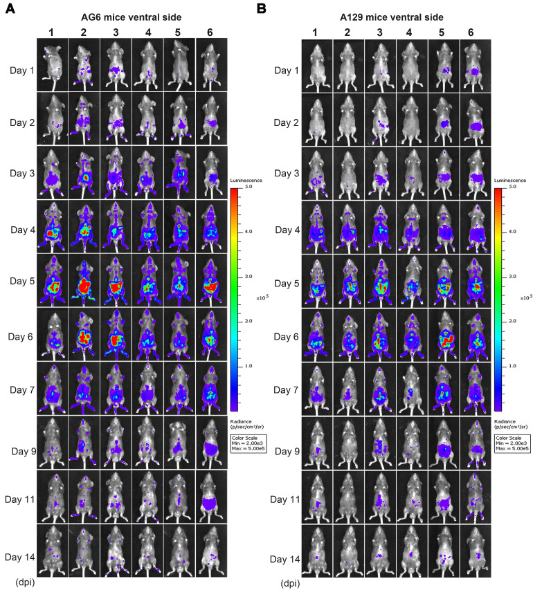
Figure 5.
Spatial and temporal progression of ZIKV-Nluc in AG6 and A129 mice in ventral views. Groups of AG6 (A) and A129 (B) mice (3-4 weeks old; n = 6) were infected with 6 × 10⁴ IFU of WT or ZIKV-Nluc via the footpad. The viral spread of ZIKV-Nluc-infected mice was monitored in real time at the indicated times.

