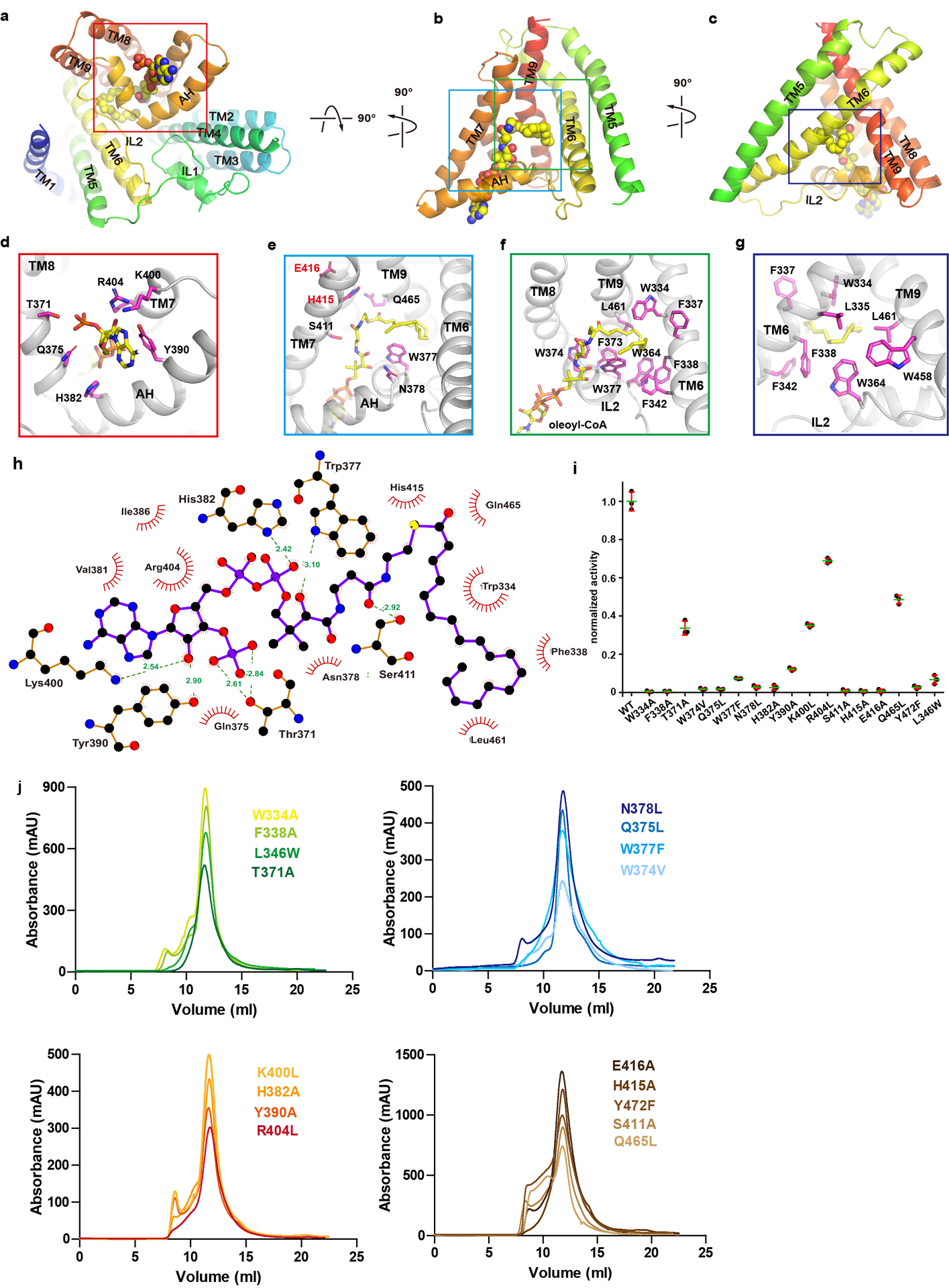
Extended Data Figure 6. Oleoyl-CoA binding site.

a-c. Oleoyl-CoA (spheres) bound to hDGAT1 protomer (cartoon) in three orientations. Details are shown in d-g. Residues coordinating oleoyl-CoA are shown as sticks with carbon atoms in magenta. h. LigPlus42,43 plot of the oleoyl-CoA binding site. i. Normalized enzymatic activity of hDGAT1 wild type and mutants. Error bars are s.e.m. derived from three independent repeats. j. Size-exclusion profiles of hDGAT1 mutants. Experiments in i were repeated independently 3 times with similar results.
