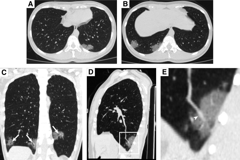
A healthy 32-year-old woman, a resident of Wuhan City, traveled to Xi’an City, which is 800 km away, to visit her husband on January 21, 2020. Five days after their reunion, her husband had a fever. On January 27, both of them had nasopharyngeal swabs collected for real-time RT-PCR assays (1, 2). On January 29, both were confirmed with coronavirus disease (COVID-19) by the Chinese Center for Disease Control and Prevention. Surprisingly, the woman was afebrile without chills and cough, and physical examination and laboratory results were unremarkable. However, the chest computed tomography (CT) scan showed bilateral subpleural ground-glass opacities in the lower lobe (Figure 1). She was immediately admitted to the isolation ward and developed a mildly productive cough with no other discomfort on January 30. After 2 days of supportive therapy, the patient recovered well and remained symptom-free until her discharge on February 17 for home isolation.
Figure 1.
The computed tomography images of an asymptomatic 32-year-old woman with coronavirus disease (COVID-19). (A and B) The axial views of the chest computed tomography examination showing bilateral subpleural areas of multifocal ground-glass opacities in the basal segment of the lower lung fields. (C–E) The coronal (C) and sagittal (D) views showing air bronchogram (C; arrows) and mildly dilated blood vessel (E; arrowhead in the partial enlarged view) within the ground-glass opacities.
Our case verified the asymptomatic infection with severe acute respiratory syndrome coronavirus 2 (SARS-CoV-2) as previously reported (3, 4) and suggested that 1) the transmission of COVID-19 seemingly could occur during the incubation period and may cause a potential threat to public health, and 2) the CT examination is very helpful for the early diagnosis of COVID-19 because the abnormalities (e.g., unilateral or bilateral subpleural multifocal ground-glass opacities of the lungs) associated with COVID-19 could be visualized on CT while subjects remain asymptomatic (5).
Supplementary Material
Footnotes
Author Contributions: Data collection and interpretation: J.Z., Y.D., L.B., and C.J. Literature search: J.Z., Y.D., and L.B. Original draft preparation: J.Z. and C.J. Review and editing: J.Z., J.P., and C.J. Supervision: J.Y. and Y.G. All authors reviewed and approved the final version of the manuscript.
Originally Published in Press as DOI: 10.1164/rccm.202002-0241IM on April 21, 2020
Author disclosures are available with the text of this article at www.atsjournals.org.
References
- 1.Corman VM, Landt O, Kaiser M, Molenkamp R, Meijer A, Chu DK, et al. Detection of 2019 novel coronavirus (2019-nCoV) by real-time RT-PCR. Euro Surveill. 2020;25:2000045. doi: 10.2807/1560-7917.ES.2020.25.3.2000045. [DOI] [PMC free article] [PubMed] [Google Scholar]
- 2.Carlos WG, Dela Cruz CS, Cao B, Pasnick S, Jamil S. Novel Wuhan (2019-nCoV) coronavirus. Am J Respir Crit Care Med. 2020;201:P7–P8. doi: 10.1164/rccm.2014P7. [DOI] [PubMed] [Google Scholar]
- 3.Hoehl S, Rabenau H, Berger A, Kortenbusch M, Cinatl J, Bojkova D, et al. Evidence of SARS-CoV-2 infection in returning travelers from Wuhan, China. N Engl J Med. 2020;382:1278–1280. doi: 10.1056/NEJMc2001899. [DOI] [PMC free article] [PubMed] [Google Scholar]
- 4.Chan JF, Yuan S, Kok KH, To KK, Chu H, Yang J, et al. A familial cluster of pneumonia associated with the 2019 novel coronavirus indicating person-to-person transmission: a study of a family cluster. Lancet. 2020;395:514–523. doi: 10.1016/S0140-6736(20)30154-9. [DOI] [PMC free article] [PubMed] [Google Scholar]
- 5.Shi H, Han X, Jiang N, Cao Y, Alwalid O, Gu J, et al. Radiological findings from 81 patients with COVID-19 pneumonia in Wuhan, China: a descriptive study. Lancet Infect Dis. 2020;20:425–434. doi: 10.1016/S1473-3099(20)30086-4. [DOI] [PMC free article] [PubMed] [Google Scholar]
Associated Data
This section collects any data citations, data availability statements, or supplementary materials included in this article.


