PLOS ONE logoLink to PLOS ONE
. 2020 May 29;15(5):e0233618. doi: 10.1371/journal.pone.0233618

A simplified method for blood feeding, oral infection, and saliva collection of the dengue vector mosquitoes

Chalida Sri-in 1,#, Shih-Che Weng 2,#, Shin-Hong Shiao 2,*, Wu-Chun Tu 1,*
Editor: Luciano Andrade Moreira3
PMCID: PMC7259494  PMID: 32469954

Abstract

A simple device using folded Parafilm-M as an artificial blood feeder was designed for studying two important dengue vector mosquitoes, Aedes aegypti and Aedes albopictus. The efficiency of the artificial blood feeder was investigated by comparing the numbers of engorged mosquitoes that fed on the artificial blood feeder versus mice as a live blood source. Significantly more engorged females Aedes aegypti fed on the artificial blood feeder than on mice. In addition, the artificial feeder could serve as a useful apparatus for oral infection via artificial blood meals, and for saliva collection in mosquitoes. Our method enabled us to collect saliva from multiple mosquitoes at once, providing sufficient infected saliva for determination of the virus titer by plaque assay analysis. Our artificial feeder has the advantage that it is simple, inexpensive, and efficient.

Introduction

The dengue vector mosquitoes, Aedes aegypti and Aedes albopictus, require blood feeding for egg maturation [1]. Anesthetized or immobilized animals such as mice, other rodents, and lagomorphs are usually used as the main blood source for maintaining mosquitoes in insectaries [25]. However, under some circumstances, it is difficult to use live animals for feeding these vectors due to a need for ethical approval, restrictions by animal care committees, and many regulations in place for the use of live animals in experiments [2, 68]. In addition, using live animals is costly and maintaining them is laborious.

Artificial blood-feeding systems are important to maintain mosquito populations in the laboratory and can be applied to replace live animals as blood sources [2]. A number of methods have been developed to feed blood-sucking insects [6,911], most of which use animal skins or thin membranes filled with warmed animal blood, that allow female mosquitoes to insert their proboscis to acquire a blood meal. These devices are generally constructed using costly materials, and are complicated to assemble [12]. Additionally, to inoculate mosquitoes with dengue virus (DENV) for various experiments, an artificial blood-feeder system is usually required for mosquitoes to take up infectious blood meals [1314].

Furthermore, DENV transmission can be determined directly by collecting infected mosquito saliva to examine the presence of virus. Several devices such as capillary tubes and suspended droplets for collecting mosquito saliva are available [1521] but it is time-consuming to collect saliva from large numbers of mosquitoes, and the saliva collected does not contain sufficient virus titer for studying DENV transmission by means of plaque assay, or by using a mouse model to examine hemorrhage development. Therefore, we developed a simple Parafilm blood feeder which has many benefits including blood feeding, oral infection, and saliva collection of mosquitoes. This apparatus has the advantage over existing artificial feeder systems that it is simple, convenient, and low cost, and requires no power supply or heater.

Materials and methods

Mosquito rearing

The Ae. aegypti UGAL/Rockefeller strain and Ae. albopictus Kaohsiung strain were maintained until adult stage at 25 ± 1°C, 80% relative humidity, and a 12:12h (Dark:Light) photoperiod as described previously [22]. Female mosquitoes 3 to 5 days after emergence were used for various experiments.

Mice rearing

Mouse-use for blood supply tests has been reviewed and approved by the Institutional Animal Care and Use Committee (approval no. 102–76), effective from October 23, 2013 to October 17, 2018. BALB/c mice were bought from Lexco Biotechnology Co., Ltd. All mice were maintained under specific-pathogen-free conditions at the Medical Entomology Laboratory, National Chung Hsing University, Taiwan. The mice were housed in accordance with local and home office regulations. Three female mice, five-week-old and specific pathogen-free, were anaesthetized using Pentobarbital sodium injection with a fixed dose of 75 mg/kg i.p. during the blood feeding experiment for 30 min. The mice used in this experiment were not euthanized.

Cell culture and dengue virus production

The Ae. albopictus C6/36 cells containing 2% heat-inactivated fetal bovine serum (FBS) and 1× penicillin–streptomycin solution were cultured in a mixture of Dulbecco’s modified Eagle’s medium (DMEM, Gibco) and Mitsuhashi and Maramorosch insect medium (MM) at 1:1 ratio. These cultured cells were infected with DENV2 strain 16681 at an infection multiplicity of 0.01. The culture supernatant was harvested 5 days after infection and subjected to a plaque assay to determine the viral titer. DENV2 at approximately 1.0 × 10⁷ PFU/ml was used to infect the mosquitoes. The plaque assay method will be further explained below.

Mosquito blood-feeding and egg laying experiments

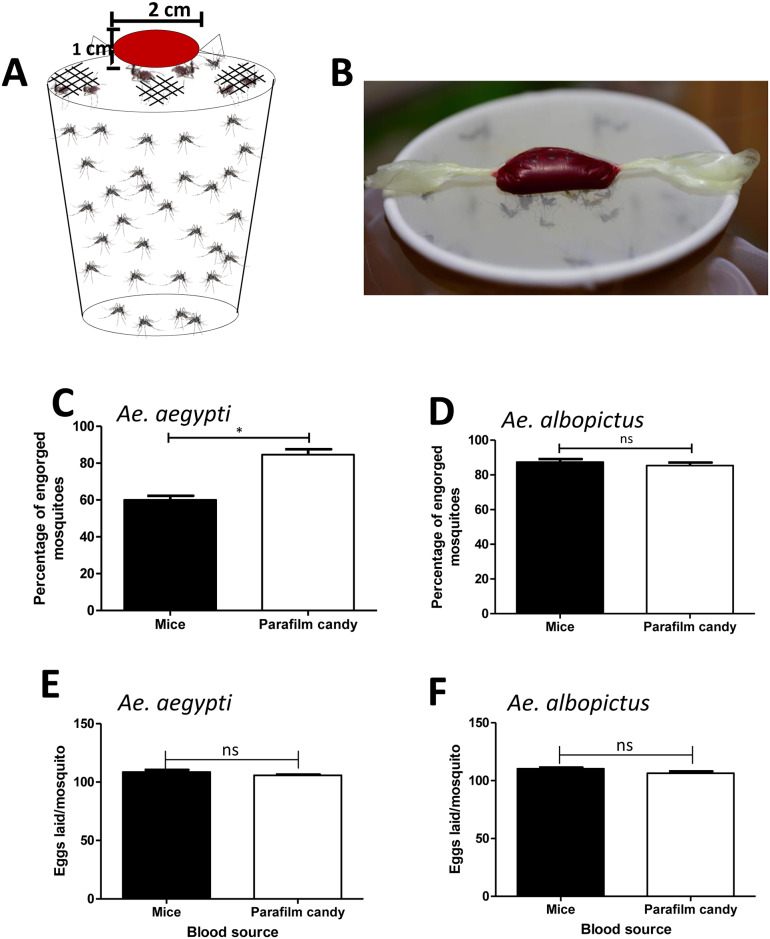
Females of Ae. aegypti and Ae. albopictus were transferred into cylindrical containers fitted with nylon mesh on top and starved through sugar-deprivation for 24 h. Mosquitoes were subsequently offered a blood meal containing 350 μl of washed sheep erythrocytes, 100 μl of 2% FBS, and 50 μl of 1 mM adenosine triphosphate (ATP). The meal was wrapped in stretched Parafilm-M membrane and warmed at 37°C, and then placed on the top of a container covered with nylon mesh (Fig 1A). Fifty mosquitoes inside the container were allowed to feed on the meal through the stretched Parafilm-M membrane (Fig 1B), or on anaesthetized mice as a control for 30 min. The surface of the mice available to the mosquitoes in the control group was limited to a 2 cm2 hole, the same diameter as the artificial blood feeder offered to the mosquitoes. Each mosquito fed either on an artificial blood meal or on a mouse was examined on a stereo microscope (Leica EZ4 HD) to determine whether it had taken a full meal and to evaluate the percentage of engorged females that their abdomens were bloated with blood after approximately one minute of feeding.

Fig 1. Mosquito blood-feeding using artificial blood feeder.

Fig 1

A. Blood meal containing 100 μl of 2% FBS, 50 μl of ATP, and 350 μl of erythrocyte was wrapped in stretched Parafilm-M membrane and put it on the top of a container covered with nylon mesh. Female mosquitoes were allowed to take up blood meal by bite wrapped blood inside parafilm. B. Mosquitoes inside the container were allowed to feed on the blood meal through the artificial blood feeder. C. To generate blood-feeding efficiency, the numbers of engorged females after 30 min of feeding on mice or artificial blood feeder were counted. Percentages of successful feeding were analyzed according to the number of engorged females divided by the total number of females allowed to feed. Data were pooled from three independent experiments and presented as mean ± SEM. *P < 0.05 (Unpaired t-test). D. To generate the effect of blood source on eggs laid, the numbers of eggs laid from engorged females that fed on mice or artificial blood feeder were counted. Each replication represented the eggs laid of 20 mosquitoes. Data were pooled from three independent experiments and presented as mean ± SEM. ns, not significant (Unpaired t-test).

All engorged females were placed in a container covered with nylon mesh with a cotton pad soaked with 10% glucose solution for feeding. On day 5 post blood meal, all engorged females were transferred and placed individually in 50-ml plastic tubes in which cotton soaked with mineral water was put on the bottom, and white paper was placed around the bottom for the females to lay eggs [23]. The oviposition tube was covered with a mesh, and a cotton pad soaked with 10% glucose solution was placed on top of each tube. The number of eggs laid by each female was counted.

Oral infection of mosquitoes with DENV2

Infection of mosquitoes was achieved through an infectious blood meal. Ae. aegypti females were transferred into cylindrical containers fitted with nylon mesh on the top and starved through sugar deprivation for 24 h. Female mosquitoes were subsequently offered an infectious blood meal prepared by mixing 200 μl of washed sheep erythrocytes, 50 μl of 1 mM ATP, and 250 μl of DENV2 16681 (2.5 x 10⁶ PFU in 250 μl) [24]. The meal was contained in a stretched Parafilm-M membrane (Fig 1A and 1B). The mosquitoes were allowed to take up the infectious blood for 1 h. Each mosquito was examined on a stereo microscope to determine whether it had taken a full meal. Mosquitoes were then transferred into cylindrical containers fitted with nylon mesh on the top and held in 10% glucose solution at 25°C and 80% humidity, according to standard rearing conditions [25].

Mosquito saliva collection and salivary protein detection

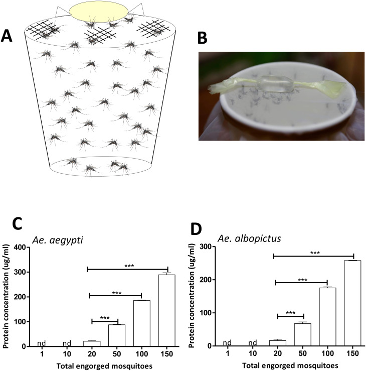
To collect saliva for measuring protein concentration, Ae. aegypti and Ae. albopictus females were starved for 24 h prior to saliva collection. On the day of saliva collection, feeding solution containing 90 μl of 1× phosphate-buffered saline (PBS) and 10 μl of 1 mM ATP at final concentration 10 μM [2627] was prepared. The feeding solution was wrapped in stretched Parafilm-M membrane (Fig 2A and 2B) and put on the top of a container covered with nylon mesh, allowing 1, 10, 20, 50, 100, and 150 mosquitoes to feed on the meal (Fig 2B). The feeding solution containing mosquito saliva was removed from the membrane and transferred to a microtube and centrifuged at 12,000 g for 1 min at 4°C. The saliva’s protein concentrations were measured using Bradford protein assays.

Fig 2. Mosquito saliva collection using artificial blood feeder.

Fig 2

A. To collect mosquito saliva using artificial blood feeder, feeding solution containing 90 ul of 1× PBS and 10 ul of 1 mM ATP was wrapped in stretched Parafilm-M membrane and put on the top of a container covered with nylon mesh. B. Mosquitoes inside the container were allowed to feed on the solution for saliva collection via the artificial feeder. C. To measure saliva protein concentration, the protein concentrations of saliva that was collected from 1, 10, 20, 50, 100, and 150 female mosquitoes were examined using Bradford protein assays. Data were pooled from three independent experiments and displayed as mean ± SEM. nd, not detected; ***P < 0.001 (Unpaired t-test).

To collect infected saliva for plaque assays, female mosquitoes were starved for 24 h prior to saliva collection. Infected mosquito saliva was examined on day 3, 5, 7, 10, and 14 post infectious blood meal. On the day of saliva collection, feeding solution containing 180 μl of 1× PBS and 20 μl of 1 mM ATP at a final concentration of 10 μM was prepared. The feeding solution was wrapped as described previously and put on the top of a container covered with nylon mesh, allowing 200 infected mosquitoes inside to feed on the meal (Fig 2A and 2B). The infected mosquitoes were allowed to feed for 30 min. The feeding solution containing mosquito saliva was removed from the membrane as described previously. This was repeated 5 times every 2-h to obtain a larger volume of saliva-containing meal. The saliva-containing meal was sterilized via passage through an Ultrafree-GV centrifugal filter 0.22 μm before transfer into a new microtube. The sterilized saliva-containing meal was concentrated through a Millipore column with filter pore size 3 kDa nominal molecular weight limit (NMWL) and centrifuged at 12,000 g for 1-h at 4°C. The solution flowing through was discarded, and the remaining solution was collected and stored at -80°C for further use.

Plaque assay analysis

A pool of infected Ae. aegypti saliva was collected at 3, 5, 7, 10, and 14 days post infection via the artificial feeder. The saliva from individual day post infection was adjusted to an equivalent protein concentration before virus titer determination by plaque assay. C6/36 cells were used for plaque assays as described previously [13, 24]. Cell monolayers were rinsed with PBS, and 200 μl of infectious mosquito saliva was added to the cell monolayers for 2-h absorption at 28°C. After absorption, 500 μl of 1% methylcellulose cell media was added, and the plates were maintained in the incubator at 28°C. After 5 days, plates were fixed with 4% formaldehyde for 1-h at room temperature. Methylcellulose overlays were then removed, and plates were stained with 1% crystal violet for 1-h.

Statistical analysis

The statistical analyses were performed using GraphPad Prism 5 software. Differences between groups were evaluated with unpaired t-tests. A P-value < 0.05 was considered to be statistically significant.

Results and discussion

Feeding efficiency is a relevant parameter that needs to be considered in the development of artificial feeders [2, 11]. To evaluate if there is better feeding efficiency of the parafilm artificial blood feeder compared to live animals, Ae. aegypti and Ae. albopictus were allowed to feed on our artificial blood feeder and mice for the same period (Fig 1A and 1B). The total number of engorged females was counted to compare the efficiency between the two different blood sources (Fig 1C and 1D and S1 Table). In Ae. aegypti, an average of 84.67% females that were fed on artificial blood feeder exhibited engorgement compared to 60% of females fed on mice (S1 Table). Furthermore, there was a significant difference in the mean percentage of engorged females between these two groups (P < 0.05, Fig 1C). However, there was not a significant difference in the mean percentage of engorged Ae. albopictus (P > 0.05, Fig 1D). An average of 87.33% females fed on the artificial blood feeder using sheep erythrocytes exhibited engorgement compared to 85.33% of females fed on mice (S1 Table). The lack of a significant difference in the mean percentage of engorged females between these two groups may be because Ae. albopictus feeds on a variety of mammals such as swine, sheep, dogs, cats, and mice [28]. In both Ae. aegypti and Ae. albopictus there was no significant difference in the number of eggs laid between the individual females fed on artificial blood feeder and mice (P > 0.05, Fig 1E and 1F and S2 Table). Therefore, these results demonstrate that our artificial blood feeder provides as efficient a blood source as mice for mosquitoes.

We suggest that the artificial feeder could be used to replace live animals for female mosquito blood feeding. The results of a simple artificial membrane-feeding method using a standard conical tube and Parafilm-M membrane reported previously did not differ significantly in the numbers of mosquitoes compared with those using the mice-feeding method [11]. Our results showed a significantly higher percentage of mosquitoes (Ae. aegypti) fed on our device than fed on live mice, demonstrating that the development of a simple artificial membrane-feeding method using Parafilm M membrane was successful. However, it is difficult to compare the feeding efficiency among different artificial feeders because of great variations in experimental conditions, such as different blood sources for the control and experimental groups. The preparation of blood sources before loading to Parafilm was also reported to be critical [11]. Moreover, the available surface of the mouse for biting by mosquito females in the control group was limited to 2 cm2, the same area as the artificial blood feeder. A higher level of feeding efficiency was observed when using the whole mouse body rather than a 2 cm2 area.

This artificial feeder can also serve as a useful apparatus for mosquito saliva collection. To collect saliva, Ae. aegypti and Ae. albopictus females were allowed to feed on the meal through the artificial feeder (Fig 2A and 2B). Our results revealed that the amount of protein detected in the artificial feeding solution increased according to the number of Ae. aegypti and Ae. albopictus that were engorged after exposure to the device (P < 0.001, Fig 2C and 2D and S3 Table). However, using the artificial feeder to collect sufficient saliva for evaluating protein concentration required at least 20 females fed on 100 μl of the feeding solution in this device (Fig 2C and 2D and S3 Table). This result suggests that our method is not suitable for collecting saliva from individual mosquitoes, but it enables the collection of saliva from multiple mosquitoes at once.

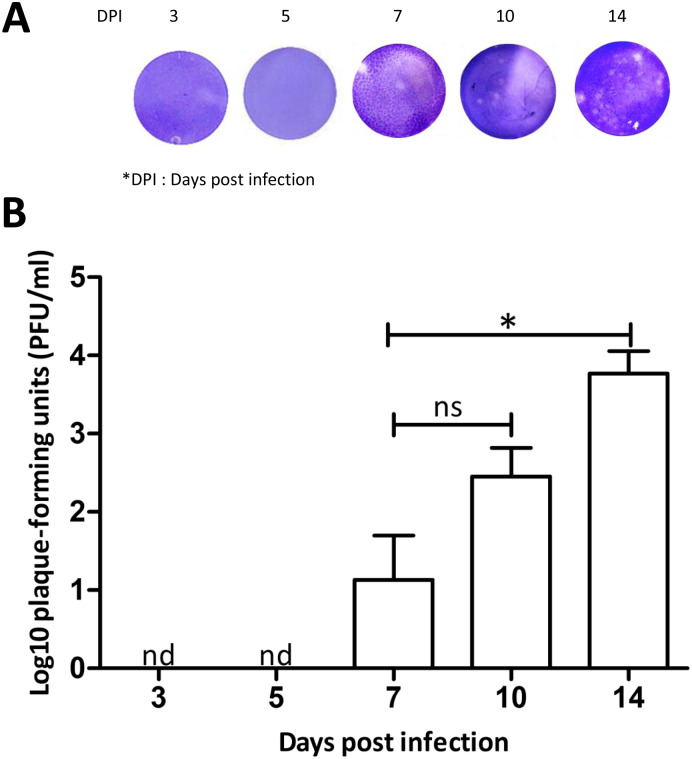
For infection with virus, Ae. aegypti consumed infectious blood using our artificial feeder loaded with washed sheep erythrocytes mixed with C6/36 cultured DENV2. We collected and evaluated DENV infection in mosquitoes’ saliva on days 3, 5, 7, 10 and 14 post infectious blood meal. From 81% to 84% of mosquitoes that fed on the saliva collection device reached full engorgement (S4 Table). Our results showed that the plaque-forming units of infected Ae. aegypti saliva could be detected from day 7 after infection (Fig 3A and 3B). Although the time post infectious blood meal to collect saliva by artificial blood feeder could increase infected saliva titers, no significant differences were observed between day 7 and 10 post infectious blood meal (P > 0.05, Fig 3B). However, significant differences were observed between day 7 and 14 post infectious blood meal, and the highest titer was found on day 14 after infectious blood meal (P < 0.05, Fig 3B). The results of DENV transmission, using our method for infected saliva collection, show that virus can be detected as early as 7 days after oral infection. During days 7 to 14, there is an increase in virus titers, but the mosquito preference to feed on our device is unchanged (S4 Table).

Fig 3. Evaluation of saliva titer collected by artificial blood feeder.

Fig 3

DENV2-infected mosquito saliva was collected at 3, 5, 7, 10, and 14 days post infection via the artificial feeder. The saliva from individual day post infection was adjusted to an equivalent protein concentration. A. Comparison of plaques produced by DENV2-infected mosquito saliva at different days post infection. The infected saliva was diluted 10 fold and assayed in C6/36 monolayer cells. B. DENV2-infected mosquito saliva titer at different days post infection was pooled from three independent experiments and presented as mean ± SEM. Total 200 mosquitoes were used for each replicate. nd, not detected; ns, not significant; *P < 0.05 (Unpaired t-test).

Using our method, the collected saliva had sufficient virus titer for studying DENV transmission by plaque assay, or by using a mouse model to determine hemorrhage development [24]. These indicate that the artificial blood feeder is an efficient method to collect DENV2-infected mosquito saliva for virus transmission study. Virus transmission using region-delimited mosquito biting in mouse ears has been studied [2930]. However, these virus transmission studies could not control the amount of saliva that the mosquito injected into the mouse, and required a high titer of infected saliva to investigate mice hemorrhage development. It is clear that methods to collect infected mosquito saliva should be developed. There is a previous study used this parafilm artificial feeder to collect DENV2-infected mosquito saliva for tests on mice, but the process and efficiency of this method were not described [24].

A previous study used a suspension feeding method to collect saliva by allowing the mosquitos to feed from a hanging blood droplet that was placed on the top of the mosquito cage, and then the droplet was tested for the presence of the virus [16]. There is a mosquito salivation device that was constructed with plexiglass as the surface on which mosquitoes were placed, and immersion oil was added to capillary tubes with a sterile pipette tip inserted into the opposite end [21]. The droplet feeding method demonstrated that virus transmission rates were consistent with the capillary tube method [31], but these saliva collection systems were based on experiments involving small sample sizes (N < 150) [16, 21, 31]. In another experiment using the midge, Culicoides nubeculosus, saliva was collected from more than 5,000 C. nubeculosus in 20 pill boxes using a modified artificial membrane feeding system in which midges, attracted by 37°C horse blood, deposited saliva proteins in the Durapore filters [32]. However, this method used inner and outer glass chambers made of costly materials and complicated to assemble. Our device provides a new simple method of mosquito saliva collection that is cost effective and useful for large sample sizes (150–200 mosquitoes in a single container). Moreover, we can increase the sample sizes by collecting mosquito saliva from many containers depending on requirements. Our experiments indicated that mosquito saliva titers and salivary protein concentrations that we collected are higher than those measured in previous studies [1721].

This artificial feeder does not require special materials or electric heaters. The defined pre-warmed temperature for blood mimics vertebrate blood temperature [11], and the meal contained FBS and ATP as a phagostimulant [33]. The artificial blood feeder specified here is inexpensive and easy to assemble with easily available common materials. Therefore, it can be considered as an alternative for blood sources from live animals to maintain mosquito populations in the laboratory. Furthermore, it is useful for oral infection, saliva collection from multiple mosquitoes at once, and studying dengue virus transmission in vector mosquitoes.

Supporting information

S1 Table. Blood-feeding efficiency of artificial blood feeder compared with mice blood-fed female mosquitoes.

(DOCX)

S2 Table. Eggs laid efficiency of individual female mosquito that fed on artificial blood feeder or mice blood-fed.

(DOCX)

S3 Table. Comparison of saliva protein concentration that collected from different number of mosquitoes using artificial feeder.

(DOCX)

S4 Table. Comparison of saliva titers collected from DENV2-infected Ae. aegypti via artificial feeder at different days post infection.

(DOCX)

Acknowledgments

The authors thank Prof. Roger F. Hou, National Chung Hsing University, for critical reading of the manuscript.

Data Availability

All relevant data are within the manuscript and its Supporting Information files.

Funding Statement

This study was supported by research grant (MOST 107-2313-B-005-027) from Ministry of Science and Technology, Executive Yuan, ROC.

References

  • 1.Clements AN (1992) The biology of mosquitoes: development, nutrition and reproduction. London: Chapman & Hall; 509 pp. [Google Scholar]
  • 2.Benedict MQ (2014) Methods in Anopheles. 4th ed. MR4. 419 pp.
  • 3.Galindo P, Srihongse S (1967) Transmission of arboviruses to hamsters by the bite of naturally infected Culex (Melanoconion) mosquitoes. Am J Trop Med Hyg 16(4):525–530. 10.4269/ajtmh.1967.16.525 [DOI] [PubMed] [Google Scholar]
  • 4.Kuo YP, Tsai KN, Luo YC, Chung PJ, Su YW, Teng Y, et al. (2018) Establishment of a mouse model for the complete mosquito-mediated transmission cycle of Zika virus. PLoS Negl Trop Dis 12(4): e0006417 10.1371/journal.pntd.0006417 [DOI] [PMC free article] [PubMed] [Google Scholar]
  • 5.French FE, West AS (1971) Skin reaction specificity of guinea pig immediate hypersensivity to bites of four mosquito species. J Parasitol 57(2):396–400. [PubMed] [Google Scholar]
  • 6.Chiang RG, Chiang JA (2010) Feeding through artificial membranes reduces blood-feeding insect, Rhodnius prolixus. Arch Insect Biochem Physiol 74(2):103–13. [DOI] [PubMed] [Google Scholar]
  • 7.Gauthier C (2008) The institutional animal care committee : Keystone of international harmonization. Japanese Soc Altern to Anim Exp 14:157–161. [Google Scholar]
  • 8.Tereza A, Filipecki P, Jose C, Machado S, Tereza A, Filipecki P, et al. (2011) The Brazilian legal framework on the scientific use of animals. ILAR e-Journal 52:e8–15. [DOI] [PubMed] [Google Scholar]
  • 9.Huebner E, Harrison RE, Yeow K (1994) A new feeding technique for experimental and routine culturing of the insect Rhodnius prolixus. Can J Zool 72(12):2244–2247. 10.1139/z94-299 [DOI] [Google Scholar]
  • 10.Waladde SM, Young AS, Morzaria SP (1996) Artificial feeding of lxodid ticks. Parasitol Today 12(7):272–278. [DOI] [PubMed] [Google Scholar]
  • 11.Costa-da-silva AL, Navarrete FR, Salvador FS, Karina-costa M, Ioshino RS, et al. (2013) Glytube : A conical tube and parafilm M-based method as a simplified device to artificially blood-feed the dengue vector mosquito, Aedes aegypti. PLoS ONE 8(1): e53816. [DOI] [PMC free article] [PubMed] [Google Scholar]
  • 12.Joanne R, Ammons D (2007) Versatile blood bags for laboratory feeding of mosquitoes. J Am Mosq Control Assoc 23(2):149–152. 10.2987/8756-971X(2007)23[149:VBBFLF]2.0.CO;2 [DOI] [PubMed] [Google Scholar]
  • 13.Das S, Garver L, Ramirez JR, Xi Z, Dimopoulos G (2008) protocol for dengue infections in mosquitoes (Ae. aegypti) and Infection Phenotype Determination. J Vis Exp (5):4–5. [DOI] [PMC free article] [PubMed] [Google Scholar]
  • 14.Salazar MI, Richardson JH, Sánchez-vargas I, Olson KE, Beaty BJ (2007) Dengue virus type 2: replication and tropisms in orally infected Aedes aegypti mosquitoes. BMC Microbiol 7: 9 10.1186/1471-2180-7-9 [DOI] [PMC free article] [PubMed] [Google Scholar]
  • 15.Hurlbut HS (1966) Mosquito salivation and virus transmission. Am Soc Trop Med Hyg 15(16):989–993. 10.4269/ajtmh.1966.15.989 [DOI] [PubMed] [Google Scholar]
  • 16.Duane J. Gubler Rosen L (1976) A simple technique for demonstrating transmission of dengue virus by mosquitoes without the use of vertebrate hosts. Am Soc Trop Med Hyg 25(1):146–150. 10.4269/ajtmh.1976.25.146 [DOI] [PubMed] [Google Scholar]
  • 17.Aitken THG (1977) An in vitro feeding technique for artificially demonstrating virus transmission by mosquitoes. Mosq News 37(1):130–133. [Google Scholar]
  • 18.Colton L, Biggerstaff BJ, Johnson A, Nasci RS (2005) Quantification of west nile virus in vector mosquito saliva. J Am Mosq Control Assoc 21(1):49–53. 10.2987/8756-971X(2005)21[49:QOWNVI]2.0.CO;2 [DOI] [PubMed] [Google Scholar]
  • 19.Smith DR, Carrara A, Aguilar PV, Weaver SC (2005) Evaluation of methods to assess transmission potential of Venezuelanequine encephalitis virus by mosquitoes and estimation of mosquito saliva titers. Am J Trop Med Hyg 73(1):33–39. [PubMed] [Google Scholar]
  • 20.Mores CN, Turell MJ, Dohm DJ, Blow JA, Carranza MT, Quintana M (2007) Experimental transmission of west nile virus by Culex nigripalpus from Honduras. Vector-Borne Zoonotic Dis 7(2):279–284. 10.1089/vbz.2006.0557 [DOI] [PubMed] [Google Scholar]
  • 21.Anderson SL, Richards SL, Smartt CT (2011) A simple method for determining arbovirus. J Am Mosq Control Assoc 26(1):108–111. [DOI] [PMC free article] [PubMed] [Google Scholar]
  • 22.Chang CH, Liu YT, Weng SC, Chen IY, Tsao PN, Shiao SH (2018) The non-canonical notch signaling is essential for the control of fertility in Aedes aegypti. PLoS Negl Trop Dis 12(3): e0006307 10.1371/journal.pntd.0006307 [DOI] [PMC free article] [PubMed] [Google Scholar]
  • 23.Ve´zilier J, Nicot A, Gandon S, Rivero A (2012) Plasmodium infection decreases fecundity and increases survival of mosquitoes. Proc R Soc B 279(August): 4033–4041. 10.1098/rspb.2012.1394 [DOI] [PMC free article] [PubMed] [Google Scholar]
  • 24.Sri-in C, Weng SC, Chen WY, Wu-hsieh BA, Tu WC, Shiao SH (2019) A salivary protein of Aedes aegypti promotes dengue-2 virus replication and transmission. Insect Biochem Mol Biol 111(May):103181 10.1016/j.ibmb.2019.103181 [DOI] [PubMed] [Google Scholar]
  • 25.Weng SC, Shiao SH (2015) Frizzled 2 is a key component in the regulation of TOR signaling- mediated egg production in the mosquito Aedes aegypti. Insect Biochem Mol Biol 61:17–24. 10.1016/j.ibmb.2015.03.010 [DOI] [PubMed] [Google Scholar]
  • 26.Brackney DE, Foy BD, Olson KE (2008) The effects of midgut serine proteases on dengue virus type 2 infectivity of Aedes aegypti. Am J Trop Med Hyg 79(2):267–274. 79/2/267 [PMC free article] [PubMed] [Google Scholar]
  • 27.Chisenhall DM, Londono BL, Christofferson RC, McCracken MK, Mores CN (2014) Effect of dengue-2 virus infection on protein expression in the salivary glands of Aedes aegypti mosquitoes. Am J Trop Med Hyg 90(3):431–437. 10.4269/ajtmh.13-0412 [DOI] [PMC free article] [PubMed] [Google Scholar]
  • 28.Ponlawath A, Harrington LC (2005) Blood feeding patterns of Aedes aegypti and Aedes albopictus in Thailand. J Med Entomol 42(5):844–9. [DOI] [PubMed] [Google Scholar]
  • 29.Cox J, Mota J, Sukupolvi-petty S, Diamond MS, Rico-hesse R (2012) Mosquito bite delivery of dengue virus enhances immunogenicity and pathogenesis in humanized mice. J Virol 86(14):7637–49. 10.1128/JVI.00534-12 [DOI] [PMC free article] [PubMed] [Google Scholar]
  • 30.Secundino NFC, Chaves BA, Orfano AS, Silveira KRD, Rodrigues NB, Campolina TB, et al. (2017) Zika virus transmission to mouse ear by mosquito bite: A laboratory model that replicates the natural transmission process. Parasit Vectors 10(1):346 10.1186/s13071-017-2286-2 [DOI] [PMC free article] [PubMed] [Google Scholar]
  • 31.Cornel AJ, Jupp PG (1989) Comparison of three methods for determining transmission rates in vector competence studies with Culex univittatus and West Nile and Sindbis viruses. J Am Mosq Control Assoc 5(1):70–72. [PubMed] [Google Scholar]
  • 32.Langner KFA, Darpel KE, Denison E, Barbara S, Leibold W, Mellor PS, et al. (2007) Collection and analysis of salivary proteins from the biting midge Culicoides nubeculosus (Diptera: Ceratopogonidae). J Med Entomol 44(2):238–48. [DOI] [PubMed] [Google Scholar]
  • 33.de Wispelaere M, Desprès P, Choumet V (2017) European Aedes albopictus and Culex pipiens are competent vectors for Japanese encephalitis virus. PLoS Negl Trop Dis 11(1): e0005294 10.1371/journal.pntd.0005294 [DOI] [PMC free article] [PubMed] [Google Scholar]

Decision Letter 0

Luciano Andrade Moreira

26 Feb 2020

PONE-D-19-36024

A simplified method for blood feeding, oral infection, and saliva collection of the dengue vector mosquitoes

PLOS ONE

Dear Dr Tu,

Thank you for submitting your manuscript to PLOS ONE. After careful consideration, we feel that it has merit but does not fully meet PLOS ONE’s publication criteria as it currently stands. Therefore, we invite you to submit a revised version of the manuscript that addresses the points raised during the review process.

Please carefully check all the aspects both reviewers have raised around your manuscript, specially related to the writing. A thorough grammar review should be performed in the end after all other points have been addressed.

We would appreciate receiving your revised manuscript by APRIL 10th. When you are ready to submit your revision, log on to https://www.editorialmanager.com/pone/ and select the 'Submissions Needing Revision' folder to locate your manuscript file.

If you would like to make changes to your financial disclosure, please include your updated statement in your cover letter.

To enhance the reproducibility of your results, we recommend that if applicable you deposit your laboratory protocols in protocols.io, where a protocol can be assigned its own identifier (DOI) such that it can be cited independently in the future. For instructions see: http://journals.plos.org/plosone/s/submission-guidelines#loc-laboratory-protocols

Please include the following items when submitting your revised manuscript:

  • A rebuttal letter that responds to each point raised by the academic editor and reviewer(s). This letter should be uploaded as separate file and labeled 'Response to Reviewers'.

  • A marked-up copy of your manuscript that highlights changes made to the original version. This file should be uploaded as separate file and labeled 'Revised Manuscript with Track Changes'.

  • An unmarked version of your revised paper without tracked changes. This file should be uploaded as separate file and labeled 'Manuscript'.

Please note while forming your response, if your article is accepted, you may have the opportunity to make the peer review history publicly available. The record will include editor decision letters (with reviews) and your responses to reviewer comments. If eligible, we will contact you to opt in or out.

We look forward to receiving your revised manuscript.

Kind regards,

Luciano Andrade Moreira, PhD

Academic Editor

PLOS ONE

Journal Requirements:

When submitting your revision, we need you to address these additional requirements:

1. Please ensure that your manuscript meets PLOS ONE's style requirements, including those for file naming. The PLOS ONE style templates can be found at http://www.plosone.org/attachments/PLOSOne_formatting_sample_main_body.pdf and http://www.plosone.org/attachments/PLOSOne_formatting_sample_title_authors_affiliations.pdf

2. Thank you for stating the following in the Acknowledgments Section of your manuscript:

"This study was supported by research grant (MOST 107-2313-B-005-027) from Ministry of Science and Technology, Executive Yuan, ROC. The authors thank Prof. Roger F. Hou, National Chung Hsing University, for critical reading of the manuscript."

We note that you have provided funding information that is not currently declared in your Funding Statement. However, funding information should not appear in the Acknowledgments section or other areas of your manuscript. We will only publish funding information present in the Funding Statement section of the online submission form.

Please remove any funding-related text from the manuscript and let us know how you would like to update your Funding Statement. Currently, your Funding Statement reads as follows:

"No, the funders had no role in study design, data collection and analysis, decision to publish, or preparation of the manuscript. "

3. Thank you for stating the following in the Methods Section of your manuscript:

"The mouse strain BALB/c was a generous from Lexco Biotechnology Co., Ltd"."

We note that you have provided funding information that is not currently declared in your Funding Statement. However, funding information should not appear in the Acknowledgments section or other areas of your manuscript. We will only publish funding information present in the Funding Statement section of the online submission form.

Please remove any funding-related text from the manuscript and let us know how you would like to update your Funding Statement. Currently, your Funding Statement reads as follows:

"No, the funders had no role in study design, data collection and analysis, decision to publish, or preparation of the manuscript. "

Additionally, because some of your funding information pertains to commercial funding, we ask you to provide an updated Competing Interests statement, declaring all sources of commercial funding.

In your Competing Interests statement, please confirm that your commercial funding does not alter your adherence to PLOS ONE Editorial policies and criteria by including the following statement: "This does not alter our adherence to PLOS ONE policies on sharing data and materials.” as detailed online in our guide for authors  http://journals.plos.org/plosone/s/competing-interests.  If this statement is not true and your adherence to PLOS policies on sharing data and materials is altered, please explain how.

Please include the updated Competing Interests Statement and Funding Statement in your cover letter. We will change the online submission form on your behalf.

[Note: HTML markup is below. Please do not edit.]

Reviewers' comments:

Reviewer's Responses to Questions

Comments to the Author

1. Is the manuscript technically sound, and do the data support the conclusions?

The manuscript must describe a technically sound piece of scientific research with data that supports the conclusions. Experiments must have been conducted rigorously, with appropriate controls, replication, and sample sizes. The conclusions must be drawn appropriately based on the data presented.

Reviewer #1: Yes

Reviewer #2: Yes

**********

2. Has the statistical analysis been performed appropriately and rigorously?

Reviewer #1: Yes

Reviewer #2: Yes

**********

3. Have the authors made all data underlying the findings in their manuscript fully available?

The PLOS Data policy requires authors to make all data underlying the findings described in their manuscript fully available without restriction, with rare exception (please refer to the Data Availability Statement in the manuscript PDF file). The data should be provided as part of the manuscript or its supporting information, or deposited to a public repository. For example, in addition to summary statistics, the data points behind means, medians and variance measures should be available. If there are restrictions on publicly sharing data—e.g. participant privacy or use of data from a third party—those must be specified.

Reviewer #1: Yes

Reviewer #2: Yes

**********

4. Is the manuscript presented in an intelligible fashion and written in standard English?

PLOS ONE does not copyedit accepted manuscripts, so the language in submitted articles must be clear, correct, and unambiguous. Any typographical or grammatical errors should be corrected at revision, so please note any specific errors here.

Reviewer #1: No

Reviewer #2: No

**********

5. Review Comments to the Author

Please use the space provided to explain your answers to the questions above. You may also include additional comments for the author, including concerns about dual publication, research ethics, or publication ethics. (Please upload your review as an attachment if it exceeds 20,000 characters)

Reviewer #1: In this manuscript, Sri-in et al propose a simple and elegant method for mosquito feeding under laboratory conditions. Different strategies, methods, and apparatus for mosquito feeding have been proposed along the past years, but usually they require a heat source or other labware. Here, the authors propose a simplified system using stretched parafilm to offer a blood or protein meal to mosquitoes. Their findings suggest that using wrapped blood inside parafilm is comparable to using mice as blood source for mosquito feeding regarding the number of engorged females and number of eggs laid per female. Also, it seems possible to recover saliva proteins and virus expelled in the saliva by infected mosquitoes using their proposed feeding system, enabling transmission experiments to be done in a simplified manner. Thus, this reviewer recommends the paper for publication with minor revision. Listed below are misunderstandings that must be fixed before publication, together with necessary clarification in methods and statistics. This reviewer will feel pleased to review the rebuttal of this manuscript upon resubmission.

Major comments:

Line 69-71 – […] “saliva collected does not contain sufficient virus titers for studying DENV transmission by means of plaque assay or mouse model.” […] – This affirmation is not entirely true. It is difficult to detect infected saliva from infected mosquitoes but is not impossible. Different studies have studied virus transmission using region-delimited mosquito biting in mouse ears (e.g. Secundino NFC et al, 2017 – doi 10.1186/s13071-017-2286-2 or Cox J et al, 2012 – doi 10.1128/JVI.00534-12). In fact, it is discussed later on in the text that their method is only suitable for collection of saliva from pools of at least 20 mosquitoes (Fig 2 C/D – Lines 210-214) thus a comparison with other methods should be discussed in the text.

Line 219-232 – Would have the authors verified the prevalence of DENV infection in individual mosquitoes upon feeding on their system comparing to a water-jacked or other previously characterized system? Their assessment of saliva detection of DENV was done in large pools of mosquitoes but could only represent a very low percentage of mosquitoes that were infected, thus representing that their system may not be efficient to infect mosquitoes. The authors even emphasize that their results “indicated that the artificial blood feeder is an efficient artificial feeder” (lines 231-232) and their results do not hold sufficient information for this affirmation. Also, it is interesting to include more details (e.g. size of mosquito pool) of the saliva DENV-detection experiment in the figure legend.

Minor comments:

For a better organization of the paper, I suggest moving Tables to supplementary information.

Line 66 – Break into a new paragraph. Ideas are not connected with previous lines.

Line 98 – Please add sentence explaining that plaque assay method will be further explained in the text.

Line 100 – Invert “laying eggs” by “egg laying”

Line 148 – “The infected mosquitoes were allowed to salivate for 30 min.” – Mosquitoes did not salivate; they were allowed to bite the candy wrap. Please rephrase.

Line 150 – “5 times” not “5 time”

Line 154 – Describe better “Millipore column measuring 3 kDa”

Line 159-160 – Is it correct that plaque assay was done using C6/36 cells? Usually these cells do not form plaques upon DENV infection, thus should not be possible to visualize using crystal violet. Please clarify.

Line 173 – change “elucidate” by “evaluate if there is”

Line 175 – “…albopictus were fed on” by “…albopictus were allowed to feed on”

Line 178 – Was it considered fully engorged females or mosquitoes that ingested any amount of blood? Please clarify.

Line 183-184 – “This result may …” This sentence brings no relevant information. Please rephrase.

Line 193 and 198 – Is reference 11 correct here?

Line 193-196 – Please clarify that only for Ae. aegypti you found significantly higher percentage of mosquitoes fed on parafilm candy.

Line 199 – The discussion starting by “In addition” is important but is lacking context on the way it’s written. Please explain or rephrase.

Line 208 – “engorged” not “engorge”

Line 215-216 – Please rephrase

Line 230 – What is high virus concentration?

Line 230-231 – Explain the sentence “Interestingly, the infected saliva collection…”

Line 233 – Lost discussion in the text. Please include context and rephrase.

Table 4 – What means “a” along the statistics represented in this table?

Reviewer #2: I feel that this is good paper describing a low cost alternative to standard artificial blood-feeder devices that are commercially available. The finding presented here validate this new method as an acceptable alternative, I believe these findings will have a great impact on the ability for labs in lower income countries to be able to perform more complex vector competence and transmission studies.

The only issue that I have found with this article is language based, there are several instances throughout the paper in which the phrasing used is confusing and hard to understand the point being made. I have highlighted these sections in my review of the article and believe that the correction of these minor issues will make for a good article.

**********

6. PLOS authors have the option to publish the peer review history of their article (what does this mean?). If published, this will include your full peer review and any attached files.

If you choose “no”, your identity will remain anonymous but your review may still be made public.

Do you want your identity to be public for this peer review? For information about this choice, including consent withdrawal, please see our Privacy Policy.

Reviewer #1: No

Reviewer #2: No

[NOTE: If reviewer comments were submitted as an attachment file, they will be attached to this email and accessible via the submission site. Please log into your account, locate the manuscript record, and check for the action link "View Attachments". If this link does not appear, there are no attachment files to be viewed.]

While revising your submission, please upload your figure files to the Preflight Analysis and Conversion Engine (PACE) digital diagnostic tool, https://pacev2.apexcovantage.com/. PACE helps ensure that figures meet PLOS requirements. To use PACE, you must first register as a user. Registration is free. Then, login and navigate to the UPLOAD tab, where you will find detailed instructions on how to use the tool. If you encounter any issues or have any questions when using PACE, please email us at figures@plos.org. Please note that Supporting Information files do not need this step.

Attachment

Submitted filename: Review Comments - Simplified Method for Blood Feeding.docx

PLoS One. 2020 May 29;15(5):e0233618. doi: 10.1371/journal.pone.0233618.r002

Author response to Decision Letter 0


13 Apr 2020

Respond to Reviewers’ Comments to the Authors

Reviewer #1:

In this manuscript, Sri-in et al propose a simple and elegant method for mosquito feeding under laboratory conditions. Different strategies, methods, and apparatus for mosquito feeding have been proposed along the past years, but usually they require a heat source or other labware. Here, the authors propose a simplified system using stretched parafilm to offer a blood or protein meal to mosquitoes. Their findings suggest that using wrapped blood inside parafilm is comparable to using mice as blood source for mosquito feeding regarding the number of engorged females and number of eggs laid per female. Also, it seems possible to recover saliva proteins and virus expelled in the saliva by infected mosquitoes using their proposed feeding system, enabling transmission experiments to be done in a simplified manner. Thus, this reviewer recommends the paper for publication with minor revision. Listed below are misunderstandings that must be fixed before publication, together with necessary clarification in methods and statistics. This reviewer will feel pleased to review the rebuttal of this manuscript upon resubmission.

Major comments:

Line 69-71 – […] “saliva collected does not contain sufficient virus titers for studying DENV transmission by means of plaque assay or mouse model.” […] – This affirmation is not entirely true. It is difficult to detect infected saliva from infected mosquitoes but is not impossible. Different studies have studied virus transmission using region-delimited mosquito biting in mouse ears (Cox J et al, 2012, NFC et al, 2017). In fact, it is discussed later on in the text that their method is only suitable for collection of saliva from pools of at least 20 mosquitoes (Fig 2 C/D – Lines 210-214) thus a comparison with other methods should be discussed in the text.

Ans: We took the reviewer’s suggestion to revise our manuscript. However, our purpose is to collect sufficient infected saliva to study virus transmission such as plaque assay and mice model (Sri-in et al., 2019). Using the parafilm candy method, we could collect sufficient infected saliva for any mosquito-borne virus transmission studies and could adjust an equivalent saliva protein concentration of every experimental sample to avoid biased results. As reviewer’s suggestion, a comparison with other methods was discussed in the text lines 224-231.”

Therefore, this paper was provided to describe a new method as an acceptable alternative to standard artificial blood-feeder and saliva collection devices that are commercially available. We believe these findings will have a helpful impact on the ability for labs to be able to perform more complex vector competence and transmission studies.

Sri-in C, Weng SC, Chen WY, Wu-hsieh BA, Tu WC, Shiao SH. A salivary protein of Aedes aegypti promotes dengue-2 virus replication and transmission. Insect Biochem Mol Biol 2019;111(May): 103181. https://doi.org/10.1016/j.ibmb.2019.103181

Line 219-232 – Would have the authors verified the prevalence of DENV infection in individual mosquitoes upon feeding on their system comparing to a water-jacked or other previously characterized system? Their assessment of saliva detection of DENV was done in large pools of mosquitoes but could only represent a very low percentage of mosquitoes that were infected, thus representing that their system may not be efficient to infect mosquitoes. The authors even emphasize that their results “indicated that the artificial blood feeder is an efficient artificial feeder” (lines 231-232) and their results do not hold sufficient information for this affirmation. Also, it is interesting to include more details (e.g. size of mosquito pool) of the saliva DENV-detection experiment in the figure legend.

Ans: We added size of mosquito pool that used in this experiment in the figure legend (line 384) and table S4. Thank you the reviewers for letting us rethink this issue, especially regarding the inability to know the individual virus content in a pool of mosquitoes, or to accurately calculate the virus power of a single mosquito individual. Our purpose is to collect the saliva from vector mosquitoes that were infected by oral infection. We had verified the saliva from individual mosquito that infected by oral infection but the saliva titer is not high enough to estimate by plaque assay. Therefore, the assessment of saliva detection of DENV was done in large pools of mosquitoes. Moreover, we had already mentioned that our method is not suitable for collecting saliva from individual mosquitoes, but it enables the collection of saliva from multiple mosquitoes at once (line 205-206).

Collecting saliva from vector mosquitoes is not an easy task. It is even more difficult to collect saliva from vector mosquitoes containing dengue virus that were infected by oral infection. We succeed to collect saliva containing virus that show in figure 3 and table S4; the saliva was diluted 10 fold and estimate by plaque assay. This indicated that the artificial blood feeder is an efficient method for collected saliva containing virus (the sentence was corrected, see line 223-224). We understand as same as reviewer that this technique cannot prove the above things. The reason why this report conducts a plaque assay is just to show this method can indeed simulate the situation of real mosquito bites. The saliva containing virus collected by this method is not contaminated by salivary gland tissue or other mosquito tissues, and these dengue viruses still have infection activity.

Secondly, our method does not require additional heat sources. In addition to the simplicity of the method, it may be helpful to prolong the time of virus infection activity. This is the advantage of using artificial methods to infect vector mosquitoes with mosquito-borne viruses.

Minor comments:

For a better organization of the paper, I suggest moving Tables to supplementary

information.

Ans: Thank you for suggestion, this has been done.

Line 66 – Break into a new paragraph. Ideas are not connected with previous lines.

Ans: The text has been revised in accordance with the opinion of the reviewer. (Line 55)

Line 98 – Please add sentence explaining that plaque assay method will be further explained in the text.

Ans: Sentence added. (Line 89)

Line 100 – Invert “laying eggs” by “egg laying”

Ans: Corrected. (Line 91)

Line 148 – “The infected mosquitoes were allowed to salivate for 30 min.” – Mosquitoes did not salivate; they were allowed to bite the candy wrap. Please rephrase.

Ans: The sentence has been reworded. (Line 139)

Line 150 – “5 times” not “5 time”

Ans: Corrected. (Line 142)

Line 154 – Describe better “Millipore column measuring 3 kDa”

Ans: Description changed from “Millipore column measuring 3 kDa” to “Millipore column with filter pore size 3 kDa nominal molecular weight limit (NMWL)”. (Line 144-146)

Line 159-160 – Is it correct that plaque assay was done using C6/36 cells? Usually these cells do not form plaques upon DENV infection, thus should not be possible to visualize using crystal violet. Please clarify.

Ans: There are many laboratories that use BHK cells for viral titration, but C6/36 cells also can be used for plaque assay upon DENV infection using the protocol from these previous studies (Line 153):

(1) Das S, Garver L, Ramirez JR, Xi Z, Dimopoulos G., 2008. Protocol for dengue infections in mosquitoes (A. aegypti) and infection phenotype determination. Journal of Visualized Experiments. (5): 4–5.

(2) Sri-in C, Weng SC, Chen WY, Wu-hsieh BA, Tu WC, Shiao SH. A salivary protein of Aedes aegypti promotes dengue-2 virus replication and transmission. Insect Biochem Mol Biol 2019;111(May): 103181. https://doi.org/10.1016/j.ibmb.2019.103181

Line 173 – change “elucidate” by “evaluate if there is”

Ans: Changed. (Line 166)

Line 175 – “…albopictus were fed on” by “…albopictus were allowed to feed on”

Ans: Corrected. (Line 168)

Line 178 – Was it considered fully engorged females or mosquitoes that ingested any amount of blood? Please clarify.

Ans: Yes, in order to ensure that the female mosquitoes will take blood meal well, the female mosquitoes were starvation treatment before the experiment. When the experiment was conducted, we observed that female mosquitoes were full of blood after one minute of feeding. The explanation was added in line 104-105.

Line 183-184 – “This result may …” This sentence brings no relevant information. Please rephrase.

Ans: Thank you for reminder. The sentences have been rephrased, refer to the relevant lines of the text. (Line 177-179)

Line 193 and 198 – Is reference 11 correct here?

Ans: We are sorry for the mistake. The references were rearranged. (See References list and line 188-194). The correct reference as follow;

Costa-da-silva L, Navarrete R, Salvador FS, Karina-costa M, Rocha R, Capurro ML. Glytube : a conical tube and parafilm M-based method as a simplified device to artificially blood-feed the dengue vector mosquito , Aedes aegypti. PLoS One. 2013;8(1):1–5.

Line 193-196 – Please clarify that only for Ae. aegypti you found significantly higher percentage of mosquitoes fed on parafilm candy.

Ans: This section has been clarified in text line 188-197.

A higher level of feeding efficiency with mice was observed when the whole body of mice rather than a 2 cm2 hole in the mouse body was used that is a mouse available surface for biting by mosquito females in the control group was limited to the same square centimeter of the artificial blood feeder. Moreover, the result that found significantly higher percentage of mosquitoes fed on parafilm candy is only for Ae. aegypti but not Ae. albopictus, may be because Ae. albopictus fed on a variety of mammals such as swine, sheep, dog, cat, and mice (line 178-179) that is different from Ae. aegypti.

Line 199 – The discussion starting by “In addition” is important but is lacking context on the way it’s written. Please explain or rephrase.

Ans: The sentence has been rephrased. See line 191-193.

Line 208 – “engorged” not “engorge”

Ans: Corrected. (Line 201)

Line 215-216 – Please rephrase

Ans: The sentence has been rephrased, refer to the relevant lines of the text. (Line 207-208).

Line 230 – What is high virus concentration?

Ans: We changed the word “virus concentration” to “virus titer” and the sentence was rephrased. (Line 221).

Line 230-231 – Explain the sentence “Interestingly, the infected saliva collection…”

Ans: Sri-in et al. (2019) had ever used parafilm candy method to collect sufficient DENV2-infected mosquito saliva for the mice test to examine hemorrhage development, but the process and efficiency of this method were not described. The sentence has been rewritten. (Line 221-223 and 229-231).

Sri-in C, Weng SC, Chen WY, Wu-hsieh BA, Tu WC, Shiao SH. A salivary protein of Aedes aegypti promotes dengue-2 virus replication and transmission. Insect Biochem Mol Biol 2019;111(May): 103181. https://doi.org/10.1016/j.ibmb.2019.103181

Line 233 – Lost discussion in the text. Please include context and rephrase.

Ans: This section has been rewritten. (Line 232)

Table 4 – What means “a” along the statistics represented in this table?

Ans: a, base values for comparison with other values; ab, not significant; b, P < 0.05. Explanation was added to Table S3 and S4.

Reviewer #2:

I feel that this is good paper describing a low cost alternative to standard artificial blood-feeder devices that are commercially available. The finding presented here validate this new method as an acceptable alternative, I believe these findings will have a great impact on the ability for labs in lower income countries to be able to perform more complex vector competence and transmission studies.

The only issue that I have found with this article is language based, there are several instances throughout the paper in which the phrasing used is confusing and hard to understand the point being made. I have highlighted these sections in my review of the article and believe that the correction of these minor issues will make for a good article.

Lines 35-37 need some revising; the current sentence structure is complicated and confusing.

Ans: We have revised these lines. (Line 26-28)

Lines 40-43 needs to be revised; again, the current sentence structure is confusing and hard to follow.

Ans: We have revised these lines. (Lines 30-33)

Line 55 consider changing to read as; …feeding these vectors due to ethical approval, …

Ans: Corrected. (Line 44)

Line 57 might read better as; …using live animals is costly and maintaining them is laborious.

Ans: Corrected. (Line 46)

Line 58 should read; Artificial blood-feeding systems are important…

Ans: Corrected. (Line 47)

Line 60 consider revising as; …feed blood-sucking insects [6-7], most of them …

Ans: Corrected. (Line 49)

Lines 61-62 consider revising as; …thin membranes filled with warmed animal blood, that allow female mosquitoes to insert their proboscis to acquire a blood meal.

Ans: Corrected. (Line 50-51)

Lines 67-74 these sentences need revising

Ans: We have revised these lines 56-64.

Line 83 consider revising as; Mouse-use for blood …

Ans: Corrected. (Line 73)

Line 85 consider revising as; BALB/c mice were obtained …

Ans: Corrected. (Line 75)

Line 90 should read; …dose of 75mg/kg i.p. during the blood feeding experiment …

Ans: Corrected. (Line 80)

Line 100 might read better as; Mosquito blood-feeding and egg laying experiments

Ans: Corrected. (Line 91)

Line 111 the authors state that they determined if mosquitoes had taken a full meal and then calculated the percentage of engorged females. Could the authors further elaborate on what is meant by a full meal and the criteria used to determine this?

Ans: In order to ensure that the female mosquitoes will take blood meal well, the female mosquitoes were starvation treatment before the experiment. When the experiment was conducted, we observed that female mosquitoes were full of blood after one minute of feeding. The explanation was added in line 104-105.

Line 124 should read as; …washed sheep erythrocytes,…

Ans: Corrected. (Line 117)

Lines 126-127 this sentence is hard to follow, please consider rephrasing

Ans: Corrected. (Lines 118-120)

Lines 150-153 are hard to follow, please revise this section

Ans: We have revised this section. (Lines 142-144)

Line 153 consider changing to; The sterilized saliva-containing …

Ans: Changed as suggested. (Lines 144-145)

Line 158 unsure what is meant by this sentence, could the authors either give a brief explanation of what is meant or revise this sentence?

Ans: The sentence has been revised. (Lines 150-152).

Line 174 change to; … artificial blood feeder compared to live animals, …

Ans: Changed as suggested. (Line 167)

Line 176 change to; …compare the efficiency between the two different blood …

Ans: Changed as suggested. (Lines 169-170)

Line 178 change to; …compared to 60% of females fed on …

Ans: Changed as suggested. (Line 171-172)

Lines 183-184 Could the authors explain what they are trying to state with this sentence? I am unsure if they mean that albopictus have a preference for human blood or if their preference is for other mammals?

Ans: The sentences have been corrected. (Line 178-179)

Line 190 should read; …for female mosquito blood feeding.

Ans: Corrected. (Line 184-185)

Lines 203-204 This sentence needs rephrasing

Ans: The sentences have been rephrased, refer to the relevant lines of the text. (Lines 194-197)

Line 208 should read; …the number of engorged Ae. aegypti…

Ans: Corrected. (Line 200-201)

Line 215 should read as; To infect with virus, Ae. aegypti consumed infectious blood using …

Ans: Corrected. (Line 207)

Lines 216-218 these sentences are hard to follow, please consider rephrasing

Ans: Corrected. (Line 208-210)

Line 229 should read; … we could collect sufficiently infected saliva…

Ans: We want to explain that the sufficient saliva containing virus will be collected when use our method. The sentence was corrected (Line 221-222)

Lines 244-246 Could the authors expound what is meant here, it is unclear to me? Are they describing limitations of other experiments are based on the sample size of mosquitoes used? Or implying that number of mosquitoes in a single container is the limiting factor?

Ans: Our device provides a new simple method of mosquito saliva collection that is cost effective and useful for large sample sizes in once (150-200 mosquitoes in a single container). Moreover, we can increase the sample sizes by collecting mosquito saliva from many containers at the same time depending on requirements. (Line 244-247)

Lines 321-322 this is confusing, please revise this sentence

Ans: We have revised these lines. (Line 355-357)

Line 345 should read; …saliva was collected at 3, 5, 7, 10, and 14 days post infection via…

Ans: Corrected. (Line 379)

Lines 347-348 should read; …DENV-2 infected mosquito saliva at different days post infection.

Ans: Corrected. (Line 381-382)

Line 349 should read; …mosquito saliva titer at different days post infection…

Ans: Corrected. (Line 383)

Attachment

Submitted filename: 20200413-Respond to reviewers.docx

Decision Letter 1

Luciano Andrade Moreira

11 May 2020

A simplified method for blood feeding, oral infection, and saliva collection of the dengue vector mosquitoes

PONE-D-19-36024R1

Dear Dr. Tu,

We are pleased to inform you that your manuscript has been judged scientifically suitable for publication and will be formally accepted for publication once it complies with all outstanding technical requirements.

Please check that are still some comments from both reviewers which might improve your final manuscript.

Within one week, you will receive an e-mail containing information on the amendments required prior to publication. When all required modifications have been addressed, you will receive a formal acceptance letter and your manuscript will proceed to our production department and be scheduled for publication.

Shortly after the formal acceptance letter is sent, an invoice for payment will follow. To ensure an efficient production and billing process, please log into Editorial Manager at https://www.editorialmanager.com/pone/, click the "Update My Information" link at the top of the page, and update your user information. If you have any billing related questions, please contact our Author Billing department directly at authorbilling@plos.org.

If your institution or institutions have a press office, please notify them about your upcoming paper to enable them to help maximize its impact. If they will be preparing press materials for this manuscript, you must inform our press team as soon as possible and no later than 48 hours after receiving the formal acceptance. Your manuscript will remain under strict press embargo until 2 pm Eastern Time on the date of publication. For more information, please contact onepress@plos.org.

With kind regards,

Luciano Andrade Moreira, PhD

Academic Editor

PLOS ONE

Additional Editor Comments (optional):

Reviewers' comments:

Reviewer's Responses to Questions

Comments to the Author

1. If the authors have adequately addressed your comments raised in a previous round of review and you feel that this manuscript is now acceptable for publication, you may indicate that here to bypass the “Comments to the Author” section, enter your conflict of interest statement in the “Confidential to Editor” section, and submit your "Accept" recommendation.

Reviewer #1: All comments have been addressed

Reviewer #2: (No Response)

**********

2. Is the manuscript technically sound, and do the data support the conclusions?

The manuscript must describe a technically sound piece of scientific research with data that supports the conclusions. Experiments must have been conducted rigorously, with appropriate controls, replication, and sample sizes. The conclusions must be drawn appropriately based on the data presented.

Reviewer #1: Yes

Reviewer #2: Yes

**********

3. Has the statistical analysis been performed appropriately and rigorously?

Reviewer #1: Yes

Reviewer #2: Yes

**********

4. Have the authors made all data underlying the findings in their manuscript fully available?

The PLOS Data policy requires authors to make all data underlying the findings described in their manuscript fully available without restriction, with rare exception (please refer to the Data Availability Statement in the manuscript PDF file). The data should be provided as part of the manuscript or its supporting information, or deposited to a public repository. For example, in addition to summary statistics, the data points behind means, medians and variance measures should be available. If there are restrictions on publicly sharing data—e.g. participant privacy or use of data from a third party—those must be specified.

Reviewer #1: Yes

Reviewer #2: Yes

**********

5. Is the manuscript presented in an intelligible fashion and written in standard English?

PLOS ONE does not copyedit accepted manuscripts, so the language in submitted articles must be clear, correct, and unambiguous. Any typographical or grammatical errors should be corrected at revision, so please note any specific errors here.

Reviewer #1: Yes

Reviewer #2: Yes

**********

6. Review Comments to the Author

Please use the space provided to explain your answers to the questions above. You may also include additional comments for the author, including concerns about dual publication, research ethics, or publication ethics. (Please upload your review as an attachment if it exceeds 20,000 characters)

Reviewer #1: The authors addressed reviewers’ comments, and the paper is sound for publication according to this reviewer. Of note, the revised manuscript with Track Changes did not match the final PDF version, which made reviewing a complicated process.

Some minor suggestions to improve clarity in the final text:

Line 200-202 – “Our results revealed that the number of engorged Ae. aegypti and Ae. albopictus fed using the artificial feeder showed significantly increased protein concentration (…)”. Suggestion: “Our results revealed that the amount of protein detected in the artificial feeding solution increased according to the number of Ae. aegypti and Ae. albopictus that were engorged after exposure to the device (…)”.

Line 208-209 – “The most appropriate time to collect infected mosquito saliva was examined on day 3, 5, 7, 10 and 14 post infectious blood meal.” – Suggestion: “We collected and evaluated DENV infection in mosquitoes’ saliva on days 3, 5, 7, 10 and 14 post infectious blood meal.”

Line 211 – “…device engorged” to “…device reached full engorgement”.

Line 221-222 – “Using our method, the saliva was collected sufficient virus titer for studying DENV transmission by plaque assay (…)”. Suggestion: “Using our method, the collected saliva had sufficient virus titer for studying DENV transmission by plaque assay (…)”.

Reviewer #2: I still feel that this paper describing a novel low cost artificial blood-feeder is good and merits publication. The authors present methods utilized to validate this alternative blood feeding system, which has the potential to greatly impact the ability for labs in lower income countries to be able to perform more complex vector competence and transmission studies.

I have found this version to be much improved compared to the original submission, with much clearer language which it made it easier to follow. I did have a few minor critiques for this version which are not critical, however there were a couple of spots that I felt needed a little more clarification to make this a more polished article. With that said I look forward to this article to be published and utilized by labs with limited financial support.

**********

7. PLOS authors have the option to publish the peer review history of their article (what does this mean?). If published, this will include your full peer review and any attached files.

If you choose “no”, your identity will remain anonymous but your review may still be made public.

Do you want your identity to be public for this peer review? For information about this choice, including consent withdrawal, please see our Privacy Policy.

Reviewer #1: No

Reviewer #2: No

Acceptance letter

Luciano Andrade Moreira

14 May 2020

PONE-D-19-36024R1

A simplified method for blood feeding, oral infection, and saliva collection of the dengue vector mosquitoes

Dear Dr. Tu:

I am pleased to inform you that your manuscript has been deemed suitable for publication in PLOS ONE. Congratulations! Your manuscript is now with our production department.

If your institution or institutions have a press office, please notify them about your upcoming paper at this point, to enable them to help maximize its impact. If they will be preparing press materials for this manuscript, please inform our press team within the next 48 hours. Your manuscript will remain under strict press embargo until 2 pm Eastern Time on the date of publication. For more information please contact onepress@plos.org.

For any other questions or concerns, please email plosone@plos.org.

Thank you for submitting your work to PLOS ONE.

With kind regards,

PLOS ONE Editorial Office Staff

on behalf of

Dr. Luciano Andrade Moreira

Academic Editor

PLOS ONE

Associated Data

    This section collects any data citations, data availability statements, or supplementary materials included in this article.

    Supplementary Materials

    S1 Table. Blood-feeding efficiency of artificial blood feeder compared with mice blood-fed female mosquitoes.

    (DOCX)

    S2 Table. Eggs laid efficiency of individual female mosquito that fed on artificial blood feeder or mice blood-fed.

    (DOCX)

    S3 Table. Comparison of saliva protein concentration that collected from different number of mosquitoes using artificial feeder.

    (DOCX)

    S4 Table. Comparison of saliva titers collected from DENV2-infected Ae. aegypti via artificial feeder at different days post infection.

    (DOCX)

    Attachment

    Submitted filename: Review Comments - Simplified Method for Blood Feeding.docx

    Attachment

    Submitted filename: 20200413-Respond to reviewers.docx

    Data Availability Statement

    All relevant data are within the manuscript and its Supporting Information files.


    Articles from PLoS ONE are provided here courtesy of PLOS

    RESOURCES