PLOS ONE logoLink to PLOS ONE
. 2020 May 29;15(5):e0232345. doi: 10.1371/journal.pone.0232345

Comparison of Alere q whole blood viral load with DBS and plasma viral load in the classification of HIV virological failure

Aabida Khan 1,¤,*, Lucia Hans 2, Nei-yuan Hsiao 1
Editor: Julie A E Nelson3
PMCID: PMC7259604  PMID: 32469947

Abstract

Background

In remote settings, timely plasma separation and transportation to testing laboratories is an impediment to the access of HIV viral load (VL) testing. Potential solutions are whole blood testing through point of care (POC) assays or dried blood spots (DBS).

Methods

We evaluated the performance of a prototype Alere q whole blood VL protocol and compared it against plasma (Abbott RealTime HIV-1) and DBS VL (Abbott RealTime HIV-1 DBS revised prototype protocol and Roche CAP/CTM HIV-1 v2.0 DBS free virus elution protocol). Virological failure (VF) was defined at >1000 copies/ml.

Results

Of 299 samples, Alere q correctly classified VF in 61% versus 87% by Abbott DBS and 76% by Roche FVE. Performance varied across plasma VL categories. Alere q showed 100% sensitivity. Below 1000 copies/ml of plasma, Alere q demonstrated over-quantification, with 19% specificity. Abbott DBS had 91% sensitivity and the best overall correlation with plasma (r2 = 0.72). Roche FVE had the best specificity of 99% but reduced sensitivity of 52%, especially between 1000–10,000 copies/ml of plasma. Correlation was best for all assays at >10,000 copies/ml.

Conclusion

Variability was prominent between the assays. Each method requires optimization to facilitate the implementation of a cut-off with optimal sensitivity and specificity for VF. Although Alere q whole blood assay exhibited excellent sensitivity, the poor specificity of only 19% would lead to unnecessary switching of regimens. Thus any VF detected would need to be confirmed by a more specific assay. Both the Abbott DBS and Roche FVE protocols showed good specificity, however sensitivity was reduced when the plasma VL was 1000–10,000 copies/ml. This could result in delays in detecting VF and accumulation of drug resistance. Field evaluation in settings that have adopted these DBS protocols are necessary.

1. Introduction

In response to the HIV pandemic threat to global health, tremendous gains have been achieved towards the UNAIDS 90-90-90 goal [1, 2]. Scaling up of HIV viral load (VL) testing has been the integral part of “the third 90” goal as plasma VL remains the best predictor of viral suppression and treatment outcome [35]. Among the challenges facing VL access, timely plasma separation and transportation to testing laboratories remain a major barrier [6]. Sampling requires collection of a venous whole blood sample that is stable at room temperature for only up to six hours therefore requiring cold chain storage and centrifugation prior to testing [7]. Sample transport networks are often suboptimal resulting in significant delays [8].

VL testing using dried blood spot (DBS) samples bypass the issue of plasma separation and the World Health Organisation (WHO) has recommended this sampling strategy to improve the reach of VL testing services [9]. New point of care (POC) technologies such as Alere q presents a potential alternative to the current plasma or DBS VL testing in specialised VL facilities. POC testing would reduce the need for expensive laboratory infrastructure and skilled laboratory workers, and also could lower the cost of testing [10]. An easy to use, reliable and affordable POC VL would facilitate timely detection of VF, allow swift clinical decision making and reduce loss to follow up [11, 12]. The main rationale for this study was to evaluate the performance of a prototype Alere q whole blood VL assay and compare it against plasma VL and DBS VL in the classification of VF, as the need remains to evaluate POC technologies, to add to the existing DBS VL data due to the variability in testing protocols, differences in methodologies affecting sensitivity and specificity, the need for standardisation and the greater need for validated assays with regulatory approval.

2. Methods

2.1 Study design and sampling

This was a laboratory-based cross-sectional study. The primary study objective was to evaluate the performance of Alere q whole blood VL assay in the classification of VF using plasma VL as the gold standard. The secondary objective was to evaluate the performance of locally available commercial VL assays (Abbott and Roche) using DBS protocols in the classification of VF using plasma VL as the gold standard. Ethical approval was received from the Human Research Ethics Committee of the Faculty of Health Sciences at the University of Cape Town (HREC REF: 819/2014). Expedited ethics approval without the need for informed consent was received as no additional samples were collected. Strict patient confidentiality was maintained and no patient identifiers were present in data analysis. Routine plasma VL and CD4 samples from public sector antiretroviral clinics across the Western Cape Province were used in this evaluation. To ensure most of the VL analytical spectrum was covered, our convenient sampling strategy randomly selected paired CD4/VL EDTA samples where at least 50 samples of plasma VL results were from each of four categories: target not detected, <40–1000 copies/ml, 1000–10,000 copies/ml and > 10,000 copies/ml. Once a plasma VL and CD4 testing were complete, the remainder of the blood sample from CD4 testing was used for whole blood Alere q test and DBS within 72 hours of sample receipt to prevent sample degradation. DBS was prepared by applying 75μL of whole blood to each spot on a Whatman 903 filter-paper card, dried and packed into plastic bags with desiccant sachets and stored at room temperature in boxes away from direct sunlight or heat. DBS VL were tested in batches within 1–2 months of preparation. Whole blood specimen that was used for the DBS and Alere q testing was stored at room temperature after CD4 testing.

2.2 Laboratory testing

The standard-of-care plasma VL testing was performed using Abbott RealTime HIV-1 assay (Abbott Laboratories, Chicago, USA) according to the manufacturer’s instructions [13]. This was used as the main reference test with which samples were chosen in their respective VL categories and results from other methodologies were evaluated. The whole blood VL on the Alere q (Alere Technologies, Jena, Germany) was performed using a prototype cartridge, which detects viral RNA from 25μL of whole blood sample. The Alere q assay evaluated in this study in 2014 and 2015 was the prototype available for evaluation during this time period. Due to the low sample volume used, the limit of detection of this prototype assay is lower than the final commercially available version known as the m-PIMA™ HIV-1/2 VL test. The whole blood assay, however, has several advantages. As a POC test, capillary whole blood can be collected from a finger/heel prick directly for testing without the need for plasma separation. Operational requirements are minimal as compared to laboratory based assays as the cartridge kits can be kept at room temperature therefore no refrigeration is required. It can be used in a non-laboratory environment and can be operated on external power, with an internal rechargeable battery protecting it against power fluctuations. It does not require any user related instrument maintenance.

DBS VL protocols performed in this study were a revised Abbott RealTime HIV-1 DBS prototype protocol (Abbott DBS) and Roche CAP/CTM HIV-1 v2.0 DBS free virus elution protocol (Roche FVE) as these are the commercial platforms available in our setting. There are several workflow improvements with the revised protocol as compared to the original ‘open mode’ protocol which includes use of one DBS spot, number of sample tubes reduced from two to one, automated DBS eluate transfer and up to 93 DBS specimens per run therefore being more cost effective [14, 15]. The one spot Abbott RealTime HIV-1 DBS protocol was WHO prequalified and CE marked in 2016 when the limit of detection was found to be 839 copies/ml compared to the prototype protocol evaluated in this study which had a limit of detection of 1000 copies/ml [14, 16]. The performance characteristics of each of the VL assays are summarized in Table 1. DBS preparation, Alere q VL and plasma VL testing were performed at Groote Schuur Virology laboratory in Cape Town while DBS protocols were performed at HIV Molecular Laboratory at Charlotte Maxeke Johannesburg Academic Hospital.

Table 1. HIV VL test methods.

Method Assay (Abbreviation) Sample Type Sample Volume Reportable Range copies/ml
1 Standard-of-care: Abbott RealTime HIV-1 [13] (Plasma VL) Plasma 600μL or 200μL 40–10 million, 150–10 million
2 Point-of-care: Alere q HIV-1/2 [17, 18] (Alere q) EDTA whole blood 25μL 2491–10 million
3 Abbott RealTime HIV-1 DBS revised prototype protocol [15] (Abbott DBS) DBS 1 x 75μL spot 1000–10 million
4 Roche COBAS AmpliPrep/COBAS TaqMan HIV-1 Test v2.0 DBS free virus elution protocol [19] (Roche FVE) DBS 1 x 75μL spot 400–10 million

Table 1 To provides a list of the assays evaluated: sample type, volume used and reportable range. Refer to cited references for details of each assay methodology.

2.3 Analysis

Sensitivity and specificity for identifying VF compared to the gold standard, plasma VL, was calculated. Plasma VL threshold of 1000 copies/ml were used as the gold standard for determining VF as this is the threshold recommended by WHO that is associated with treatment failure. Linear correlation and Bland-Altman plots were used to compare VL methodologies from a quantitative perspective using Microsoft Excel 2016 (Microsoft, USA), while sensitivity, specificity and correct classification of VF were calculated using Stata version 10 (StataCorp, USA). VL results are reported in copies/ml or log10 transformed copies/ml.

3. Results

3.1 Samples and patient characteristics

Two hundred and ninety-nine patient samples were selected for the study. The patients in each plasma VL categories were as follows: 94 (32%) were target not detected, 52 (17%) between <40–1000 copies/ml, 52 (17%) 1000–10,000 copies/ml, 53 (18%) 10,000–100,000 copies/ml and 48 (16%) >100,000 copies/ml. The median age of the patients was 35 years (interquartile range 28–41) with majority (93%) of samples from adults.

3.2 Overall performance and classification of VF

Sensitivity for identifying VF when plasma VL>1000 copies/ml: Alere q had the best sensitivity of 100% (95% CI 97–100%), followed by 91% (95% CI 85–95%) with Abbott DBS, while Roche FVE had a reduced sensitivity of 52% (95% CI 44–61%). Roche FVE showed 0% sensitivity when plasma VL was 1000–10,000 copies/ml but had 80% sensitivity when plasma VL was >10,000 copies/ml.

Specificity for identifying absence of VF when plasma VL<1000 copies/ml: Roche FVE had the best specificity of 99% (95% CI 95–100%), followed by 84% (95% CI 77–89%) with Abbott DBS, while Alere q had a reduced specificity of 19% (95% CI 13–27%).

Overall Alere q, Abbott DBS and Roche FVE correctly classified VF in 61%, 87% and 76% of samples respectively. Abbott DBS had the best correlation with plasma, followed by Roche FVE and Alere q (Figs 1 and 2). The performance varied across plasma VL categories (Table 2 and Figs 1 and 2). The greatest variability in classifying VF was observed in the <40–1000 copies/ml category.

Fig 1. Correlation plots between plasma VL and Alere q/DBS assays in log10 copies/ml.
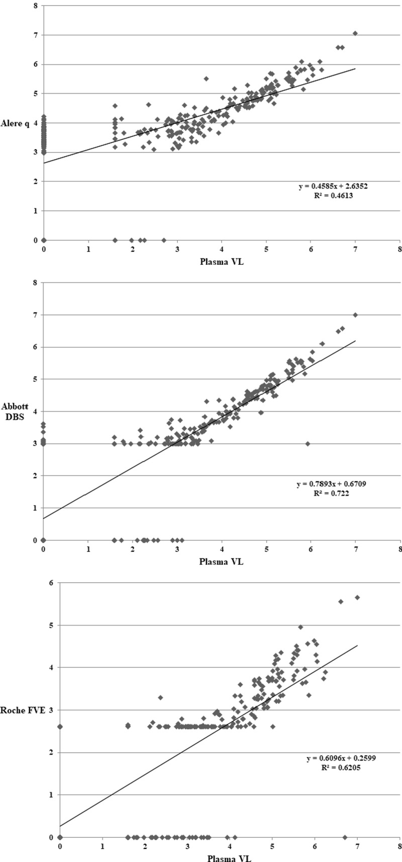
Fig 1

Correlation plots between plasma VL and 1a) Alere qR2 = 0.46. 1b) Abbott DBSR2 = 0.72. 2.9log10 has been used on plot for samples <1000 copies/ml (lower limit of quantification). 1c) Roche FVE—R2 = 0.62. 2.6log10 has been used on plot for Roche FVE samples <400 copies/ml (lower limit of quantification). Results below the detectable limit are plotted as 0 for all the plots.

Fig 2. Bland Altman plots between plasma VL and Alere q/DBS assays in log10 copies/ml.
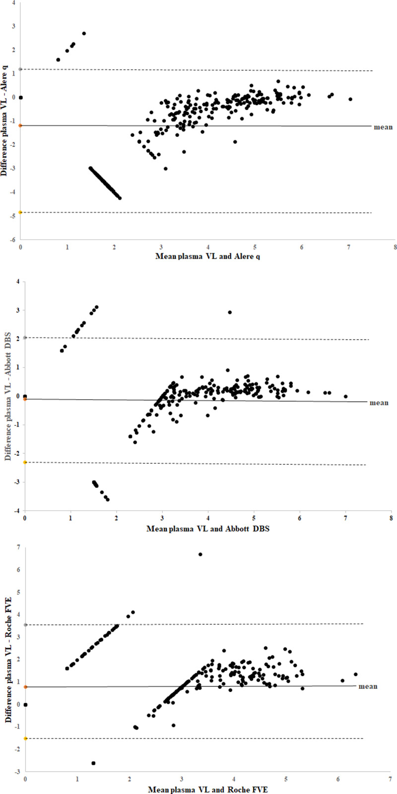
Fig 2

Overall mean log10 copies/ml difference between plasma VL and 2a) Alere q = -1.18 2b) Abbott DBS = -0.11 2c) Roche FVE = 0.78. Viral load that was below the detectable limit was plotted as 0 on graph, <1000 copies/ml for Abbott DBS was plotted as 2.9 log10, and <400 copies/ml for Roche FVE was plotted as 2.6 log10. The horizontal lines represent the mean (solid line) and the ± 1.96 standard deviations (dashed lines).

Table 2. Percentage of correct classification of VF by Alere q and DBS methods according to plasma VL categories.

Plasma VL categories Target not detected <40–1000 copies/ml Specificity 1000–10,000 copies/ml >10,000 copies/ml Sensitivity
Alere q 22% (21/94) 14% (7/51) 19% (28/145) (95% CI 13–27%) 100% (52/52) 100% (101/101) 100% (153/153) (95% CI 97–100%)
Abbott DBS 89% (84/94) 75% (38/51) 84% (122/145) (95% CI 77–89%) 75% (39/52) 99% (97/98) 91% (136/150) (95% CI 85–95%)
Roche FVE 100% (90/90) 98% (49/50) 99% (139/140) (95% CI 95–100%) 0% (0/50) 80% (76/95) 52% (76/145) (95% CI 44–61%)

Definition of correct classification of VF by Alere q and DBS methods according to plasma VL categories using the VF threshold of 1000 copies/ml: when plasma VL < 1000 copies/ml (both target not detected and <40-1000copies/ml categories) then Alere q/DBS VL <1000 copies/ml (specificity in identifying absence of VF), when plasma VL > = 1000 copies/ml (both 1000–10,000 and >10,000 copies/ml categories) then Alere q/DBS VL > = 1000 copies/ml (sensitivity in identifying VF). Alere q showed 100% sensitivity, however there was reduced specificity in both the target not detected and <40–1000 copies/ml plasma VL categories.(22% and 14% respectively). For Abbott DBS, sensitivity was best above 10,000 copies/ml, however sensitivity was 75% in the 1000–10,000 copies/ml category, and specificity was 75% in the <40–1000 copies/ml category. Roche FVE demonstrated the opposite performance with 0% sensitivity in the 1000–10,000 copies/ml category but had the best specificity. Correct classification was best for all methods at >10,000 copies/ml.

4. Discussion

Our study compared several different approaches adopted by manufacturers in an attempt to provide alternatives to plasma VL testing. These methodologies differ in terms of sample treatment, nucleic acid extraction, amplification targets, detection methods and software cutoff algorithms, and thus the cause of the varied performances is likely multi-factorial.

Our study is one of the preliminary studies evaluating Alere q whole blood VL. Its advantages are POC test, ease of use, minimal training required, small portable instrument, small sample volume, built in controls, results within an hour and excellent sensitivity. These advantages were confirmed in early infant diagnosis field study in South Africa where good performance was shown and use reported as highly acceptable [20].

Both Alere q whole blood VL and Abbott DBS measures cell associated HIV RNA as both extraction methods are RNA selective. The over-quantification was significantly greater with Alere q, probably due to other differences such as assay target, detection methodology, in conjunction with cell associated HIV RNA. However, the inclusion of cell-associated HIV RNA can vary greatly from one clinical sample to the next depending on clinical factors such as duration of viral suppression, CD4 count, haematocrit and other factors such as HIV DNA copy number in cells, as well as presence of spliced and unspliced HIV RNA in cells (low levels of unspliced RNA in patients suppressed on ART) [10, 21].

A study on Alere q using finger-prick capillary whole blood samples in a primary health care centre in Mozambique had similar results to ours albeit with better specificity, sensitivity was 96.83% and specificity was 47.8% [22]. Jani and colleagues suggested that a raised cut-off of 10,000 copies/ml would provide a better predictor of VF for Alere q and our independent ROC analysis confirms this finding [22]. Its low throughput and reduced specificity warrant further investigation to identify its niche use in large ART programmes. In a programmatic setting, individuals with VL’s within this range would either require confirmatory plasma VL testing at a reference laboratory, or have further POC VL’s to monitor the whole blood VL trajectory before a clinical decision can be made. Both of these approaches would result in delay and thus needs to be weighed against the risk of misclassification. The WHO 2016 consolidated guidelines on the use of antiretroviral drugs for treating and preventing HIV infection recommend the threshold of 1000 copies/ml for VF using DBS as well as a sensitivity and specificity >85%, therefore Alere q using whole blood does not fulfil this acceptability criteria.

Although the limit of detection of Alere q using whole blood is 2491 copies/ml, patients with VF have a plasma VL >1000 copies/ml and use of whole blood would include measurement of cell associated HIV RNA therefore elevating the viral load. Our analysis showed 100% sensitivity in detection of VF using Alere q including plasma VL’s between 1000–3000 copies/ml.

Although DBS methodologies compares favourably in overall proportion of correctly identified VF, their lack of sensitivity and the variability between methodologies detected in our data is a concern. Reduced sensitivity of DBS is attributed to smaller volume of blood processed, reduced extraction efficiency and presence of PCR inhibitors [21, 23]. The DBS protocols had the tendency to lower VL quantification when the plasma VL was 1000–10,000 copies/ml, as demonstrated by the reduced sensitivity in this category, as compared to a good sensitivity seen at >10,000 copies/ml plasma VL. The reduced sensitivity of Roche FVE, in particular, may be related to the inefficiency of the free virus elution and the lower input volume of DBS. Increasing the software correction factor as shown by Pollack et al. or using two or more spots may help to improve the sensitivity [24]. Pooled estimates of sensitivity and specificity based on a technical evaluation meta-analysis for Roche FVE was 95% sensitivity and 94% specificity while for Abbott DBS one spot was 88% sensitivity and 99% specificity [25]. Our study found a lower sensitivity (52%) with use of Roche FVE but specificity was comparable [19, 26, 27]. An evaluation of Roche FVE by Taieb et al. reported 55% sensitivity and 100% specificity, similar to our findings [28]. The sensitivity and specificity of Abbott DBS in our study is comparable to other studies evaluating the one spot or two spot protocol [14, 2932].

A study evaluating DBS for VF by Sawadogo and colleagues concluded that the recommendations should not treat different assays and extraction methods as homogeneous and explicit recommendations for each should be available [33]. Inzaule and colleagues argue that the DBS cut-off of 1000 copies/ml is too stringent for VF based on their analysis of 79,566 VL results from patients receiving ART in western Kenya and will result in unnecessary repeat tests and regimen switches [34]. These studies, in conjunction with our finding of great variability between methodologies, suggest either some form of harmonization of the whole blood and DBS VL methodologies and interpretation is required, or other alternatives should be explored. In particular, the use of field plasma separation device and plasma separation cards could be a preferred method for performing VL in remote settings [25, 35].

Our study has the following limitations. Most significantly, DBS preparation and POC testing was performed in the laboratory in a controlled environment by skilled laboratory workers and not by the intended POC health care workers, as a result venous whole blood samples and not capillary blood was used for the POC and DBS assays in this evaluation. However, study that is conducted in the field would introduce even more variance and likely amplify the discrepancies between various technologies that we have found. The cross-sectional nature of the study precludes any follow-up analysis and with limited clinical and laboratory parameters, it is difficult gain insight into the exact cause of the inter-assay VL variance. Appropriately powered longitudinal studies would be required to address this issue.

5. Conclusion

Although various factors lead to the variability in whole blood and DBS VL quantification, the crucial advantage advocating their use is increasing access to VL. The strength of Alere q is its use as a POC test using whole blood and the excellent sensitivity of 100%, however this was offset by the specificity of only 19%. The over estimation of VL would lead to unnecessary re-testing and switching regimens. Optimisation of Alere q whole blood VL is required to facilitate the implementation of a cut-off with optimal sensitivity and specificity for VF. Both the Abbott DBS and Roche FVE protocols showed good specificity, however sensitivity was reduced when the plasma VL was 1000–10,000 copies/ml. This could result in delays in detecting VF and accumulation of drug resistance. Field evaluation in settings that have adopted these DBS protocols are necessary to gauge its real world performance.

Supporting information

S1 File. Receiver operating characteristic (ROC) curve analysis.

(DOCX)

Acknowledgments

The authors would like to thank Dr Sergio Carmona for the inter-laboratory collaboration and the staff at National Health Laboratory Service in Groote Schuur Hospital and Charlotte Maxeke Johannesburg Academic Hospital who assisted with the laboratory work.

Poster presentation of initial study findings at 8th IAS Conference on HIV Pathogenesis, Treatment & Prevention, 19–22 July 2015, Vancouver, Canada

Data Availability

All relevant data are within the manuscript and its Supporting Information files.

Funding Statement

The author(s) received no specific funding for this work.

References

  • 1.UNAIDS. 90–90–90—An Ambitious Treatment Target To Help End The AIDS Epidemic 2014 [Available from: http://www.unaids.org/en/resources/documents/2014/90-90-90.
  • 2.UNAIDS Data 2019 [Available from: https://www.unaids.org/en/resources/documents/2019/2019-UNAIDS-data.
  • 3.Lecher S, Ellenberger D, Kim AA, Fonjungo PN, Agolory S, Yolande BM, et al. Scale-up of HIV viral load monitoring—seven sub-Saharan African countries MMWR Morb Mortal Wkly Rep. 2015. November 27;64(46):1287–90 2015 [Available from: 10.15585/mmwr.mm6446a3 [DOI] [PubMed] [Google Scholar]
  • 4.World Health Organisation Consolidated guidelines on the use of antiretroviral drugs for treating and preventing HIV infection Recommendations for a public health approach—Second edition 2016 [Available from: http://www.who.int/hiv/pub/arv/arv-2016/en/. [PubMed]
  • 5.Keiser O, Chi BH, Gsponer T, Boulle A, Orrell C, Phiri S, et al. Outcomes of antiretroviral treatment in programmes with and without routine viral load monitoring in Southern Africa. AIDS (London, England). 2011;25(14):1761. [DOI] [PMC free article] [PubMed] [Google Scholar]
  • 6.Stevens WS, Scott LM, Crowe SM. Quantifying HIV for monitoring antiretroviral therapy in resource-poor settings. Journal of Infectious Diseases. 2010;201(Supplement_1):S16–S26. [DOI] [PubMed] [Google Scholar]
  • 7.World Health Organization Technical and operational considerations for implementing HIV viral load testing 2014 [Available from: http://www.who.int/hiv/pub/arv/viral-load-testing-technical-update/en/.
  • 8.Stevens WS, Marshall TM. Challenges in implenting HIV load testing in South Africa. Journal of Infectious Diseases. 2010;201(Supplement_1):S78–S84. [DOI] [PubMed] [Google Scholar]
  • 9.Peter T, Ellenberger D, Kim AA, Boeras D, Messele T, Roberts T, et al. Early antiretroviral therapy initiation: access and equity of viral load testing for HIV treatment monitoring. The Lancet Infectious Diseases. 2017;17(1):e26–e9. [DOI] [PMC free article] [PubMed] [Google Scholar]
  • 10.UNITAID HIV/AIDS Diagnostics Technology Landscape 5th edition 2015 [Available from: https://aidsfree.usaid.gov/sites/default/files/hivaids_diag_tech.pdf.
  • 11.Shafiee H, Wang S, Inci F, Toy M, Henrich TJ, Kuritzkes DR, et al. Emerging technologies for point-of-care management of HIV infection. Annual review of medicine. 2015;66:387–405. [DOI] [PubMed] [Google Scholar]
  • 12.Peeling R, Mabey D. Point-of-care tests for diagnosing infections in the developing world. Clinical microbiology and infection. 2010;16(8):1062–9. [DOI] [PubMed] [Google Scholar]
  • 13.Abbott Molecular: Abbott RealTime HIV-1 package insert 2007 [Available from: http://www.fda.gov/downloads/BiologicsBloodVaccines/BloodBloodProducts/ApprovedProducts/PremarketApprovalsPMAs/UCM091193.pdf.
  • 14.Tang N, Pahalawatta V, Frank A, Bagley Z, Viana R, Lampinen J, et al. HIV-1 viral load measurement in venous blood and fingerprick blood using Abbott RealTime HIV-1 DBS assay. Journal of Clinical Virology. 2017;92:56–61. [DOI] [PubMed] [Google Scholar]
  • 15.Shihai Huang SC BE, John Salituro, Chadwick Dunn, Livhuwani Nxumalo, Jeffrey Wuitschick, Jens Dhein. Improved Performance of the New Prototype Automated Abbott RealTime HIV-1 DBS Viral Load Assay: Potential Use in Expanded Viral Load Testing in Resource Limited Setting. ASLM2014.
  • 16.WHO prequalification of diagnostics programme public report Product: Abbott RealTime HIV-1 (m2000sp) April 2018, version 8.0 [Internet]. Available from: https://www.who.int/diagnostics_laboratory/evaluations/pq-list/hiv-vrl/180423_amended_final_pqpr_0145_027_00_v11.pdf?ua=1.
  • 17.Ullrich T, Ermantraut E, Schulz T, Steinmetzer K. Competitive Reporter Monitored Amplification (CMA)-Quantification of Molecular Targets by Real Time Monitoring of Competitive Reporter Hybridization. PLoS One. 2012;7(4):e35438. [DOI] [PMC free article] [PubMed] [Google Scholar]
  • 18.Alere q HIV-1/2 User guide/package insert [Available from: https://www.alere.com/en/home/product-details/alere-q-analyser.html.
  • 19.Wu X, Crask M, Ramirez H, Landas T, Do TD, Honisch C, et al. A simple method to elute cell-free HIV from dried blood spots improves their usefulness for monitoring therapy. Journal of Clinical Virology. 2015;65:38–40. [DOI] [PubMed] [Google Scholar]
  • 20.Dunning L, Kroon M, Hsiao N-y, Myer L. Field evaluation of HIV point-of-care testing for early infant diagnosis in Cape Town, South Africa. PloS one. 2017;12(12):e0189226. [DOI] [PMC free article] [PubMed] [Google Scholar]
  • 21.Parkin NT. Measurement of HIV-1 viral load for drug resistance surveillance using dried blood spots: literature review and modeling of contribution of DNA and RNA. AIDS reviews. 2014;16(3):160–71. [PubMed] [Google Scholar]
  • 22.Jani IV, Meggi B, Vubil A, Sitoe NE, Bhatt N, Tobaiwa O, et al. Evaluation of the whole-blood Alere Q NAT point-of-care RNA assay for HIV-1 viral load monitoring in a primary health care setting in Mozambique. Journal of clinical microbiology. 2016;54(8):2104–8. [DOI] [PMC free article] [PubMed] [Google Scholar]
  • 23.Smit PW, Sollis KA, Fiscus S, Ford N, Vitoria M, Essajee S, et al. Systematic review of the use of dried blood spots for monitoring HIV viral load and for early infant diagnosis. PloS one. 2014;9(3):e86461. [DOI] [PMC free article] [PubMed] [Google Scholar]
  • 24.Pollack TM, Duong HT, Truong PT, Pham TT, Do CD, Colby D. Sensitivity and specificity of two dried blood spot methods for HIV-1 viral load monitoring among patients in Hanoi, Vietnam. PloS one. 2018;13(1). [DOI] [PMC free article] [PubMed] [Google Scholar]
  • 25.World Health Organisation HIV molecular diagnostics toolkit to improve access to viral load testing and infant diagnosis 2019 [Available from: https://www.who.int/hiv/pub/vct/hiv-molecular-diagnostic/en/.
  • 26.Makadzange AT, Boyd FK, Chimukangara B, Masimirembwa C, Katzenstein D, Ndhlovu CE. A simple phosphate-buffered-saline-based extraction method improves specificity of HIV viral load monitoring using dried blood spots. Journal of clinical microbiology. 2017;55(7):2172–9. [DOI] [PMC free article] [PubMed] [Google Scholar]
  • 27.Pannus P, Claus M, Gonzalez MMP, Ford N, Fransen K. Sensitivity and specificity of dried blood spots for HIV-1 viral load quantification: A laboratory assessment of 3 commercial assays. Medicine. 2016;95(48). [DOI] [PMC free article] [PubMed] [Google Scholar]
  • 28.Taieb F, Tram TH, Ho HT, Pham VA, Nguyen HL, Pham BH, et al., editors. Evaluation of two techniques for viral load monitoring using dried blood spot in routine practice in Vietnam (French National Agency for AIDS and Hepatitis Research 12338). Open forum infectious diseases; 2016: Oxford University Press. [DOI] [PMC free article] [PubMed]
  • 29.Erba F, Brambilla D, Ceffa S, Ciccacci F, Luhanga R, Sidumo Z, et al. Measurement of viral load by the automated Abbott real-time HIV-1 assay using dried blood spots collected and processed in Malawi and Mozambique. South African Medical Journal. 2015;105(12). [DOI] [PubMed] [Google Scholar]
  • 30.Rutstein SE, Kamwendo D, Lugali L, Thengolose I, Tegha G, Fiscus SA, et al. Measures of viral load using Abbott RealTime HIV-1 Assay on venous and fingerstick dried blood spots from provider-collected specimens in Malawian District Hospitals. Journal of Clinical Virology. 2014;60(4):392–8. [DOI] [PMC free article] [PubMed] [Google Scholar]
  • 31.Zeh C, Ndiege K, Inzaule S, Achieng R, Williamson J, Chang JC-W, et al. Evaluation of the performance of Abbott m2000 and Roche COBAS Ampliprep/COBAS Taqman assays for HIV-1 viral load determination using dried blood spots and dried plasma spots in Kenya. PloS one. 2017;12(6):e0179316. [DOI] [PMC free article] [PubMed] [Google Scholar]
  • 32.Taieb F, Hong TT, Ho HT, Thanh BN, Phuong TP, Ta DV, et al. First field evaluation of the optimized CE marked Abbott protocol for HIV RNA testing on dried blood spot in a routine clinical setting in Vietnam. PloS one. 2018;13(2). [DOI] [PMC free article] [PubMed] [Google Scholar]
  • 33.Sawadogo S, Shiningavamwe A, Chang J, Maher AD, Zhang G, Yang C, et al. Limited utility of dried-blood- and plasma spot-based screening for antiretroviral treatment failure with Cobas Ampliprep/TaqMan HIV-1 version 2.0. J Clin Microbiol. 2014;52(11):3878–83. [DOI] [PMC free article] [PubMed] [Google Scholar]
  • 34.Inzaule SC, Hamers RL, Zeh CE, de Wit TFR. Stringent HIV viral load threshold for virological failure using dried blood spots: is the perfect the enemy of the good? JAIDS Journal of Acquired Immune Deficiency Syndromes. 2016;71(1):e30–e3. [DOI] [PubMed] [Google Scholar]
  • 35.Carmona S, Seiverth B, Magubane D, Hans L, Hoppler M. Separation of Plasma from Whole Blood by Use of the cobas Plasma Separation Card: a Compelling Alternative to Dried Blood Spots for Quantification of HIV-1 Viral Load. Journal of clinical microbiology. 2019;57(4):e01336–18. [DOI] [PMC free article] [PubMed] [Google Scholar]

Decision Letter 0

Julie AE Nelson

18 Dec 2019

PONE-D-19-29100

Comparison of Alere q whole blood viral load with DBS and plasma viral load in the classification of HIV virological failure

PLOS ONE

Dear Dr Khan,

Thank you for submitting your manuscript to PLOS ONE. After careful consideration, we feel that it has merit but does not fully meet PLOS ONE’s publication criteria as it currently stands. Therefore, we invite you to submit a revised version of the manuscript that addresses the points raised during the review process.

Two reviewers have raised serious issues with the manuscript as written, and each of their concerns must be addressed. Careful attention should be paid to use of "specificity" and "sensitivity" in the correct context, and provide sufficient detail of how the samples were created and how they were tested.

We would appreciate receiving your revised manuscript by Feb 01 2020 11:59PM. When you are ready to submit your revision, log on to https://www.editorialmanager.com/pone/ and select the 'Submissions Needing Revision' folder to locate your manuscript file.

If you would like to make changes to your financial disclosure, please include your updated statement in your cover letter.

To enhance the reproducibility of your results, we recommend that if applicable you deposit your laboratory protocols in protocols.io, where a protocol can be assigned its own identifier (DOI) such that it can be cited independently in the future. For instructions see: http://journals.plos.org/plosone/s/submission-guidelines#loc-laboratory-protocols

Please include the following items when submitting your revised manuscript:

  • A rebuttal letter that responds to each point raised by the academic editor and reviewer(s). This letter should be uploaded as separate file and labeled 'Response to Reviewers'.

  • A marked-up copy of your manuscript that highlights changes made to the original version. This file should be uploaded as separate file and labeled 'Revised Manuscript with Track Changes'.

  • An unmarked version of your revised paper without tracked changes. This file should be uploaded as separate file and labeled 'Manuscript'.

Please note while forming your response, if your article is accepted, you may have the opportunity to make the peer review history publicly available. The record will include editor decision letters (with reviews) and your responses to reviewer comments. If eligible, we will contact you to opt in or out.

We look forward to receiving your revised manuscript.

Kind regards,

Julie AE Nelson, PhD

Academic Editor

PLOS ONE

Journal Requirements:

1.

When submitting your revision, we need you to address these additional requirements.

Please ensure that your manuscript meets PLOS ONE's style requirements, including those for file naming. The PLOS ONE style templates can be found at

http://www.journals.plos.org/plosone/s/file?id=wjVg/PLOSOne_formatting_sample_main_body.pdf and http://www.journals.plos.org/plosone/s/file?id=ba62/PLOSOne_formatting_sample_title_authors_affiliations.pdf

2. In ethics statement in the manuscript and in the online submission form, please provide additional information about the patient records/samples used in your retrospective study. Specifically, please ensure that you have discussed whether all data/samples were fully anonymized before you accessed them and/or whether the IRB or ethics committee waived the requirement for informed consent. If patients provided informed written consent to have data/samples from their medical records used in research, please include this information.

[Note: HTML markup is below. Please do not edit.]

Reviewers' comments:

Reviewer's Responses to Questions

Comments to the Author

1. Is the manuscript technically sound, and do the data support the conclusions?

The manuscript must describe a technically sound piece of scientific research with data that supports the conclusions. Experiments must have been conducted rigorously, with appropriate controls, replication, and sample sizes. The conclusions must be drawn appropriately based on the data presented.

Reviewer #1: Partly

Reviewer #2: No

**********

2. Has the statistical analysis been performed appropriately and rigorously?

Reviewer #1: No

Reviewer #2: No

**********

3. Have the authors made all data underlying the findings in their manuscript fully available?

The PLOS Data policy requires authors to make all data underlying the findings described in their manuscript fully available without restriction, with rare exception (please refer to the Data Availability Statement in the manuscript PDF file). The data should be provided as part of the manuscript or its supporting information, or deposited to a public repository. For example, in addition to summary statistics, the data points behind means, medians and variance measures should be available. If there are restrictions on publicly sharing data—e.g. participant privacy or use of data from a third party—those must be specified.

Reviewer #1: Yes

Reviewer #2: No

**********

4. Is the manuscript presented in an intelligible fashion and written in standard English?

PLOS ONE does not copyedit accepted manuscripts, so the language in submitted articles must be clear, correct, and unambiguous. Any typographical or grammatical errors should be corrected at revision, so please note any specific errors here.

Reviewer #1: Yes

Reviewer #2: Yes

**********

5. Review Comments to the Author

Please use the space provided to explain your answers to the questions above. You may also include additional comments for the author, including concerns about dual publication, research ethics, or publication ethics. (Please upload your review as an attachment if it exceeds 20,000 characters)

Reviewer #1: The manuscript by Khan and colleagues presents a laboratory evaluation of the new Alere q point of care (POC) machine to measure HIV viral load from whole blood as compared to plasma which is the gold standard; interestingly, they also compare its performances to those of DBS, again as compared to plasma. This kind of evaluation is essential to help scaling-up of HIV viral load monitoring in resource-limited settings which still is scarce in many settings. In that respect, I am favorable to accept this manuscript if the modifications are conducted.

1) In the method section, the authors mention that the samples originate from routine care. The plasma is intended for HIV viral load monitoring, while the whole blood sample was intended for CD4 viral load measurement. Due to the sampling strategy, HIV viral load on plasma needed to be performed first before DBS samples were prepared and before a sample dedicated to Alere q was saved. Some details about the delay between whole blood sampling and DBS preparation, about the condition of storage of the whole blood, and about the condition and duration of storage of DBS would be welcome. Could the authors also mention if the CD4 measurement was performed or not.

2) In table 1, the reportable HIV viral load range of each technique is indicated. It is surprising to see that the range is 2491 copies/mL to 10 million copies/mL for the Alere q POC, and 1000 copies/mL to 10 million copies/mL for the Abbott DBS technique. This is not correct for the Abbott DBS technique. Moreover, this does not make sense given the evaluation they conduct to identify virological failure at the threshold of 1000 copies/mL. It is also surprising to read that each spot is 75µL as the manufacturers recommend 70µL/spot; 70µL is also mentioned in the reference 14 chosen by the authors in table 1, I was unable to identify reference 13. Tang et al. in Journal of clinical virology 2017 is probably a better reference.

3) In the “analysis” subsection of the “methods” section, it should be clearly stated that correlations and Bland-Altman analysis aim at comparing the methodologies from a quantitative perspective. The ROC curve analysis is mentioned in this section but never presented in the result section and briefly mentioned in the discussion; this is however not of prime interest in this study. If the authors insisted to present the results from the ROC curve, it should come after mentioning that the main goal of the evaluation is a qualitative evaluation, and estimates the ability of the different techniques to identify patients in virological failure defined at the threshold of 1000 copies/mL, when this threshold is used for both the technique evaluated and the plasma. This evaluation relies on the estimation of sensitivity and specificity of each technique as compared to plasma. These terms never appear in the method section while they are much more important than the “proportions of correctly classified”; indeed, the WHO guidelines state that DBS techniques should present a sensitivity and specificity >85% in order to be an acceptable option for HIV viral load monitoring, it is clearly not a criteria based simply on the proportion of correctly classified. The “analysis” subsection must be written in a more intelligible way, and the “results” section should clearly state the sensitivities and specificities as this is not done.

4) In the “results” section, nearly all the data from the table 2 are already in the text. Consider removing the table. Figure 1 provides no informative data; consider removing. The section 3.2 alternates between quantitative comparisons and qualitative comparisons. This should be rewritten for better clarity.

5) In the “results” section, the authors mention indifferently specificity or upward misclassification on the one hand, and sensitivity or downward misclassification on the other hand; this makes the reading very complex and confusing. The standard statistical method to compare a test to a gold standard relies on estimation of sensitivity and specificity. The authors should stick to these terms for the sake of comparison. In table 3, it would be very valuable to present not only % but numbers as well. Two columns could also be added to present sensitivity and specificity with their confidence intervals. This section suffers from repetition.

6) The Bland-Altman is supposed to be a representation of the mean of two techniques on the x-axis against the difference between the two techniques on the y-axis. The caption on the x-axis seem to indicate that the Bland-Altman analysis is not correctly conducted. A legend for the x-axis should be reported. Moreover, this analysis requires to represent three horizontal lines: the mean difference and the 95% confidence interval of the difference. None of these appear in the figures. This must be addressed as it is difficult with the current figures to judge the quality of the techniques.

7) In the discussion, the authors mention the DBS protocols had the tendency to lower viral load quantification. I disagree with this statement. It might be very true for the Roche protocol with a sensitivity to detect failure of 76%; this is also obvious on the scatter plot (especially if the authors add the line with equation y=x). This is less true with the Abbott protocol with sensitivity and specificity of 95% and 92%, respectively. The under-estimation of the viral load level with the Roche technique has already been described in the literature, and this should be reported here. The low sensitivity of the Roche DBS technique is particularly worrying as about 25% of those in failure would not be identified as such. The authors state that Alere q has a very good sensitivity, which is indeed 100%. However, the specificity was particularly bad (61%) which should be discussed. It looks like they attempt to discuss it by mentioning that in case of “false virological failure”, the patients would undergo confirmatory plasma VL testing at a laboratory of further POC VL monitoring. But this is not totally clear. The discussion needs clarification and should be put in perspectives with other similar studies as well as with the WHO recommendations.

Minor comments:

Throughout the manuscript, ul is written instead of µL.

IQR should be written in full at the first appearance

ART should be written in full at the first appearance, and not at its second appearance

Mention of “Alere q” is not homogeneous throughout the manuscript

Reviewer #2: The present study aims to address a cotemporary, globally important issue for HIV treatment and care; however, the manuscript in its current format needs significant improvement in order to reach publication standard. Following are some comments hopefully can help

Introduction

- The argument that “quality of evidence is low due to insufficient published data….” (Lines 45-47 page 3) is not well supported by existing evidence: According to a recent WHO report there were 40 technical evaluations of DBS across 25 countries examining 6 different commercially available VL technologies resulted in more than 10,000 paired DBS-plasma points. It suggested “sufficient evidence has been generated on the performance of DBS specimens for viral load testing to support the initiation of scale-up” (HIV molecular diagnostics toolkit to improve access to viral load testing and infant diagnostics, July 2019). The rationale for the present study need to be revised and strengthen

- Detailed descriptions of the POC VL test under investigation are needed. The Alere q POC viral load had been on the market (?) for a while (previous study published in 2016 – reference 19) why there is still “prototype” whole blood protocol? More details on operational requirements of the technologies are also needed

- Clear statement of the study objectives: primary and secondary objectives are recommended: which was the index test, which was the reference, and comparators

Methods

- Lines 60-61: … Lower than detection limit (LDL) of which technology? There were 3 different tech/protocols mentioned. Please explain why these specific technologies/protocol were selected? Was Sample size calculation performed?

- As this is a diagnostic accuracy study (as it appears in the introduction of the manuscript) , I’m not quite sure why was it needed/authors wanted) to collect data to “determine potential predictors of VL method discrepancies” ??? How did it come even before knowing there were “discrepancies” across measures in this study? As paired samples were used/tested

- VL threshold of 1000 copies/ml was used so detailed analysis for diagnostic accuracy of this cut-off needed to be described (sensitivity, specificity…). Same as for Bland-Altman analysis for quantitative measures

Results

- Lines 98-100 page 5 CD4 count, ART regimen/history not directly relevant/necessary for the main purpose of this study

- Lines 105-118 page 6: presentation of the results is not clear and somewhat repetitive. Suggest to separate out caparisons for primary and secondary objectives Alere q vs. plasma and Alere q vs DBS

- Lines 121-133 page 7 repeated results (with different expressions). Lines 146-154 page 8 not necessary/relevant

Discussion

- Lines 159-161 “the tendency of Alere q… most likely due to cell associated HIV RNA…” this is also true for DBS (which is also dried whole blood sample). Should/can the Alere q run on fresh plasma sample processing at site using mini centrifuge?

- Discussions of clinical factors influencing the variability in VL measures across technologies are not necessary. Potential reasons for reduced accuracy of Roche DBS/FVE has been extensively discussed in literature

- The recommendation of increasing correction factor or using more spots are too general and prompt the question of why this challenge has not been addressed even though is has been identified previously (low specificity)?

- The “excellent sensitivity” note (of Alere q whole blood) is somewhat misleading without an equal highlighted note/discussion of “low specificity” of <50%, which is consistent with previous research and suggested that the technology has not been improved over last few years??? This has an important implication for field implementation as it indicates very high/unacceptable over estimation of HIV VL leading to unnecessary retesting/confirmatory testing of treatment failure and switching regimen

- Lines 199-200 please check the references

Conclusion

- Not clear on key messages. Again highlighting the “excellent sensitivity” without noting low specificity is misleading and based on results of this study the use of “prototype” whole blood protocol Alere q for HIV VL testing should not be “warranted” without further technology improvement and validation

Figures 3a/b BA plots should be revised clearly shown 95%CI of the mean deferences & LoAs

**********

6. PLOS authors have the option to publish the peer review history of their article (what does this mean?). If published, this will include your full peer review and any attached files.

If you choose “no”, your identity will remain anonymous but your review may still be made public.

Do you want your identity to be public for this peer review? For information about this choice, including consent withdrawal, please see our Privacy Policy.

Reviewer #1: No

Reviewer #2: Yes: Minh D Pham

[NOTE: If reviewer comments were submitted as an attachment file, they will be attached to this email and accessible via the submission site. Please log into your account, locate the manuscript record, and check for the action link "View Attachments". If this link does not appear, there are no attachment files to be viewed.]

While revising your submission, please upload your figure files to the Preflight Analysis and Conversion Engine (PACE) digital diagnostic tool, https://pacev2.apexcovantage.com/. PACE helps ensure that figures meet PLOS requirements. To use PACE, you must first register as a user. Registration is free. Then, login and navigate to the UPLOAD tab, where you will find detailed instructions on how to use the tool. If you encounter any issues or have any questions when using PACE, please email us at figures@plos.org. Please note that Supporting Information files do not need this step.

PLoS One. 2020 May 29;15(5):e0232345. doi: 10.1371/journal.pone.0232345.r002

Author response to Decision Letter 0


7 Feb 2020

5. Review Comments to the Author

Please use the space provided to explain your answers to the questions above. You may also include additional comments for the author, including concerns about dual publication, research ethics, or publication ethics. (Please upload your review as an attachment if it exceeds 20,000 characters)

Reviewer #1: The manuscript by Khan and colleagues presents a laboratory evaluation of the new Alere q point of care (POC) machine to measure HIV viral load from whole blood as compared to plasma which is the gold standard; interestingly, they also compare its performances to those of DBS, again as compared to plasma. This kind of evaluation is essential to help scaling-up of HIV viral load monitoring in resource-limited settings which still is scarce in many settings. In that respect, I am favorable to accept this manuscript if the modifications are conducted.

1) In the method section, the authors mention that the samples originate from routine care. The plasma is intended for HIV viral load monitoring, while the whole blood sample was intended for CD4 viral load measurement. Due to the sampling strategy, HIV viral load on plasma needed to be performed first before DBS samples were prepared and before a sample dedicated to Alere q was saved. Some details about the delay between whole blood sampling and DBS preparation, about the condition of storage of the whole blood, and about the condition and duration of storage of DBS would be welcome. Could the authors also mention if the CD4 measurement was performed or not.

Answer: To ensure most of the VL analytical spectrum was covered, our convenient sampling strategy randomly selected paired CD4/VL EDTA samples where at least 50 samples of plasma VL results were from each of four categories: <40 copies/ml, 40–1000 copies/ml, 1000 – 10,000 copies/ml and > 10,000 copies/ml. Once a plasma VL and CD4 testing were complete, the remaining paired CD4 sample was used for whole blood Alere q test and DBS within 72 hours of sample receipt to prevent sample degradation. DBS was prepared by applying 75µL of whole blood to each spot on a Whatman 903 filter-paper card, dried and packed into plastic bags with desiccant sachets and stored at room temperature in boxes away from direct sunlight or heat. DBS VLs were tested in batches within 1-2 months of preparation. Whole blood specimen that was used for the DBS and Alere q testing was stored at room temperature after CD4 testing.

2) In table 1, the reportable HIV viral load range of each technique is indicated. It is surprising to see that the range is 2491 copies/mL to 10 million copies/mL for the Alere q POC, and 1000 copies/mL to 10 million copies/mL for the Abbott DBS technique. This is not correct for the Abbott DBS technique. Moreover, this does not make sense given the evaluation they conduct to identify virological failure at the threshold of 1000 copies/mL. It is also surprising to read that each spot is 75µL as the manufacturers recommend 70µL/spot; 70µL is also mentioned in the reference 14 chosen by the authors in table 1, I was unable to identify reference 13. Tang et al. in Journal of clinical virology 2017 is probably a better reference.

Answer: Alere q requires 25µL whole blood, due to the smaller sample volume as compared to DBS and plasma the limit of detection is therefore lower. According to the package insert and the WHO Prequalification of In Vitro Diagnostics public report on Alere™ q HIV-1/2 Detect, which was designed as a qualitative nucleic acid amplification test for the detection of Human Immunodeficiency Virus (HIV) type 1 groups M/N and O and type 2, the limit of detection for HIV-1 group M is 2491 copies/ml. (Available from: https://www.who.int/diagnostics_laboratory/evaluations/pq-list/hiv-vrl/160613PQPublicReport_0226-032-00AlereHIVDetect_v2.pdf.). Patients with virological failure have a plasma VL >1000 copies/ml. Use of whole blood would include measurement of cell associated HIV RNA therefore elevating the VL. Our analysis showed 100% sensitivity in detection of virological failure using Alere q including plasma VL’s between 1000-3000 copies/ml.

In reference 17, “Smit PW, Sollis KA, Fiscus S, Ford N, Vitoria M, Essajee S, et al. Systematic review of the use of dried blood spots for monitoring HIV viral load and for early infant diagnosis. PloS one. 2014;9(3):e86461” – thirteen studies evaluated DBS HIV viral load and the volume of blood per DBS spot ranged from 50–100µL. Most manufacturers do recommend 70µL/spot. To optimize sensitivity and facilitate full coverage of spot, 75µL of whole blood was applied to each spot.

The Abbott DBS protocol that was evaluated is a revised prototype protocol that uses one DBS spot (reportable range 1000-10 million copies/ml) as compared to two DBS spots used by the Abbott RealTime HIV-1 original ‘open mode’ protocol (reportable range 550-10 million copies/ml). Reference 13, “Shihai Huang SC, Brian Erickson, John Salituro, Chadwick Dunn, Livhuwani Nxumalo, Jeffrey Wuitschick, Jens Dhein. Improved Performance of the New Prototype Automated Abbott RealTime HIV-1 DBS Viral Load Assay: Potential Use in Expanded Viral Load Testing in Resource Limited Setting. ASLM2014” was presented as an oral and poster presentation at the African Society for Laboratory Medicine in 2014(From: https://www.eiseverywhere.com/docs/4466/ScientificProgrammeBook). There are several workflow improvements with the revised prototype protocol as compared to the original ‘open mode’ protocol which includes use of one DBS spot, number of sample tubes reduced from two to one, automated DBS eluate transfer and up to 93 DBS specimens per run therefore being more cost effective. We had also evaluated the original ‘open mode’ protocol however chose not to include the data as the results from both protocols were similar.

Have added the Tang et al. in Journal of clinical virology 2017, as it also evaluates the revised Abbott DBS protocol and is more recent.

3) In the “analysis” subsection of the “methods” section, it should be clearly stated that correlations and Bland-Altman analysis aim at comparing the methodologies from a quantitative perspective. The ROC curve analysis is mentioned in this section but never presented in the result section and briefly mentioned in the discussion; this is however not of prime interest in this study. If the authors insisted to present the results from the ROC curve, it should come after mentioning that the main goal of the evaluation is a qualitative evaluation, and estimates the ability of the different techniques to identify patients in virological failure defined at the threshold of 1000 copies/mL, when this threshold is used for both the technique evaluated and the plasma. This evaluation relies on the estimation of sensitivity and specificity of each technique as compared to plasma. These terms never appear in the method section while they are much more important than the “proportions of correctly classified”; indeed, the WHO guidelines state that DBS techniques should present a sensitivity and specificity >85% in order to be an acceptable option for HIV viral load monitoring, it is clearly not a criteria based simply on the proportion of correctly classified. The “analysis” subsection must be written in a more intelligible way, and the “results” section should clearly state the sensitivities and specificities as this is not done.

Answer: See revised manuscript for re-written analysis section of methods as per reviewer’s comments.

4) In the “results” section, nearly all the data from the table 2 are already in the text. Consider removing the table. Figure 1 provides no informative data; consider removing. The section 3.2 alternates between quantitative comparisons and qualitative comparisons. This should be rewritten for better clarity.

Answer: Removed table 2 and figure 1 as per reviewer’s recommendation. Re-written section 3.2.

5) In the “results” section, the authors mention indifferently specificity or upward misclassification on the one hand, and sensitivity or downward misclassification on the other hand; this makes the reading very complex and confusing. The standard statistical method to compare a test to a gold standard relies on estimation of sensitivity and specificity. The authors should stick to these terms for the sake of comparison. In table 3, it would be very valuable to present not only % but numbers as well. Two columns could also be added to present sensitivity and specificity with their confidence intervals. This section suffers from repetition.

Answer: To use the terms sensitivity and specificity as recommended and remove upward/downward misclassification. Table amended as per reviewer recommendations.

6) The Bland-Altman is supposed to be a representation of the mean of two techniques on the x-axis against the difference between the two techniques on the y-axis. The caption on the x-axis seem to indicate that the Bland-Altman analysis is not correctly conducted. A legend for the x-axis should be reported. Moreover, this analysis requires to represent three horizontal lines: the mean difference and the 95% confidence interval of the difference. None of these appear in the figures. This must be addressed as it is difficult with the current figures to judge the quality of the techniques.

Answer: See revised Bland-Altman analysis as per reviewer recommendations.

7) In the discussion, the authors mention the DBS protocols had the tendency to lower viral load quantification. I disagree with this statement. It might be very true for the Roche protocol with a sensitivity to detect failure of 76%; this is also obvious on the scatter plot (especially if the authors add the line with equation y=x). This is less true with the Abbott protocol with sensitivity and specificity of 95% and 92%, respectively. The under-estimation of the viral load level with the Roche technique has already been described in the literature, and this should be reported here. The low sensitivity of the Roche DBS technique is particularly worrying as about 25% of those in failure would not be identified as such. The authors state that Alere q has a very good sensitivity, which is indeed 100%. However, the specificity was particularly bad (61%) which should be discussed. It looks like they attempt to discuss it by mentioning that in case of “false virological failure”, the patients would undergo confirmatory plasma VL testing at a laboratory of further POC VL monitoring. But this is not totally clear. The discussion needs clarification and should be put in perspectives with other similar studies as well as with the WHO recommendations.

Answer: Performance of DBS VL is highly variable. The DBS protocols had the tendency to lower viral load quantification when the plasma VL was 1000-10,000 copies/ml, as demonstrated by the reduced sensitivity in this category, as compared to a good sensitivity seen at >10,000 copies/ml plasma VL. Other studies have evaluated the Roche free virus elution (FVE) protocol. An evaluation by Wu et al. had a 90% sensitivity and 100% specificity from paired DBS and plasma samples from 196 patients, with plasma VLs ranging from undetectable to 106 copies/ml. Another study by Makadzange et al. had a 92.7% sensitivity and 100% specificity and found that DBS viral load values were 1 log10 copies/ml lower than those obtained by plasma. Pannus et al. evaluated both Roche FVE and Abbott DBS which had 80.8% and 76% sensitivity and 87.3% and 89.7% specificity respectively. Pooled estimates of sensitivity and specificity based on published data up to June 2015 in the WHO HIV treatment guidelines 2016 for Roche FVE was 85% sensitivity and 94% specificity while for Abbott DBS was 95% sensitivity and 92% specificity. Our study found a lower sensitivity (52%) with use of Roche FVE but specificity was comparable. The sensitivity and specificity of Abbott DBS in our study is comparable to other studies (Erba et al, Rutstein et al, Zeh et al.).

The reduced specificity of Alere q is most likely due to the inclusion of cell associated HIV RNA in its methodology. The inclusion of cell-associated HIV RNA can vary greatly from one clinical sample to the next depending on clinical factors such as duration of viral suppression, CD4 count, haematocrit and other factors such as HIV DNA copy number in cells, as well as presence of spliced and unspliced HIV RNA in cells. A study on Alere q POC VL using finger-prick capillary whole blood samples in a primary health care centre in Mozambique had similar results to ours albeit with better specificity, sensitivity was 96.83% and specificity was 47.8%. Jani and colleagues suggested that a raised cut-off of 10,000 copies/ml would provide a better predictor of VF for Alere q and our independent ROC analysis confirms this finding. However the WHO 2016 consolidated guidelines on the use of antiretroviral drugs for treating and preventing HIV infection recommend the threshold of 1000 copies/ml for virological failure using DBS as well as a sensitivity and specificity >85%.

Minor comments:

Throughout the manuscript, ul is written instead of µL. Answer: Changed to µL

IQR should be written in full at the first appearance Answer: Amended

ART should be written in full at the first appearance, and not at its second appearance Answer: Amended

Mention of “Alere q” is not homogeneous throughout the manuscript Answer: Amended

Reviewer #2: The present study aims to address a cotemporary, globally important issue for HIV treatment and care; however, the manuscript in its current format needs significant improvement in order to reach publication standard. Following are some comments hopefully can help

Introduction

- The argument that “quality of evidence is low due to insufficient published data….” (Lines 45-47 page 3) is not well supported by existing evidence: According to a recent WHO report there were 40 technical evaluations of DBS across 25 countries examining 6 different commercially available VL technologies resulted in more than 10,000 paired DBS-plasma points. It suggested “sufficient evidence has been generated on the performance of DBS specimens for viral load testing to support the initiation of scale-up” (HIV molecular diagnostics toolkit to improve access to viral load testing and infant diagnostics, July 2019). The rationale for the present study need to be revised and strengthen

Answer: The main rationale for this study was to evaluate the performance of a prototype Alere q HIV VL assay using whole blood and compare it against plasma VL and DBS VL in the classification of virological failure, as the need remains to evaluate POC technologies, to add to the existing DBS VL data due to the variability in testing protocols, differences in methodologies affecting sensitivity and specificity, the need for standardisation and the greater need for validated assays with regulatory approval. New POC technologies such as Alere q presents a potential alternative to the current plasma or DBS VL testing in specialised VL facilities. POC testing would reduce the need for expensive laboratory infrastructure and highly skilled laboratory workers, and also could lower the cost of testing. Rapid, reliable and affordable POC VL, with minimal equipment and training requirements, would have a major impact on treatment outcomes by facilitating timely detection of virological failure and swift clinical decision making as well as reduce loss to follow up.

The 2016 WHO consolidated guidelines on the use of antiretroviral drugs for treating and preventing HIV infection have recommended the use of DBS in settings where plasma VL cannot be done due to logistical, infrastructural or operational barriers. Only two commercial assays have received CE-IVD and WHO prequalification regulatory approval for viral load using DBS (Abbott RealTime HIV-1 and bioMérieux NucliSENS EasyQ® HIV-1). The systematic review and meta-analysis on the performance of using DBS for viral load testing which includes the 40 technical evaluations from 25 countries is currently unpublished. The sample size evaluated for Abbott DBS and Roche FVE was only 2704 and 3076 respectively.

- Detailed descriptions of the POC VL test under investigation are needed. The Alere q POC viral load had been on the market (?) for a while (previous study published in 2016 – reference 19) why there is still “prototype” whole blood protocol? More details on operational requirements of the technologies are also needed

Answer: The prototype Alere q HIV-1/2 assay was evaluated in this study as specimens were tested in 2014 and 2015 using the prototype available for evaluation during this time period. The Alere™ q HIV-1/2 Detect has undergone WHO prequalification as a qualitative nucleic acid amplification test for the detection of HIV in plasma and whole blood in 2016. Abbott Diagnostics is now the supplier of Alere, and offer the viral load as the m-PIMA™ HIV-1/2 VL test which is commercially available, and has received CE-IVD marking and WHO prequalification using plasma in 2019. Its advantages are point of care test, ease of use, minimal training required, small portable instrument, small sample volume, built in controls, results within an hour and excellent sensitivity. The whole blood assay however is more advantageous as a point of care test as capillary whole blood can be collected from a finger/heel prick directly for testing. Operational requirements are minimal as compared to laboratory based assays as the cartridge kits can be kept at room temperature therefore no refrigeration is required. It can be used in a non-laboratory environment and can be operated on external power, with an internal rechargeable battery protecting it against power fluctuations. It does not require any user related instrument maintenance.

- Clear statement of the study objectives: primary and secondary objectives are recommended: which was the index test, which was the reference, and comparators

Answer: Study objective

To evaluate the performance of a prototype Alere q whole blood VL POC assay in the classification of virological failure using plasma VL as the gold standard.

To evaluate the performance of locally available commercial VL assays (Abbott and Roche) using DBS protocols in the classification of virological failure using plasma VL as the gold standard.

Methods

- Lines 60-61: … Lower than detection limit (LDL) of which technology? There were 3 different tech/protocols mentioned. Please explain why these specific technologies/protocol were selected? Was Sample size calculation performed?

Answer: Plasma VL was used as the gold standard. Plasma specimens routinely tested by the Abbott RealTime HIV-1 assay were selected. To ensure most of the VL analytical spectrum was covered, our convenient sampling strategy randomly selected plasma VL results to include at least 50 specimens from each of four categories: <40 copies/ml, 40–1000 copies/ml, 1000 – 10,000 copies/ml and > 10,000 copies/ml.

The prototype Alere q whole blood VL point of care assay was available for evaluation as an attractive means to increase access to VL testing. The DBS protocols were selected as Abbott and Roche plasma viral load platforms are locally available in our setting.

To ensure most of the VL analytical spectrum was covered, our convenient sampling strategy randomly selected plasma VL results to include at least 50 specimens from each of four categories listed above. Testing was then done based on the number of Alere q whole blood VL cartridges available, to include a total of 299 with results.

- As this is a diagnostic accuracy study (as it appears in the introduction of the manuscript) , I’m not quite sure why was it needed/authors wanted) to collect data to “determine potential predictors of VL method discrepancies” ??? How did it come even before knowing there were “discrepancies” across measures in this study? As paired samples were used/tested

Answer: It was anticipated from previous studies on DBS VL and WHO guidelines that discrepancies would occur as plasma measures cell free virus only while whole blood/DBS also includes cell associated virus. The inclusion of cell-associated HIV RNA can vary greatly from one clinical sample to the next depending on clinical factors such as duration of viral suppression, CD4 count, haematocrit and other factors such as HIV DNA copy number in cells, as well as presence of spliced and unspliced HIV RNA in cells (low levels of unspliced RNA in patients suppressed on ART). The volume of blood per DBS spot can range 50-100µL therefore can lead to reduced sensitivity as compared to plasma VL that tests 200-600µL. These factors were anticipated to impact on sensitivity and specificity.

- VL threshold of 1000 copies/ml was used so detailed analysis for diagnostic accuracy of this cut-off needed to be described (sensitivity, specificity…). Same as for Bland-Altman analysis for quantitative measures

Answer: See revised manuscript for sensitivity and specificity. See revised Bland-Altman analysis as per reviewer recommendations.

Results

- Lines 98-100 page 5 CD4 count, ART regimen/history not directly relevant/necessary for the main purpose of this study

Answer: Removed CD4 count and ART history.

- Lines 105-118 page 6: presentation of the results is not clear and somewhat repetitive. Suggest to separate out caparisons for primary and secondary objectives Alere q vs. plasma and Alere q vs DBS

Answer: See revised manuscript for amended results.

- Lines 121-133 page 7 repeated results (with different expressions). Lines 146-154 page 8 not necessary/relevant

Answer: Figure 1 removed. Table amended as per reviewer 1 recommendations. Can remove lines 146-154 page 8.

Discussion

- Lines 159-161 “the tendency of Alere q… most likely due to cell associated HIV RNA…” this is also true for DBS (which is also dried whole blood sample). Should/can the Alere q run on fresh plasma sample processing at site using mini centrifuge?

Answer: Yes, both Alere q whole blood assay and Abbott DBS protocols measures cell associated HIV RNA, both extraction methods are RNA selective. The over-quantification was significantly greater with Alere q, probably due to other differences such as assay target, detection methodology, in conjunction with cell associated HIV RNA. The Roche FVE measures only extracellular HIV RNA similar to plasma therefore it demonstrated the best specificity.

Abbott Diagnostics is now the supplier of Alere, and offer the viral load as the m-PIMA™ HIV-1/2 VL test which is commercially available, and has received CE-IVD marking and WHO prequalification using plasma in 2019. It requires the collection of an EDTA whole blood sample which requires centrifugation so that 50μL of plasma can be used for testing.

- Discussions of clinical factors influencing the variability in VL measures across technologies are not necessary. Potential reasons for reduced accuracy of Roche DBS/FVE has been extensively discussed in literature

Answer: Removed section 3.3.

- The recommendation of increasing correction factor or using more spots are too general and prompt the question of why this challenge has not been addressed even though is has been identified previously (low specificity)?

Answer: Pooled estimates of sensitivity and specificity based on published data up to June 2015 in the WHO HIV treatment guidelines 2016 for Roche FVE was 85% sensitivity and 94% specificity. Our study found a lower sensitivity (52%) with use of Roche FVE but specificity was comparable. The results from the technical evaluation meta-analysis in the WHO HIV molecular diagnostics toolkit to improve access to viral load testing and infant diagnostics 2019, showed a sensitivity of 95% and specificity of 94% for Roche FVE. This has shown a significant improvement since previous estimates. The meta-analysis is unpublished and there is no indication of a change in testing methodology or processing.

- The “excellent sensitivity” note (of Alere q whole blood) is somewhat misleading without an equal highlighted note/discussion of “low specificity” of <50%, which is consistent with previous research and suggested that the technology has not been improved over last few years??? This has an important implication for field implementation as it indicates very high/unacceptable over estimation of HIV VL leading to unnecessary retesting/confirmatory testing of treatment failure and switching regimen

Answer: Have amended discussion - Alere q whole blood assay had an excellent sensitivity of 100%, however this was offset by the specificity of only 19%. The over estimation of HIV VL would lead to unnecessary retesting/confirmatory testing of treatment failure and switching regimens. The WHO 2016 consolidated guidelines on the use of antiretroviral drugs for treating and preventing HIV infection recommend a sensitivity and specificity >85% using DBS for virological failure.

- Lines 199-200 please check the references

Answer: Checked reference 22 “Carmona S, Seiverth B, Magubane D, Hans L, Hoppler M. Separation of Plasma from Whole Blood by Use of the cobas Plasma Separation Card: a Compelling Alternative to Dried Blood Spots for Quantification of HIV-1 Viral Load. Journal of clinical microbiology. 2019;57(4):e01336-18” – evaluation of plasma separation card therefore kept.

Removed reference 23 “Zeh C, Ndiege K, Inzaule S, Achieng R, Williamson J, Chang JC-W, et al. Evaluation of the performance of Abbott m2000 and Roche COBAS Ampliprep/COBAS Taqman assays for HIV-1 viral load determination using dried blood spots and dried plasma spots in Kenya. PloS one. 2017;12(6):e0179316” removed as plasma separation device/card not used to prepare dried plasma spot. Reference changed to WHO HIV molecular diagnostics toolkit to improve access to viral load testing and infant diagnostics July 2019 which includes use of plasma separation device/card.

Conclusion

- Not clear on key messages. Again highlighting the “excellent sensitivity” without noting low specificity is misleading and based on results of this study the use of “prototype” whole blood protocol Alere q for HIV VL testing should not be “warranted” without further technology improvement and validation

Answer: See revised manuscript for revised conclusion based on recommendations.

Figures 3a/b BA plots should be revised clearly shown 95%CI of the mean deferences & LoAs

Answer: See revised Bland-Altman analysis as per reviewer recommendations.

Attachment

Submitted filename: Response to Reviewers PONE final.docx

Decision Letter 1

Julie AE Nelson

25 Feb 2020

PONE-D-19-29100R1

Comparison of Alere q whole blood viral load with DBS and plasma viral load in the classification of HIV virological failure

PLOS ONE

Dear Dr Khan,

Thank you for submitting your manuscript to PLOS ONE. After careful consideration, we feel that it has merit but does not fully meet PLOS ONE’s publication criteria as it currently stands. Therefore, we invite you to submit a revised version of the manuscript that addresses the points raised during the review process.

Remaining issues to address include those of Reviewer 1 below. In particular, the study needs to include data from after 2015. While it is true that the data presented are from the earlier time, more studies have been published and the pooled results must be addressed to make this manuscript relevant to 2020. 

The authors must also address the following issues. The samples included in the testing were all from HIV-infected individuals, although some were not detectable by the Abbott gold standard assay. The Abbott assay has both <40cp/mL detected and <40cp/mL not detected as result options, so it must be addressed if these two categories were combined as not detected or if they were split, putting the detected ones with the 40-1000cp/mL group. Overall, there must be a better description of how the data were compared. There is a description of the VF level, but it is unclear how that was applied to the data from samples with <1000cp/mL as the plasma VL. The authors must be very specific: how were the data from each of the assays treated to decide if they had the correct classification? In particular, Table 2 is confusing for this reason: for the <40 sample category, was it that 22% were correctly not detected by Alere (and the others were incorrectly detected) or that 22% were correctly detected by Alere (and the other were incorrectly not detected)? For the other groups, what was considered "correct classification", detected at any number or detected at a number in the same range (such as 1000-10,000)? Or were they correctly classified if they were above 1000cp/mL as the VF threshold? There are no figure legends to help describe the figures, and there are no differences in markers between the groups to see how the data in the table fit with the graphs. The authors must add more transparency to their results and calculations to make their data useful to the wider research community.

We would appreciate receiving your revised manuscript by Apr 10 2020 11:59PM. When you are ready to submit your revision, log on to https://www.editorialmanager.com/pone/ and select the 'Submissions Needing Revision' folder to locate your manuscript file.

If you would like to make changes to your financial disclosure, please include your updated statement in your cover letter.

To enhance the reproducibility of your results, we recommend that if applicable you deposit your laboratory protocols in protocols.io, where a protocol can be assigned its own identifier (DOI) such that it can be cited independently in the future. For instructions see: http://journals.plos.org/plosone/s/submission-guidelines#loc-laboratory-protocols

Please include the following items when submitting your revised manuscript:

  • A rebuttal letter that responds to each point raised by the academic editor and reviewer(s). This letter should be uploaded as separate file and labeled 'Response to Reviewers'.

  • A marked-up copy of your manuscript that highlights changes made to the original version. This file should be uploaded as separate file and labeled 'Revised Manuscript with Track Changes'.

  • An unmarked version of your revised paper without tracked changes. This file should be uploaded as separate file and labeled 'Manuscript'.

Please note while forming your response, if your article is accepted, you may have the opportunity to make the peer review history publicly available. The record will include editor decision letters (with reviews) and your responses to reviewer comments. If eligible, we will contact you to opt in or out.

We look forward to receiving your revised manuscript.

Kind regards,

Julie AE Nelson, PhD

Academic Editor

PLOS ONE

[Note: HTML markup is below. Please do not edit.]

Reviewers' comments:

Reviewer's Responses to Questions

Comments to the Author

1. If the authors have adequately addressed your comments raised in a previous round of review and you feel that this manuscript is now acceptable for publication, you may indicate that here to bypass the “Comments to the Author” section, enter your conflict of interest statement in the “Confidential to Editor” section, and submit your "Accept" recommendation.

Reviewer #1: (No Response)

**********

2. Is the manuscript technically sound, and do the data support the conclusions?

The manuscript must describe a technically sound piece of scientific research with data that supports the conclusions. Experiments must have been conducted rigorously, with appropriate controls, replication, and sample sizes. The conclusions must be drawn appropriately based on the data presented.

Reviewer #1: Yes

**********

3. Has the statistical analysis been performed appropriately and rigorously?

Reviewer #1: Yes

**********

4. Have the authors made all data underlying the findings in their manuscript fully available?

The PLOS Data policy requires authors to make all data underlying the findings described in their manuscript fully available without restriction, with rare exception (please refer to the Data Availability Statement in the manuscript PDF file). The data should be provided as part of the manuscript or its supporting information, or deposited to a public repository. For example, in addition to summary statistics, the data points behind means, medians and variance measures should be available. If there are restrictions on publicly sharing data—e.g. participant privacy or use of data from a third party—those must be specified.

Reviewer #1: Yes

**********

5. Is the manuscript presented in an intelligible fashion and written in standard English?

PLOS ONE does not copyedit accepted manuscripts, so the language in submitted articles must be clear, correct, and unambiguous. Any typographical or grammatical errors should be corrected at revision, so please note any specific errors here.

Reviewer #1: Yes

**********

6. Review Comments to the Author

Please use the space provided to explain your answers to the questions above. You may also include additional comments for the author, including concerns about dual publication, research ethics, or publication ethics. (Please upload your review as an attachment if it exceeds 20,000 characters)

Reviewer #1: The authors have made great efforts to answer to the comments, and the article has improved considerably. I still have some minor comments however before the manuscript is deemed suitable for publication.

1. In the method section, the very first sentence is in contradiction with the following. Indeed, the authors state that the goals are to compare HIV viral load results obtained with Alere q POC with plasma HIV viral load results on the one hand, and with DBS viral load results on the other hand. However, two sentences later thy mention that secondary objectives are to compare DBS viral load results to plasma viral load results.

I guess the first sentence can be removed.

2. Line 73, when the authors say “the remaining paired CD4 sample was used for whole blood Alere q test…” I think that the terminlogy “CD4 sample was used” is not correct. Wouldn’t it be more accurate to say something like “the remainis of the blood sample for CD4 measurement”?

3. In current reference 14 (as cited in Table 1) , the limit of detection of the Abbott technique was found to be 839 copies/mL. This is also the threshold mentioned in the WHO pre-qualification document (1) This is also the technique specified in Taieb et al (PLOS one 2018) following the manufacturer’s information.

4. In the statistical method section, reference to ROC curve appears but no such result is presented. Consider removing the sentence referring to ROC curves.

5. In the results section, lines 140-144 are strangely placed. Consider moving them before the paragraph currently starting line 132.

6. In the discussion, I do not understand why the authors specify that they stopped their literature search in June 2015. This does not make sense in 2020. Moreover, some articles evaluating the DBS techniques of interest have been published since. These articles should also be regarded especially since some articles fou,d similar results with the Roche technique (e.g. Taieb et al. OFID 2016).

7. I still have issues with the Bland-Altman analysis. The figure should include the 95% confidence limits of the differences. I doubt the two horizontal grey lines correspond to these limits. The figures must be amended.

(1) https://www.who.int/diagnostics_laboratory/evaluations/pq-list/hiv-vrl/170830_amended_final_pr_0145_027_00.pdf

**********

7. PLOS authors have the option to publish the peer review history of their article (what does this mean?). If published, this will include your full peer review and any attached files.

If you choose “no”, your identity will remain anonymous but your review may still be made public.

Do you want your identity to be public for this peer review? For information about this choice, including consent withdrawal, please see our Privacy Policy.

Reviewer #1: No

[NOTE: If reviewer comments were submitted as an attachment file, they will be attached to this email and accessible via the submission site. Please log into your account, locate the manuscript record, and check for the action link "View Attachments". If this link does not appear, there are no attachment files to be viewed.]

While revising your submission, please upload your figure files to the Preflight Analysis and Conversion Engine (PACE) digital diagnostic tool, https://pacev2.apexcovantage.com/. PACE helps ensure that figures meet PLOS requirements. To use PACE, you must first register as a user. Registration is free. Then, login and navigate to the UPLOAD tab, where you will find detailed instructions on how to use the tool. If you encounter any issues or have any questions when using PACE, please email us at figures@plos.org. Please note that Supporting Information files do not need this step.

PLoS One. 2020 May 29;15(5):e0232345. doi: 10.1371/journal.pone.0232345.r004

Author response to Decision Letter 1


6 Apr 2020

6. Review Comments to the Author

Please use the space provided to explain your answers to the questions above. You may also include additional comments for the author, including concerns about dual publication, research ethics, or publication ethics. (Please upload your review as an attachment if it exceeds 20,000 characters)

Reviewer #1: The authors have made great efforts to answer to the comments, and the article has improved considerably. I still have some minor comments however before the manuscript is deemed suitable for publication.

1. In the method section, the very first sentence is in contradiction with the following. Indeed, the authors state that the goals are to compare HIV viral load results obtained with Alere q POC with plasma HIV viral load results on the one hand, and with DBS viral load results on the other hand. However, two sentences later thy mention that secondary objectives are to compare DBS viral load results to plasma viral load results.

I guess the first sentence can be removed.

Answer: removed as per reviewer comment.

2. Line 73, when the authors say “the remaining paired CD4 sample was used for whole blood Alere q test…” I think that the terminlogy “CD4 sample was used” is not correct. Wouldn’t it be more accurate to say something like “the remainis of the blood sample for CD4 measurement”?

Answer: amended as per reviewer comment.

3. In current reference 14 (as cited in Table 1) , the limit of detection of the Abbott technique was found to be 839 copies/mL. This is also the threshold mentioned in the WHO pre-qualification document (1) This is also the technique specified in Taieb et al (PLOS one 2018) following the manufacturer’s information.

Answer: During the period of evaluation in 2014-2015, the Abbott RealTime HIV-1 DBS revised prototype protocol was under research use and not WHO pre-qualified. The limit of detection was reported at 1000 copies/ml (Shihai Huang SC, Brian Erickson, John Salituro, Chadwick Dunn, Livhuwani Nxumalo, Jeffrey Wuitschick, Jens Dhein. Improved Performance of the New Prototype Automated Abbott RealTime HIV-1 DBS Viral Load Assay: Potential Use in Expanded Viral Load Testing in Resource Limited Setting. ASLM2014) . Detectable results below the limit of detection were reported as <1000 copies/ml. WHO pre-qualification and CE marking of the one spot Abbott RealTime HIV-1 DBS protocol was done in 2016 in which the limit of detection was found to be 839 copies/ml (WHO prequalification of diagnostics programme public report 2016 and Tang N, Pahalawatta V, Frank A, Bagley Z, Viana R, Lampinen J, et al. HIV-1 viral load measurement in venous blood and fingerprick blood using Abbott RealTime HIV-1 DBS assay. Journal of Clinical Virology. 2017;92:56-61). Field evaluation by Taieb et al (PLOS one 2018) was done in 2017. Will remove reference Tang et al from Table 1 and discuss it under section 2.2 laboratory testing.

4. In the statistical method section, reference to ROC curve appears but no such result is presented. Consider removing the sentence referring to ROC curves.

Answer: Removed sentence referring to ROC curves.

5. In the results section, lines 140-144 are strangely placed. Consider moving them before the paragraph currently starting line 132.

Answer: Lines 140-144 are notes summarizing table 2. Can move it as per reviewer’s comment.

6. In the discussion, I do not understand why the authors specify that they stopped their literature search in June 2015. This does not make sense in 2020. Moreover, some articles evaluating the DBS techniques of interest have been published since. These articles should also be regarded especially since some articles fou,d similar results with the Roche technique (e.g. Taieb et al. OFID 2016).

Answer: Have included more recent studies and references as suggested. Pooled estimates of sensitivity and specificity have been changed to include more recent estimates reported in the World Health Organisation HIV molecular diagnostics toolkit to improve access to viral load testing and infant diagnosis 2019.

7. I still have issues with the Bland-Altman analysis. The figure should include the 95% confidence limits of the differences. I doubt the two horizontal grey lines correspond to these limits. The figures must be amended.

Answer: Have amended figures as advised.

(1) https://www.who.int/diagnostics_laboratory/evaluations/pq-list/hiv-vrl/170830_amended_final_pr_0145_027_00.pdf

Attachment

Submitted filename: PONE response to reviewers 2nd revision.docx

Decision Letter 2

Julie AE Nelson

14 Apr 2020

Comparison of Alere q whole blood viral load with DBS and plasma viral load in the classification of HIV virological failure

PONE-D-19-29100R2

Dear Dr. Khan,

We are pleased to inform you that your manuscript has been judged scientifically suitable for publication and will be formally accepted for publication once it complies with all outstanding technical requirements.

Within one week, you will receive an e-mail containing information on the amendments required prior to publication. When all required modifications have been addressed, you will receive a formal acceptance letter and your manuscript will proceed to our production department and be scheduled for publication.

Shortly after the formal acceptance letter is sent, an invoice for payment will follow. To ensure an efficient production and billing process, please log into Editorial Manager at https://www.editorialmanager.com/pone/, click the "Update My Information" link at the top of the page, and update your user information. If you have any billing related questions, please contact our Author Billing department directly at authorbilling@plos.org.

If your institution or institutions have a press office, please notify them about your upcoming paper to enable them to help maximize its impact. If they will be preparing press materials for this manuscript, you must inform our press team as soon as possible and no later than 48 hours after receiving the formal acceptance. Your manuscript will remain under strict press embargo until 2 pm Eastern Time on the date of publication. For more information, please contact onepress@plos.org.

With kind regards,

Julie AE Nelson, PhD

Academic Editor

PLOS ONE

Additional Editor Comments (optional):

Reviewers' comments:

Reviewer's Responses to Questions

Comments to the Author

1. If the authors have adequately addressed your comments raised in a previous round of review and you feel that this manuscript is now acceptable for publication, you may indicate that here to bypass the “Comments to the Author” section, enter your conflict of interest statement in the “Confidential to Editor” section, and submit your "Accept" recommendation.

Reviewer #1: All comments have been addressed

**********

2. Is the manuscript technically sound, and do the data support the conclusions?

The manuscript must describe a technically sound piece of scientific research with data that supports the conclusions. Experiments must have been conducted rigorously, with appropriate controls, replication, and sample sizes. The conclusions must be drawn appropriately based on the data presented.

Reviewer #1: Yes

**********

3. Has the statistical analysis been performed appropriately and rigorously?

Reviewer #1: Yes

**********

4. Have the authors made all data underlying the findings in their manuscript fully available?

The PLOS Data policy requires authors to make all data underlying the findings described in their manuscript fully available without restriction, with rare exception (please refer to the Data Availability Statement in the manuscript PDF file). The data should be provided as part of the manuscript or its supporting information, or deposited to a public repository. For example, in addition to summary statistics, the data points behind means, medians and variance measures should be available. If there are restrictions on publicly sharing data—e.g. participant privacy or use of data from a third party—those must be specified.

Reviewer #1: Yes

**********

5. Is the manuscript presented in an intelligible fashion and written in standard English?

PLOS ONE does not copyedit accepted manuscripts, so the language in submitted articles must be clear, correct, and unambiguous. Any typographical or grammatical errors should be corrected at revision, so please note any specific errors here.

Reviewer #1: Yes

**********

6. Review Comments to the Author

Please use the space provided to explain your answers to the questions above. You may also include additional comments for the author, including concerns about dual publication, research ethics, or publication ethics. (Please upload your review as an attachment if it exceeds 20,000 characters)

Reviewer #1: (No Response)

**********

7. PLOS authors have the option to publish the peer review history of their article (what does this mean?). If published, this will include your full peer review and any attached files.

If you choose “no”, your identity will remain anonymous but your review may still be made public.

Do you want your identity to be public for this peer review? For information about this choice, including consent withdrawal, please see our Privacy Policy.

Reviewer #1: No

Acceptance letter

Julie AE Nelson

15 May 2020

PONE-D-19-29100R2

Comparison of Alere q whole blood viral load with DBS and plasma viral load in the classification of HIV virological failure

Dear Dr. Khan:

I am pleased to inform you that your manuscript has been deemed suitable for publication in PLOS ONE. Congratulations! Your manuscript is now with our production department.

If your institution or institutions have a press office, please notify them about your upcoming paper at this point, to enable them to help maximize its impact. If they will be preparing press materials for this manuscript, please inform our press team within the next 48 hours. Your manuscript will remain under strict press embargo until 2 pm Eastern Time on the date of publication. For more information please contact onepress@plos.org.

For any other questions or concerns, please email plosone@plos.org.

Thank you for submitting your work to PLOS ONE.

With kind regards,

PLOS ONE Editorial Office Staff

on behalf of

Dr. Julie AE Nelson

Academic Editor

PLOS ONE

Associated Data

    This section collects any data citations, data availability statements, or supplementary materials included in this article.

    Supplementary Materials

    S1 File. Receiver operating characteristic (ROC) curve analysis.

    (DOCX)

    Attachment

    Submitted filename: Response to Reviewers PONE final.docx

    Attachment

    Submitted filename: PONE response to reviewers 2nd revision.docx

    Data Availability Statement

    All relevant data are within the manuscript and its Supporting Information files.


    Articles from PLoS ONE are provided here courtesy of PLOS

    RESOURCES