PLOS ONE logoLink to PLOS ONE
. 2020 May 29;15(5):e0233558. doi: 10.1371/journal.pone.0233558

Evaluation of infrared thermography as a non-invasive method of measuring the autonomic nervous response in sheep

Mhairi A Sutherland 1,*, Gemma M Worth 1,¤a,#, Suzanne K Dowling 1, Gemma L Lowe 1, Vanessa M Cave 1, Mairi Stewart 1,¤b,#
Editor: I Anna S Olsson2
PMCID: PMC7259670  PMID: 32469977

Abstract

Eye temperature measured using infrared thermography (IRT) can be used as a non-invasive measure of autonomic nervous system (ANS) activity in cattle. The objective of this study was to evaluate if changes in eye temperature (measured using IRT) can be used to non-invasively measure ANS activity in sheep. Twenty, 2 to 4-year-old, Romney ewes were randomly assigned to receive either epinephrine (EPI) or physiological saline (SAL) for 5 min administered via jugular catheter (n = 10 ewes/treatment). Eye temperature (°C) was recorded continuously using IRT for approximately 25 min before and 20 min after the start of infusion. Heart rate and heart rate variability, measured using the root mean square of successive differences (RMSSD) and the standard deviation of all inter-beat intervals (SDNN), were recorded for 5 min before and up to 10 min after the start of infusion. Blood samples were taken before and after the infusion period to measure plasma epinephrine, norepinephrine, cortisol and packed cell volume (PCV) concentrations. During the infusion period, maximum eye temperature was on average higher (P<0.05) in sheep that received epinephrine than those that received saline. On average, heart rate was higher (SAL: 87.5 beats/min, EPI: 123.2 beats/min, SED = 7.07 beats/min; P<0.05), and RMSSD (SAL: 55.3 ms, EPI: 17.3 ms, SED = 14.18 ms) and SDNN (SAL: 54.3 ms, EPI: 21.5 ms, SED = 10.00 ms) lower (P<0.05) in ewes during the 5 min post-infusion period compared with ewes that received saline. An infusion of epinephrine resulted in higher geometric mean epinephrine (P<0.05) and cortisol (P<0.05) but not norepinephrine (P>0.05) concentrations in ewes compared to an infusion of saline. PCV concentrations were higher (P<0.001) by 7 ± 1.0% (mean±SED) in ewes after an epinephrine infusion. These results suggest that heart rate variability is a sensitive, non-invasive method that can be used to measure ANS activity in sheep, whereas change in eye temperature measured using IRT is a less sensitive method.

Introduction

Animals respond to stressors both behaviourally and physiologically, which enable them to adapt to these challenges; these changes can be measured and used to assess an animal’s welfare. Physiological responses to stress include activation of the autonomic nervous system (ANS) and hypothalamic-pituitary adrenal axis. Activation of the ANS is the initial response to stress, also known as the fight/flight response, and provides more information about acute stress and the fear response than the slower responding hypothalamic-pituitary adrenal axis [1]. The ANS can be measured in farm animals based on changes in catecholamine concentrations, heart rate, heart rate variability (HRV), and stress-induced hyperthermia [1]. Catecholamines (e.g., epinephrine and norepinephrine concentrations) can be challenging to measure due to their short half-life in plasma and the need to handle animals in order to collect blood samples, which in itself can stimulate the release of catecholamines. Heart rate reflects both the sympathetic and parasympathetic activity of the nervous systems, whereas HRV provides information on the balance between the activity of the sympathetic and parasympathetic branches of the ANS [2]. Though heart rate and HRV are useful measures of ANS activity in animals, their measurement is reliant on animals being fitted with monitoring devices (e.g., heart rate monitors); systems that require animals to carry equipment could potentially cause changes in physiology and behaviour [3]. Changes in core body and surface temperatures can also be used as an indicator of acute stress in animals [4] and these changes in temperature can be measured non-invasively using infrared thermography (IRT). Therefore, IRT can potentially be used as a non-invasive method to measure stress in animals [5].

Infrared thermography is a non-invasive method of detecting the amount of infrared energy an object radiates [6] and based on a change in peripheral blood flow [7] can be used to non-invasively measure changes in an animal’s surface temperature [5]. Infrared thermography has been used to detect changes in surface temperature across a range of anatomical regions, and over a range of different species (as reviewed by Cook and Schaefer [5]). Temperature of the eye region has commonly been measured in many species because this area is not affected by the presence of hair [7]. Additionally, the lacrimal caruncle region of the eye has a rich capillary bed innervated by the sympathetic nervous system [8]. Therefore, the eye offers itself as an ideal location for measuring changes in blood flow relating to the ANS [7]. Changes in eye temperature, measured using IRT, have been shown in response to a range of stressors in a variety of species [816].

Throughout their lifetime sheep may be exposed to different types of stressors. These stressors can be both physical and psychological, and stem from factors such as inclement weather, malnutrition, handling, painful husbandry procedures and transport [17]. As with other species, there are few non-invasive measures of the ANS available to researchers for use in sheep. Previous research suggests that IRT could potentially be used to measure stress in sheep: lacrimal caruncle temperature was higher during restraint and a voluntary approach test in sheep [15], however, a 30 min noxious ischaemic (painful) stimulus caused no changes in eye temperature in sheep [18]. Due to the inconsistent stress related changes in eye temperature reported in sheep to date, we wanted to validate whether eye temperature (measured using IRT) could be used to assess ANS activity in sheep. To achieve this, we used epinephrine to stimulate an increase in the ANS response in sheep based on methodology described in Stewart et al [19]. An epinephrine infusion was used rather than a biological stressor, so as not to confound the physiological with the psychological aspects of the stress response. The objectives of our study were to first, determine whether stimulation of the ANS response using epinephrine would cause changes in eye temperature in sheep and second, compare these changes in eye temperature with validated measures of ANS activity (e.g., heart rate, HRV, packed cell volume).

Materials and methods

The study was undertaken in April 2012 at the Ruakura Research Centre, located in Hamilton, Waikato region, New Zealand. All procedures involving animals were approved by the Ruakura Animal Ethics Committee (protocol no. 12594) under the New Zealand Animal Welfare Act 1999.

Animals and treatments

Twenty, 2 to 4-year-old Romney ewes (68.6 ± 5 kg, mean ± SD), were used in this study. Prior to the start of the study, ewes were managed outdoors on pasture under normal farm practice. Two weeks prior to the start of the study, ewes were moved into the research facility and acclimated to intensive handling and the facilities where the procedures would take place. During this time ewes were also acclimatised to restraint in a modified calf chute with a head catch (Cattlemaster calf bail, Te Pari Products, Oamaru, New Zealand), the IRT camera and to wearing heart rate monitors. At the end of the 2-week acclimation period, all animals appeared habituated to the research facility and the restraint chute, however no data was collected to confirm this. The trial took place over 5 days, with four ewes sampled each day (two ewes per treatment), one at a time.

One day prior to sampling, ewes were shorn down the left side of the body around the girth area to allow maximum contact for the heart rate monitors and down both sides of the neck to facilitate insertion of the jugular catheters. Bilateral indwelling jugular catheters were inserted, one for infusion and one for blood sampling. The catheters were removed immediately after sampling the following day.

Ewes were randomly assigned to one of two treatments (n = 10 ewes/treatment): epinephrine HCL (EPI: 6 μg/kg per min; Cat. No. E4642, Sigma Aldrich, St Louis, MO) dissolved in approximately 60 mL of physiological saline (SAL: Baxter Healthcare, New South Wales, Australia) or an equivalent volume of physiological saline. Infusions were delivered using a Baxter Flo-Gard 6201 volumetric infusion pump (Baxter Healthcare, Deerfield, IL). Ewes were given 1 h in a holding pen to settle before sampling started. Approximately 10 min before sampling started, each ewe was restrained manually, fitted with a heart rate monitor and then moved into the chute where it was restrained for infusion and sampling. Baseline data was collected for approximately 25 min followed by the infusion period (average duration: 05:50 ± 00:04 mins), after which post-treatment responses were monitored for a further 15 min. Over this period, cardiac activity and eye temperature was recorded and blood samples were collected (see below for actual timing of sampling and recording periods for each measure).

Infrared thermography

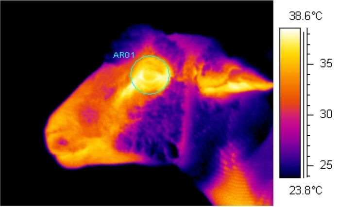
Using an infrared camera (ThermaCam S60, FLIR Systems AB, Danderyd, Sweden), continuous infrared recordings were collected at a rate ~60 frames/s and a resolution of 640 x 480 pixels (S1 Movie). This was achieved through the use of image analysis interface software (ThermaCam Researcher, version 2.10, FLIR Systems AB, Danderyd, Sweden) which connected the camera to a laptop. Whilst restrained in the chute, infrared recordings of the eye region were collected from the left side of the ewe with the infrared camera positioned on a tripod at a set distance of 1 m and at a 90° angle from the direction the ewe was facing. The camera was set to an emissivity of ε = 0.98, based on the generally accepted emissivity of an animal’s body [20] and as used previously in sheep [15, 21]. Ambient temperature (°C) and relative humidity (%) were recorded in close proximity to the chute in which sheep were held every hour using a handheld logger (Kestrel 3000, Nielsen-Kellerman, Boothwyn, PA) and entered into the infrared camera to adjust for changes in atmospheric conditions. Infrared recordings of the eye region were collected continuously for approximately 25 min before until 20 min after the start of the infusion period. From the continuous recordings, image analysis software (ThermaCam Researcher, version 2.7, FLIR Systems AB, Danderyd, Sweden) was used to select individual images (frames) comprising the recordings at 20 s intervals. Only images which were in focus and in which the eye was fully open were used as images which do not meet these requirements can affect the accuracy of the results. Maximum eye temperature (°C) was determined from each of these selected images by tracing a circle around the eye region as to include the medial, posterior palpebral border of the lower eyelid and the lacrimal caruncle (Fig 1).

Fig 1. Example infrared image.

Fig 1

Image shows the area traced around the eye region including the medial, posterior palpebral border of the lower eyelid and the lacrimal caruncle during analysis to determine the maximum eye temperature (°C).

Heart rate

The inter-beat interval (IBI) was recorded every second using Polar heart rate monitors (RS800CX™, Polar Electro Oy, Helsinki, Finland), and heart rate and HRV data were analysed over the 5 min period prior to and up to 10 min after the start of the infusion period. Monitors were strapped firmly around the thorax immediately behind the forelimbs, with electrode contact points on areas from which wool had been removed and ultrasound transmission gel (Aquasonic 100, Parker Laboratories Inc., Fairfield, NJ) applied. At the end of each sampling period the stored heart rate was downloaded via a serial interface to a computer for analysis. The heart rate and IBI data were extracted using Polar ProTrainer 5 (version 5.35, Polar Electro Oy, Helsinki, Finland). Heart rate (recorded every second) was averaged over 1-min intervals, and IBI data were used to calculate HRV parameters, the standard deviation of all inter-beat intervals (SDNN) and the root mean square of successive differences (RMSSD). For SDNN and RMSSD analysis, short segments of data containing 256 beats were examined for 5 min before and up to 10 min after the start of the infusion period. Before analysis, a correction function within the Polar software (Polar Precision Performance Software, version 4.03, Polar Electro Oy, Helsinki, Finland), set on default parameters, was used to correct for any artefacts and only data with an error rate of less than 5% were included in the analysis.

Blood chemistry

To measure epinephrine and norepinephrine concentrations, continuous blood samples (6 mL) were collected at 30 s intervals into pre-chilled EDTA tubes (Becton Dickinson, Plymouth, UK) using a peristaltic pump starting -2 min and ending 10 min relative to the start of the infusion period (time 0). Additional blood samples (6 mL) were collected into tubes containing lithium heparin (Becton Dickinson) to measure cortisol and pack cell volume (PCV) concentrations. The maximum amount of blood taken per sheep was 250 mL. Blood samples were immediately placed on ice and centrifuged (2500 rpm) at 4°C for 10 min, within 10 min of collection. Plasma was stored at –80°C until analysed. Samples collected at −2, 0, 2, 4, 6, 8 and 10 min were assayed for epinephrine and norepinephrine concentrations using methodology described in Stewart et al. [19]. Briefly, 1 mL of plasma was extracted on alumina and the extracted catecholamines were separated and measured using reverse-phase HPLC. Cortisol concentrations were measured at –10, –5 and 15 min using a commercially available antibody radioimmunoassay kit (Coat-A-Count® Cortisol, Siemens; Los Angeles, CA). Packed cell volume was measured in samples taken at -5 and 5 min, using an ADVIA 120 Haematology system (Siemens Healthcare GmbH, Erlangen, Germany).

Statistical analysis

The Bayesian mixed model smoothing programme, Flexi 5.60 (Upsdell, [22]), was used to determine the relationship between maximum eye temperature and treatment over time. The model forced the two treatment groups (epinephrine and saline) to be the same at baseline but allowed them to vary during the infusion and post-infusion periods. Individual animal differences and correlations between measurements taken on the same animal over time were allowed for. Fisher's least significant differences at the 5% level were used to compare the smoothed curves for the epinephrine and saline treatments over time.

For HR and HRV parameters, due to equipment malfunction, data were only collected from 10 ewes that received epinephrine and eight that received saline. For each animal, average HR, RMSSD and SNDD was calculated during infusion and over the 5-min periods before (baseline) and after infusion (post-infusion). These were analysed by repeated measures formulated as a linear mixed model and fitted using Residual Maximum Likelihood (REML). Correlations between measurements taken on the same animal over time were modelled using a power model of order 1. The significance of the treatment by time interaction (treatment x time) was assessed using an F-test and unprotected Fisher's least significant differences at the 5% level were used to compare the treatment means at each time period.

The epinephrine, norepinephrine and cortisol concentration data were similarly analysed, however prior to analysis, the concentrations were log-transformed to stabilise the variance. Furthermore, orthogonal contrasts were used to compare the mean change in log concentration following infusion between treatments.

Mean change in PCV (post-pre) between the two treatment groups were compared using one-way ANOVA. For each treatment group, a t-test was used to assess whether mean change was significantly different from zero.

Unless otherwise stated, all statistical analyses were performed using Genstat 19 (VSN International [23]). Residual diagnostic plots were inspected for evidence of departures from the assumptions of normality and homogeneity of variance.

Results

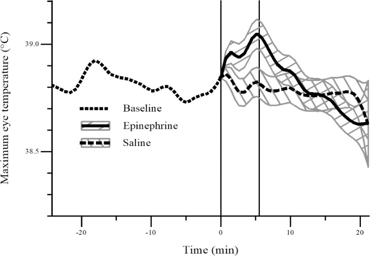
During the infusion period, maximum eye temperature was higher on average (P < 0.05) in sheep that received epinephrine than those that received saline (Fig 2). As mentioned, due to the rich capillary bed located at the lacrimal caruncle, the maximum eye temperatures were typically recorded at this location within the eye region. Mean (± SD) maximum eye temperature before, during and after infusion: Baseline: SAL: 38.9 ± 0.17°C, EPI: 38.9 ± 0.33°C; Infusion: SAL: 38.9 ± 0.19°C, EPI: 39.0 ± 0.42°C; Post-infusion: SAL: 38.8 ± 0.20°C, EPI: 38.8 ± 0.50°C. Range of maximum eye temperature before, during and after infusion: Baseline: SAL: 38.4–39.4°C, EPI: 38.0–39.4°C; Infusion: SAL: 38.4–39.2°C, EPI: 37.9–39.9°C; Post-infusion: SAL: 38.4–39.2°C, EPI: 37.8–40.3°C.

Fig 2. Average maximum eye temperature (°C) in sheep following an infusion (0 to 5 min) of epinephrine (n = 10) or saline (n = 10).

Fig 2

The plot gives the estimated smooth curve at baseline (dotted), and for the epinephrine (solid) and saline (dashed) treatment groups post-baseline. The grey bands are Fisher's least significant differences between treatments. The two treatments are significantly different at the 5% level where the bands do not overlap. The left-hand and right-hand vertical lines denote the start and end of the infusion period, respectively.

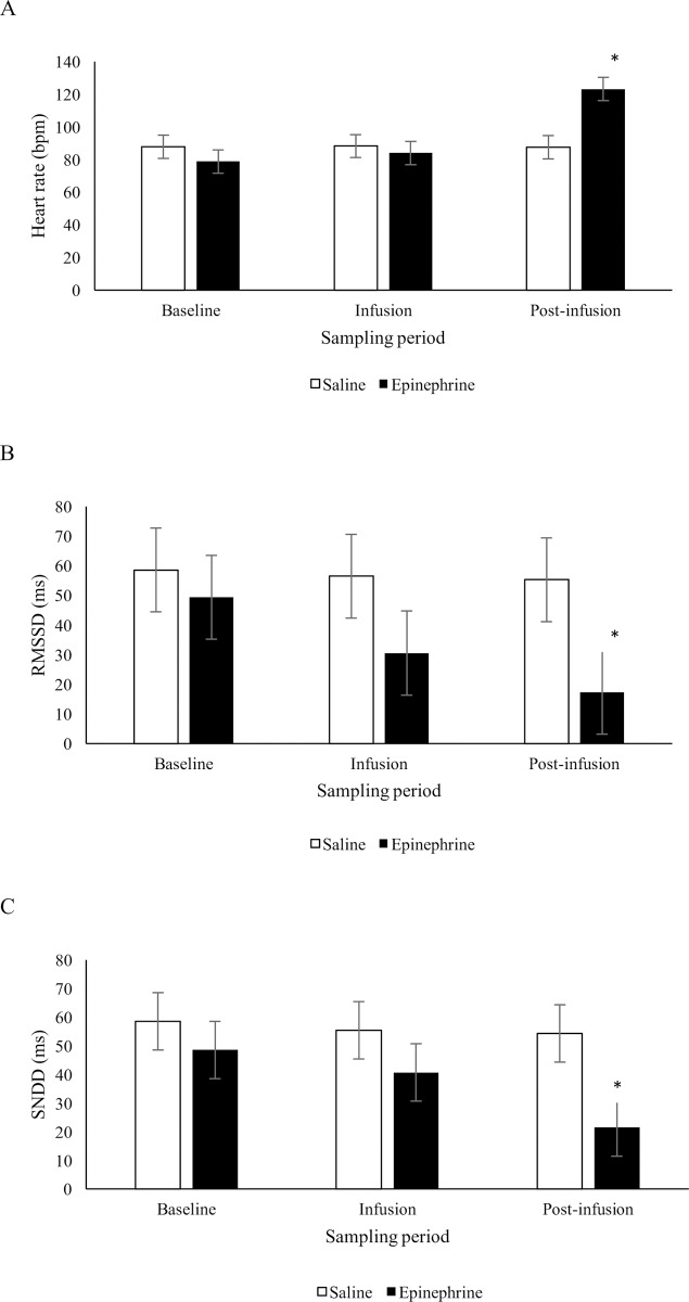
There was no statistical evidence that heart rate differed between sheep that received epinephrine or saline during the infusion period, but during the 5 min post-infusion period, heart rate was on average higher (P < 0.05) in sheep that received epinephrine than those that received saline (F2,33.5 = 27.98, treatment x time: P < 0.001; Fig 3A). There was a tendency for a treatment x time effect on RMSSD (F2,33.3 = 2.65, P = 0.085; Fig 3B) and a statistically significant treatment x time effect on SDNN (F2,33.8 = 3.41, P = 0.045; Fig 3C). During the infusion period, average RMSSD and SDNN was not significantly different (P > 0.05) between saline and epinephrine infused sheep. During the post-infusion period, RMSSD and SDNN were on average lower (P < 0.05) in sheep that received epinephrine than those that received saline. There was no evidence of a difference in baseline values for HR, RMSSD and SDNN between sheep that received epinephrine or saline.

Fig 3.

Fig 3

Mean (± SED) heart rate (bpm) (A), RMSSD (ms) (B) and SDNN (ms) (C) in sheep during a 5 min period before (baseline), during (infusion) and following (post-infusion) an infusion with epinephrine (n = 10) or saline (n = 8). *Treatments differ at P < 0.05.

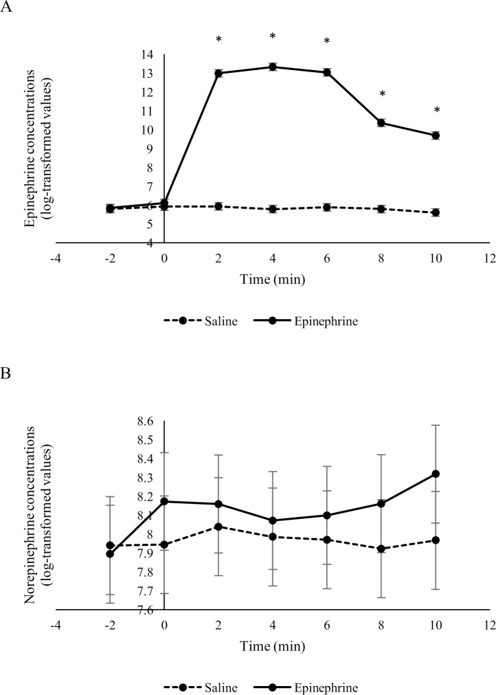
Overall, the epinephrine infusion affected epinephrine concentrations (F2,94.5 = 390.7, treatment x time: P < 0.001) but not norepinephrine concentrations (F2,95.8 = 0.9, treatment x time: P = 0.508) (Fig 4). Geometric mean epinephrine concentrations were higher (P < 0.05) in sheep 2, 4, 6, 8 and 10 min after receiving an infusion of epinephrine compared to sheep that received an infusion of saline. Geometric mean norepinephrine concentrations were similar (P > 0.05) between sheep that received epinephrine or saline at all time points during the infusion and post-infusion periods.

Fig 4.

Fig 4

Epinephrine (A) and norepinephrine (B) concentrations (log-transformed means ± SED) in sheep in response to an infusion of epinephrine or saline (n = 10 / treatment). The infusion period started at time 0 and ended after 5 minutes. *Treatments differ at P < 0.05.

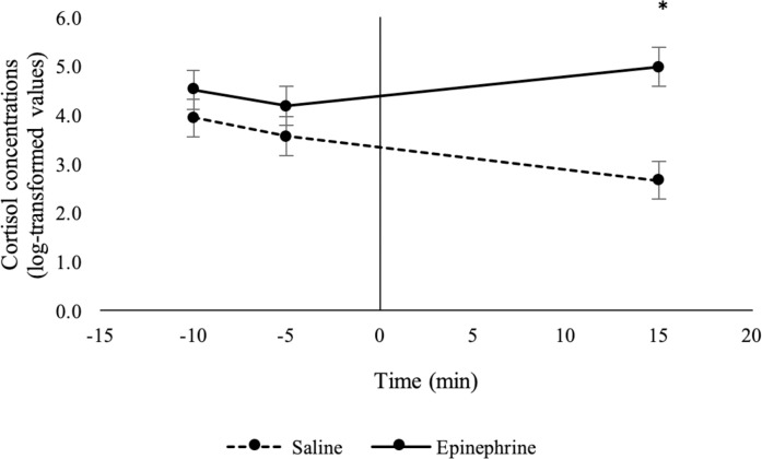
Epinephrine infusion affected cortisol concentrations in sheep (F2,31.3 = 21.1, treatment x time: P < 0.001). Geometric mean cortisol concentrations were higher (P < 0.05) in sheep that received epinephrine than those that received saline 15 min after the start of the infusion period (Fig 5). Cortisol concentrations did not differ (P > 0.05) between sheep that received epinephrine or saline at 10 or 5 min prior to the start of the infusion period (baseline period).

Fig 5. Cortisol concentrations (log-transformed means ± SED) in sheep in response to an infusion of epinephrine or saline (n = 10 / treatment).

Fig 5

The infusion period started at time 0 and ended after 5 minutes. *Treatments differ at P < 0.05.

The PCV increased in sheep that received an epinephrine infusion but decreased in sheep that were given saline (mean change in PCV: epinephrine: 7%, saline: -1%, SED = 1.0%; F1,16 = 58.3, treatment: P < 0.001).

Discussion

Eye temperature, heart rate and HRV, and plasma epinephrine, cortisol and PCV concentrations all changed in response to an epinephrine infusion in sheep in the present study. Interestingly, eye temperature increased in the present study but decreased in cattle in response to a similar 5-min epinephrine challenge [19]. Acute psychological stress stimulates sympathetic-mediated cutaneous vasoconstriction, causing a rapid drop in skin temperature. This drop is then followed by a rise in core body temperature and a subsequent rise in peripheral temperatures as vasodilation occurs to dissipate excess heat [14]. This characteristic decrease in surface temperature and increase in core body temperature following exposure to a psychological stressor is termed stress-induced hyperthermia [4]. Numerous studies measuring eye temperature in response to stress show this characteristic response; eye temperature drops rapidly in response to a stressor and then increases back to baseline levels, sometimes increasing above baseline levels. For example, this pattern was observed in cattle in response to an epinephrine challenge [19], disbudding [9], startle and an electric prod [12] and in chickens in response to restraint [13]. Conversely, eye temperature has been found to increase in response to castration [11] and catheterisation [8] in cattle, clipping [16] and noseband tightening [24] in horses, restraint in chickens [14] and sheep [15] and in response to a veterinary examination in dogs [25]. It is unclear why eye temperature sometimes increases in response to stress; however, it is hard to directly compare these studies due to differences in timing of data collection. For example, in some studies, IRT images were only taken before and after exposure to the stressor, hence the drop-in eye temperature could have been missed. The types of stressors also differed markedly among studies. Furthermore, when making comparisons between studies it is important to consider how the eye region was defined. Differences in this definition could result in the eye region comprising of different tissues and this could alter the temperature change observed in response to a stressor. However, it is important to note that many of the studies mentioned here used the same definition of the eye region [8, 9, 11, 12, 16, 19, 25] and opposing changes in eye temperature in response to a stressor were still observed.

In addition to the eye region, sympathetic-mediated cutaneous vasoconstriction stimulated by acute stress can be measured using other peripheral body regions. For example, IRT has also been used to detect changes in skin temperature in response to stress on the dorsal surface of deer [26], comb and wattle in poultry [14, 27] and nasal region in rhesus monkeys [28] and pigs [29]. Continuous change in temperature of the head region of sheep in response to an epinephrine infusion recorded in the present study (S1 Movie), suggests that other regions of the head, such as the ears, could potentially provide a more sensitive region to measure temperature changes in response to stress in sheep. However, it was beyond the scope of our study to measure temperature changes of other body regions.

Sheep and cattle appeared to react differently during the 5-min epinephrine infusion. In the present study, eye temperature tended to be higher and HRV numerically lower in sheep given epinephrine during the infusion period compared with sheep that received saline, but epinephrine had no effect on heart rate or norepinephrine concentrations. Conversely in cattle, eye temperature, heart rate and norepinephrine concentrations decreased and HRV increased during the epinephrine infusion period [19]. Lower RMSSD and SDNN suggests activation of the sympathetic nervous system in sheep in response to a 5-min epinephrine challenge, conversely, the increase in RMSSD and reduction in norepinephrine concentrations suggests that epinephrine altered the parasympathetic tone in cattle. An epinephrine challenge may have differentially altered the sympathetic/parasympathetic balance in sheep and cattle, leading to the difference in the HRV and eye temperature response between species. It would therefore be of interest to evaluate if the ANS response of sheep to more biologically relevant stressors, such as painful husbandry procedures or handling, would differ from cattle.

During the post-infusion period, heart rate, cortisol and PCV levels were higher and HRV was lower in sheep that received epinephrine, compared with sheep that received saline in the present study. These results are similar to those found in cattle in response to a 5-min epinephrine infusion challenge [19]. Similar to cattle, saline did not cause an increase in cortisol concentrations suggesting that handling and the infusion procedure did not stimulate the hypothalamic-pituitary adrenal response in the present study. The increase in cortisol and PCV in sheep suggests that epinephrine stimulated the sympathetic nervous system resulting in splenic contractions and activation of the hypothalamic-pituitary adrenal axis respectively. Moreover, lower RMSSD and SDNN during the post-infusion period further supports increased sympathetic tone in sheep in response to an epinephrine challenge.

Heart rate, HRV and PCV were measured in conjunction with eye temperature to validate that changes in eye temperature were associated with other measures of ANS activity in the present study. The dose of epinephrine used was sufficient to cause a marked increase in plasma epinephrine concentrations as well as an increase in heart rate and PCV concentrations, and a reduction in HRV compared with sheep given saline. An increase in PCV is an indicator of splenic contractions during endogenous epinephrine release [30], and heart rate and HRV is influenced by both sympathetic and parasympathetic activity [2], therefore these results suggest that the epinephrine infusion was sufficient to activate the ANS in sheep. However, in the present study, there was only weak evidence that eye temperature increased in response to an epinephrine infusion which is in contrast to the marked change in eye temperature found in cattle in response to an epinephrine challenge. Our results are similar to Stubsjoen et al. [18] who reported that eye temperature, measured using IRT, did not change but SDNN and RMSSD tended to decrease in sheep in response to a noxious ischaemic stimulus. However, Cannas et al. [15] found that eye temperature in sheep was higher during restraint and a voluntary approach test. Once again, differences in data collection methodology between studies, such as timing of sampling, may account for these differences.

Conclusions

Changes in HRV and eye temperature in response to an epinephrine infusion suggests, that in sheep, HRV is a more sensitive measure of ANS activity than eye temperature. However, further research is needed to confirm this finding using biologically relevant acute stressors. Only a small increase in eye temperature was found, however, further studies evaluating changes in temperature of other body regions, such as the ear, could be worthwhile to determine whether IRT would be a useful non-invasive method to measure stress and welfare in sheep.

Supporting information

S1 Movie. Example of a continuous infrared recording.

Movie shows a continuous infrared recording collected at a rate ~60 frames/s and a resolution of 640 x 480 pixels of the head of a sheep given an epinephrine infusion. White pixels represent temperatures exceeding 38.9°C.

(MOV)

S1 Dataset. Dataset of study evaluating the effect of an epinephrine challenge on the autonomic response in sheep.

(XLSX)

Acknowledgments

Technical assistance from AgResearch staff was greatly appreciated, in particular Martin Upsdell for his assistance with the Flexi analysis.

Data Availability

All relevant data are within the manuscript and its Supporting Information files.

Funding Statement

This work was funded by the New Zealand Ministry of Business, Innovation and Employment (formerly the New Zealand Foundation for Research, Science and Technology; Contract C10X0813; Wellington, New Zealand). MBIE and AgResearch had no role in the study design, data collection and analysis, interpretation of data, preparation of the manuscript or decision to publish. AgResearch Ltd. (Hamilton, New Zealand) provided support in the form of salaries for authors [MAS, GMW, SKD, GLL, VMC, MS] and research materials. The specific roles of these authors are articulated in the ‘author contributions’ section.

References

  • 1.Moberg GP. Biological responses to stress: Implications for animal welfare In: Moberg GP, Mench JA, editors. The Biology of Animal Stress; CABI Publishing: Wallingford, UK, 2007; p. 1–121. [Google Scholar]
  • 2.von Borell E, Langbein J, Després G, Hansen S, Leterrier C, Marchant-Forde J, et al. Heart rate variability as a measure of autonomic regulation of cardiac activity for assessing stress and welfare in farm animals—A review. Phys. Behav. 2007;92:293–316. [DOI] [PubMed] [Google Scholar]
  • 3.Stewart M, Webster JR, Schaefer AL, Cook NJ, Scott SL. Infrared thermography as a non-invasive tool to study animal welfare. Anim. Wel. 2005;14(4):319–25. [Google Scholar]
  • 4.Bouwknecht JA, Olivier B, Paylor RE. The stress-induced hyperthermia paradigm as a physiological animal model for anxiety: A review of pharmacological and genetic studies in the mouse. Neurosci. Biobehav. Rev. 2007;31(1):41–59. 10.1016/j.neubiorev.2006.02.002 [DOI] [PubMed] [Google Scholar]
  • 5.Cook NJ, Schaefer AL. Infrared thermography and disease surveillance, In: Luzi F, Mitchell M, Nanni Costa L, Redaelli V, editors. Thermography: Current status and advances in livestock animals and in veterinary medicine. Brescia, Fondazione Iniziative Zooprofilattiche. 2013;92:41–6. [Google Scholar]
  • 6.Tattersall GJ. Infrared thermography: A non-invasive window into thermal physiology. Comp. Biochem. Physiol. A Mol. Integr. Physiol. 2016;202:78–98. [DOI] [PubMed] [Google Scholar]
  • 7.Dai F, Cogi NH, Heinzl EUL, Dalla Costa E, Canali E, Minero M. Validation of a fear test in sport horses using infrared thermography. J. Vet. Behav. Clin. Appl. Res. 2015;10:128–36. [Google Scholar]
  • 8.Stewart M, Webster JR, Verkerk GA, Schaefer AL, Colyn JJ, Stafford KJ. Non-invasive measurement of stress in dairy cows using infrared thermography. Physiol. Behav. 2007;92:520–5. [DOI] [PubMed] [Google Scholar]
  • 9.Stewart M, Stafford KJ, Dowling SK, Schaefer AL, Webster JR. Eye temperature and heart rate variability of calves disbudded with or without local anaesthetic. Phys. Behav. 2008;93(4–5):789–97. 10.1016/j.physbeh.2007.11.044 [DOI] [PubMed] [Google Scholar]
  • 10.Stewart M, Stookey JM, Stafford KJ, Tucker CB, Rogers AR, Dowling SK, et al. Effects of local anesthetic and a non-steroidal anti-inflammatory drug on pain responses of dairy calves to hot-iron dehorning. J. Dairy Sci. 2009;92:1512–9. [DOI] [PubMed] [Google Scholar]
  • 11.Stewart M, Verkerk GA, Stafford KJ, Schaefer AL, Webster JR. Noninvasive assessment of autonomic activity for evaluation of pain in calves, using surgical castration as a model. J. Dairy Sci. 2010;93:3602–9. [DOI] [PubMed] [Google Scholar]
  • 12.Stewart M, Schaefer AL, Haley DB, Colyn JJ, Cook NJ, Stafford KJ, et al. Infrared thermography as a non-invasive method for detecting fear-related responses of cattle to different handling procedures. Anim. Wel. 2008;17:387–93. [Google Scholar]
  • 13.Edgar JL, Nicol CJ, Pugh CA, Paul ES. Surface temperature changes in response to handling in domestic chickens. Physiol. Behav. 2013;119:195–200. 10.1016/j.physbeh.2013.06.020 [DOI] [PubMed] [Google Scholar]
  • 14.Moe RO, Bohlin J, Flø A, Vasdal G, Stubsjøen SM. Hot chicks, cold feet. Physiol. Behav. 2017;179:42–8. 10.1016/j.physbeh.2017.05.025 [DOI] [PubMed] [Google Scholar]
  • 15.Cannas S, Palestrini C, Canali E, Cozzi B, Ferri N, Heinzi E, et al. Thermography as a non-invasive measure of stress and fear of humans in sheep. Animals 2018;8:146. [DOI] [PMC free article] [PubMed] [Google Scholar]
  • 16.Yarnell K, Hall C, Billett E. An assessment of the aversive nature of an animal management procedure (clipping) using behavioral and physiological measures. Physiol. Behav. 2013;118:32–9. 10.1016/j.physbeh.2013.05.013 [DOI] [PubMed] [Google Scholar]
  • 17.Dwyer CM. Welfare of sheep: Providing for welfare in an extensive environment. Sm. Rum. Res. 2009;86(1–3):14–21. 10.1016/j.smallrumres.2009.09.010 [DOI] [Google Scholar]
  • 18.Stubsjøen SM, Flø AS, Moe RO, Janczak AM, Skjerve E, Valle PS, et al. Exploring non-invasive methods to assess pain in sheep. Phys. Behav. 2009;98(5):640–8. 10.1016/j.physbeh.2009.09.019 [DOI] [PubMed] [Google Scholar]
  • 19.Stewart M, Webster JR, Stafford KJ, Schaefer AL, Verkerk GA. Technical note: Effects of an epinephrine infusion on eye temperature and heart rate variability in bull calves. J. Dairy Sci. 2010;93(11):5252–7. 10.3168/jds.2010-3448 [DOI] [PubMed] [Google Scholar]
  • 20.Redaelli V, Ludwig N, Costa LN, Crosta L, Riva J, Luzi F. Potential application of thermography (IRT) in animal production and for animal welfare. A case report of working dogs. Ann Ist Super Sanità. 2014;50:147–52. [DOI] [PubMed] [Google Scholar]
  • 21.Byrne DT, Perry DP, Esmonde H, McGivern F, Creighton P, McHugh N. Infrared thermography as a tool to detect hoof lesions in sheep. Transl. Anim. Sci. 2019;3:577–88. [DOI] [PMC free article] [PubMed] [Google Scholar]
  • 22.Upsdell MP. Bayesian smoothers as an extension of nonlinear regression. NZ. Stat. 1994;29:66–81. [Google Scholar]
  • 23.Genstat for Windows 19th Edition VSN International, Hemel Hempstead, UK: 2017. [Google Scholar]
  • 24.Fenner K, Yoon S, White P, Starling M, McGreevy P. The Effect of Noseband Tightening on Horses’ Behavior, Eye Temperature, and Cardiac Responses. PLoS ONE 2016;11(5): e0154179 10.1371/journal.pone.0154179. [DOI] [PMC free article] [PubMed] [Google Scholar]
  • 25.Travain T, Colombo ES, Heinzl E, Bellucci D, Previde EP, Valsecchi P. Hot dogs: Thermography in the assessment of stress in dogs (Canis familiaris)—A pilot study. J. Vet. Behav. 2015;10(1):17–23. 10.1016/j.jveb.2014.11.003 [DOI] [Google Scholar]
  • 26.Cook NJ, Schaefer AL. Stress responses of Wapiti (Cervus elaphus canadensis) to removal of velvet antler. Can. J. Anim. Sci. 2002;82:11–7. [Google Scholar]
  • 27.Herborn KA, Graves JL, Jerem P, Evans NP, Nager R, McCafferty DJ, et al. Skin temperature reveals the intensity of acute stress. Phys. Behav. 2015;152(Part A):225–30. 10.1016/j.physbeh.2015.09.032 [DOI] [PMC free article] [PubMed] [Google Scholar]
  • 28.Nakayama K, Goto S, Kuraoka K, Nakamura K. Decrease in nasal temperature of rhesus monkeys (Macaca mulatta) in negative emotional state. Phys. Behav. 2005;84(5):783–90. 10.1016/j.physbeh.2005.03.009 [DOI] [PubMed] [Google Scholar]
  • 29.Sutherland M, Dowling S, Backus B, Stewart M. The use of infrared thermography to assess stress in pigs. Proc. 7th Euro. Conf. Prec. Livest. Farm. 2015; p. 691–9.
  • 30.Potocnik SJ, Wintour EM. Development of the spleen as a red blood cell reservoir in lambs. Reprod. Fertil. Dev. 1996;8(3):311–5. [DOI] [PubMed] [Google Scholar]

Decision Letter 0

I Anna S Olsson

12 Mar 2020

PONE-D-19-33285

Evaluation of infrared thermography to measure the autonomic nervous response in sheep­­­­

PLOS ONE

Dear Dr. Sutherland,

Thank you for submitting your manuscript to PLOS ONE. After careful consideration, we feel that it has merit but does not fully meet PLOS ONE’s publication criteria as it currently stands. Therefore, we invite you to submit a revised version of the manuscript that addresses the points raised during the review process.

You will find detailed comments from the two independent reviewers below. Please take them into consideration when revising the paper. I would like to draw special attention to the detailed comments by Reviewer 1.

In addition, based on my own reading of the manuscript, I ask that you revise the discussion so that it focuses primarily on the results of your study and how they relate to what has been published previously. As both reviewers point out, there is presently too much overlap with the literature review in the introduction section, and parts of the discussion are not well linked to your own results. 

A minor detail

Lines 73-74 I think you intend to say "Moreover, eye temperature has commonly been measured in many species because this area is not affected by the presence of hair"

We would appreciate receiving your revised manuscript by Apr 26 2020 11:59PM. When you are ready to submit your revision, log on to https://www.editorialmanager.com/pone/ and select the 'Submissions Needing Revision' folder to locate your manuscript file.

If you would like to make changes to your financial disclosure, please include your updated statement in your cover letter.

To enhance the reproducibility of your results, we recommend that if applicable you deposit your laboratory protocols in protocols.io, where a protocol can be assigned its own identifier (DOI) such that it can be cited independently in the future. For instructions see: http://journals.plos.org/plosone/s/submission-guidelines#loc-laboratory-protocols

Please include the following items when submitting your revised manuscript:

  • A rebuttal letter that responds to each point raised by the academic editor and reviewer(s). This letter should be uploaded as separate file and labeled 'Response to Reviewers'.

  • A marked-up copy of your manuscript that highlights changes made to the original version. This file should be uploaded as separate file and labeled 'Revised Manuscript with Track Changes'.

  • An unmarked version of your revised paper without tracked changes. This file should be uploaded as separate file and labeled 'Manuscript'.

Please note while forming your response, if your article is accepted, you may have the opportunity to make the peer review history publicly available. The record will include editor decision letters (with reviews) and your responses to reviewer comments. If eligible, we will contact you to opt in or out.

We look forward to receiving your revised manuscript.

Kind regards,

I Anna S Olsson, Ph.D.

Academic Editor

PLOS ONE

Journal Requirements:

When submitting your revision, we need you to address these additional requirements.

1) Please ensure that your manuscript meets PLOS ONE's style requirements, including those for file naming. The PLOS ONE style templates can be found at

http://www.journals.plos.org/plosone/s/file?id=wjVg/PLOSOne_formatting_sample_main_body.pdf and http://www.journals.plos.org/plosone/s/file?id=ba62/PLOSOne_formatting_sample_title_authors_affiliations.pdf

2) Thank you for stating the following in the Financial Disclosure section:

[This study was funded by the New Zealand Foundation for Research, Science and

Technology (C10X0813). The funders had no role in study design, data collection and

analysis, decision to publish, or preparation of the manuscript.].   

We note that one or more of the authors are employed by a commercial company: AgResearch Ltd.

i. Please provide an amended Funding Statement declaring this commercial affiliation, as well as a statement regarding the Role of Funders in your study. If the funding organization did not play a role in the study design, data collection and analysis, decision to publish, or preparation of the manuscript and only provided financial support in the form of authors' salaries and/or research materials, please review your statements relating to the author contributions, and ensure you have specifically and accurately indicated the role(s) that these authors had in your study. You can update author roles in the Author Contributions section of the online submission form.

Please also include the following statement within your amended Funding Statement.

“The funder provided support in the form of salaries for authors [insert relevant initials], but did not have any additional role in the study design, data collection and analysis, decision to publish, or preparation of the manuscript. The specific roles of these authors are articulated in the ‘author contributions’ section.”

If your commercial affiliation did play a role in your study, please state and explain this role within your updated Funding Statement.

ii. Please also provide an updated Competing Interests Statement declaring this commercial affiliation along with any other relevant declarations relating to employment, consultancy, patents, products in development, or marketed products, etc. 

Within your Competing Interests Statement, please confirm that this commercial affiliation does not alter your adherence to all PLOS ONE policies on sharing data and materials by including the following statement: "This does not alter our adherence to  PLOS ONE policies on sharing data and materials.” (as detailed online in our guide for authors http://journals.plos.org/plosone/s/competing-interests) . If this adherence statement is not accurate and  there are restrictions on sharing of data and/or materials, please state these. Please note that we cannot proceed with consideration of your article until this information has been declared.

Please include both an updated Funding Statement and Competing Interests Statement in your cover letter. We will change the online submission form on your behalf.

Please know it is PLOS ONE policy for corresponding authors to declare, on behalf of all authors, all potential competing interests for the purposes of transparency. PLOS defines a competing interest as anything that interferes with, or could reasonably be perceived as interfering with, the full and objective presentation, peer review, editorial decision-making, or publication of research or non-research articles submitted to one of the journals. Competing interests can be financial or non-financial, professional, or personal. Competing interests can arise in relationship to an organization or another person. Please follow this link to our website for more details on competing interests: http://journals.plos.org/plosone/s/competing-interests

[Note: HTML markup is below. Please do not edit.]

Reviewers' comments:

Reviewer's Responses to Questions

Comments to the Author

1. Is the manuscript technically sound, and do the data support the conclusions?

The manuscript must describe a technically sound piece of scientific research with data that supports the conclusions. Experiments must have been conducted rigorously, with appropriate controls, replication, and sample sizes. The conclusions must be drawn appropriately based on the data presented.

Reviewer #1: Yes

Reviewer #2: Partly

**********

2. Has the statistical analysis been performed appropriately and rigorously?

Reviewer #1: Yes

Reviewer #2: No

**********

3. Have the authors made all data underlying the findings in their manuscript fully available?

The PLOS Data policy requires authors to make all data underlying the findings described in their manuscript fully available without restriction, with rare exception (please refer to the Data Availability Statement in the manuscript PDF file). The data should be provided as part of the manuscript or its supporting information, or deposited to a public repository. For example, in addition to summary statistics, the data points behind means, medians and variance measures should be available. If there are restrictions on publicly sharing data—e.g. participant privacy or use of data from a third party—those must be specified.

Reviewer #1: Yes

Reviewer #2: Yes

**********

4. Is the manuscript presented in an intelligible fashion and written in standard English?

PLOS ONE does not copyedit accepted manuscripts, so the language in submitted articles must be clear, correct, and unambiguous. Any typographical or grammatical errors should be corrected at revision, so please note any specific errors here.

Reviewer #1: Yes

Reviewer #2: No

**********

5. Review Comments to the Author

Please use the space provided to explain your answers to the questions above. You may also include additional comments for the author, including concerns about dual publication, research ethics, or publication ethics. (Please upload your review as an attachment if it exceeds 20,000 characters)

Reviewer #1: This is an interesting paper investigating changes in eye region temperature as captured with IRT. The work is sound and adds to the growing literature on the potential value of surface temperature measures in stress assessment. However the authors must provide a stronger justification for the use of an epinephrine infusion rather than a relevant stressor. I also have various questions about methodology. In particular I would like reassurance on the effectiveness of habituation and a better description of the experimental procedure with regard to other possible sources of stress.

Introduction

The relevant literature is nicely reviewed, but I expect a stronger explanation for the rationale of using epinephrine infusion rather than a biologically relevant acute stressor. Almost all other studies in this field have used relevant stressful events to examine surface temperature responses – why was this approach not taken? I suspect there was some activation of stress due to experimental procedures in any case (see comments below) but this was not the aim.

Methods

The experimental procedures are not at clear in several places and more detail is needed. Specifically:

What handling was required to administer the treatments and collect blood samples?

What evidence (behavioural, HR, hormonal?) was there that habitation to handling and restraint was effective prior to the experiment starting? Otherwise possible influences on the stress response must be expected and fully discussed.

The total duration of thermal recording is not provided – was this as for blood sampling or for longer? Where were the animals during this time? Were they restrained? How was distance from the camera managed if the animals were not restrained? The description ‘camera positioned at a distance of approximately 1 m’ is problematic, as even small distance changes can affect accuracy – what was the range?

The IRT variable might be more accurately referred to as ‘eye region temperature’ since structures other than the eye itself were included.

Again what evidence is there that habituation to wearing HR monitors was successful? What was the duration of habituation? I ask these questions because for a study on acute stress it is crucial to know to what extent the experimental procedure affected the phenomenon under study.

Movie – this is useful but consider rescaling the temperature bar to the right – areas around the eye appear as white which is not in the calibration bar (presumably in excess of 38 degrees C).

Results

A mixture of P= and P< is reported – suggest consistency with P= except for P<0.001. This applies to the abstract as well.

Why are there so few sampling points for cortisol? These are so few as to be rather uninformative. The upward trajectory of the epinephrine treatment line suggests that further increase was possible, particularly given the fact that 15 mins is quite a short timescale for a cortisol response – this should be acknowledged.

Given that eye region temperature change is the focus of the paper, it would be good to see some more detailed data rather than a single figure. At least provide mean and SD temperature increase and range – this would allow more ready comparison with other work. Currently this information is difficult/impossible to glean accurately from the figure.

Can the authors report which region of the eye was usually the position of their maximum temperature recording? This may be relevant to interpretation (see below).

Discussion

In the discussion of why some studies show eye temp increases in response to stress while others show decreases, the authors mention differences in timing of recoding (which is fair enough and it is true that the initial drop can be very rapid and so missed) but it is also important to mention that different studies use different definitions of ‘eye’ when reporting temperature change, such that some are imaging the cornea, while others are imaging the whole eye region (and taking maximums from various sub regions thereof). These tissues differ markedly in various characteristics including peripheral circulation and its control by the ANS – this should be reflected in the authors discussion.

The authors notice that body surface areas other than the eye may reveal SIH – but say this is beyond the scope of the current study. Fair enough, but this seems a shame since at least one ear seems to be visible at all times in the movie and surface temperature analysis would be feasible.

The authors conclude that the HPA axis was not stimulated, but based on the figure I think care should be taken here – see comment above that the upward trajectory of the epinephrine treatment line suggests that further increase was possible. It should be acknowledged that 15 mins is a short timescale for a cortisol response which may well have continued/appeared beyond this time.

Lines 345-362 – many relevant factors for effective IRT data collection are discussed, but without adequate reference to the current study – do any of these limitations apply or if not how were they overcome and how do we know that? As it is it is rather generic and does not add much to the discussion.

Conclusion

Suggest rewording to “Changes in HRV and eye temperature in response to an epinephrine infusion suggests that, in sheep, HRV is a more sensitive non-invasive method than IRT to measure ANS activity (as induced by epinephrine infusion).” – since we don’t know if the same results would have been seen had a biologically relevant acute stressor been applied.

This point should also be indicated in the abstract.

Reviewer #2: This manuscript approaches an interesting scientific question to the assessment of autonomic nervous response with non-invasive methods. Considering the conclusion “These results suggest that heart rate variability is a sensitive, non-invasive method that can be used to measure ANS activity in sheep, whereas change in eye temperature measured using IRT is a less sensitive method”, authors may consider a change in focus, starting from the title, and put more emphasis to heart rate variability throughout the paper. If temperature remains as part of the title and the objective, authors should give its numerical values in the abstract and in the results section in a more explicit manner. It is important to improve the introduction, to better present to readers the relevance of this work. Please provide an explicit and complete statement of the objective of the work in the end of the introduction. There is excessive repetition of text between introduction and discussion.

A major issue with the manuscript is the ambiguity on the effects of treatment on eye temperature, as detailed below. If authors have the thermographic images of the head, they could verify the hypothesis they bring that other regions, such as ears, may be more responsive in terms of superficial temperature. Additionally, the discussion needs considerable improvement through a more extensive review of the literature and increased complexity.

Specific Comments

L81: Please refine link to previous paragraph, which presented a list of stressful situations.

L83: Please explain kind of noxious stimulus and animal species involved, to improve the flow of reading.

L219-220: what are the numeric values?

L245, 257: Do you mean “affected”?

L304-305: Do you have the data on temperature on other body regions? It could significantly improve this manuscript.

L274: Was it only a tendency for temperature increase in this study? But how come in the abstract it is stated that there was a difference (P<0.05, L31-32)? Then again in the conclusion it is understandable in the first phrase that there were changes in the eye temperature (L364).

**********

6. PLOS authors have the option to publish the peer review history of their article (what does this mean?). If published, this will include your full peer review and any attached files.

If you choose “no”, your identity will remain anonymous but your review may still be made public.

Do you want your identity to be public for this peer review? For information about this choice, including consent withdrawal, please see our Privacy Policy.

Reviewer #1: No

Reviewer #2: No

[NOTE: If reviewer comments were submitted as an attachment file, they will be attached to this email and accessible via the submission site. Please log into your account, locate the manuscript record, and check for the action link "View Attachments". If this link does not appear, there are no attachment files to be viewed.]

While revising your submission, please upload your figure files to the Preflight Analysis and Conversion Engine (PACE) digital diagnostic tool, https://pacev2.apexcovantage.com/. PACE helps ensure that figures meet PLOS requirements. To use PACE, you must first register as a user. Registration is free. Then, login and navigate to the UPLOAD tab, where you will find detailed instructions on how to use the tool. If you encounter any issues or have any questions when using PACE, please email us at figures@plos.org. Please note that Supporting Information files do not need this step.

PLoS One. 2020 May 29;15(5):e0233558. doi: 10.1371/journal.pone.0233558.r002

Author response to Decision Letter 0


22 Apr 2020

The authors would like to thank the editor and the reviewers for their thoughtful and very helpful comments on this manuscript. The authors have considered all the comments listed below, have responded to these comments individually and made the appropriate changes to the manuscript.

Editor

I ask that you revise the discussion so that it focuses primarily on the results of your study and how they relate to what has been published previously. As both reviewers point out, there is presently too much overlap with the literature review in the introduction section, and parts of the discussion are not well linked to your own results.

Au: Based on your comments the discussion is now more focused on the results of our study and how they relate to previous studies. We have reduced the level or repetition between the introduction and discussion.

Lines 73-74 I think you intend to say "Moreover, eye temperature has commonly been measured in many species because this area is not affected by the presence of hair"

AU: This sentence has now been changed to ‘Temperature of the eye region has commonly been measured in many species because this area is not affected by the presence of hair’.

Requested removal of “an anatomical region”

AU: Changed as requested.

REVIEWER 1

Expects stronger explanation for the rationale for using epinephrine infusion rather than a biologically relevant acute stressor. Almost all studies in the field have used relevant stressful events to examine surface temp responses- why was this approach not taken?

AU: This study was designed as a validation study so we wanted to make sure that we activated the ANS hence we used an epinephrine infusion. A similar study design was successfully used in cattle to validate IRT as a non-invasive measure of the ANS. As this was a validation study the purpose was not to measure how an animal responds to a particular biological stressor. However, this is an important next step and we have mentioned this in the discussion. Also, we have clarified the explanation of the objectives in the introduction.

I suspect there was some activation of stress due to experimental procedures in any case but this was not the aim.

AU: Yes, it is very likely that there was some activation of the stress response in these sheep, but to help reduce this we habituated the sheep to the facilities and equipment for 2 weeks before the start of the study. We also included a control group (saline), to take into account any changes in response to stress that was not associated with the epinephrine infusion.

What evidence (behavioural, HR or hormonal?) was there that habituation to handling and restraint was effective prior to the experiment starting? Otherwise possible influences on the stress response must be expected and fully discussed.

Again what evidence is there that habituation to wearing HR monitors was successful? What was the duration of habituation? I ask these questions because for a study on acute stress it is crucial to know to what extent the experimental procedure affected the phenomenon under study.

AU: Sheep were acclimated to the research facility and equipment for 2 weeks prior to the start of the study. This information has been clarified in the methods section. We would expect after this period of time that all animals would be habituated, and they did appear so, however we took no measures to prove this. This has been clarified in the methods section. We also had a control group in this study that only received saline to take into account for any ANS changes not related to the epinephrine.

What handling was required to administer treatments and collect blood samples?

AU: Animals were manually restrained to put the heart rate monitors on and then strained in the chute for the other measures. This has been clarified in the methods section.

The total duration of the thermal imaging is not provided- was this as for blood sampling or longer? Where were the animals during this time? Were they restrained? How was camera distance managed if the animals were not restrained?

AU: Within this section we have clarified the duration of the infrared recordings as being 25 mins both before and following the start of the infusion period. We have also explained that infrared recordings were collected while the animal was restrained in the chute and have clarified how distance and angle were kept consistent.

The description “camera positioned at a distance of approximately 1m” is problematic, as even small distance changes can affect accuracy- what was the range?

AU: The word “approximately” has been removed as the distance and angle of the camera from the animal was keep constant throughout the duration of the study by the infrared camera being used with a tripod that was placed at a marked location 1m away from the animal. This has been clarified in the methods section.

This is useful but consider rescaling the temperature bar to the right- areas around the eye appear as white which is not in the calibration bar (presumably in excess of 38 C)

AU: We do not have the ability to rescale the temperature bar in this example video. However, for clarity we have provided an explanation in the figure legend that the white pixels represent those in excess of 38.9C.

A mixture of P= and P< is reported- suggest consistency with P= except for P<0.001. this applies to the abstract as well.

AU: The significance of the treatment by time interaction (treatment x time) was assessed using an F-test and the exact p-values have been included in the text. The unprotected Fisher's least significant differences at the 5% level were used to compare the treatment means at each time period, hence for these comparisons we only have p-values of < or > 0.05.

Why are there so few sampling points for cortisol? These are so few as to be rather uninformative. The upward trajectory of the epinephrine treatment line suggests that further increase was possible, particularly given the fact that 15 mins is quite a short timescale for a cortisol response- this should be acknowledged.

AU: Yes, the cortisol response to epinephrine may have increased further after 15 minutes, however, blood samples were only collected up to 15 minutes after the start the infusion. This is because we were predominantly interested in the acute ANS response (the 5-minute period during and after the epinephrine infusion) not the HPA response.

Given that eye region temperature change is the focus of the paper, it would be good to see some more detailed data rather than a single figure. At least provide mean and SD temperature increase and range- this would allow more ready comparison to other work. Currently this information is difficult/impossible to glean accurately from the figure.

AU: As requested we have included additional information e.g., mean and SD and ranges relating to the maximum eye temperature at the different stages of the experiment for the epinephrine and saline treatments.

Can the authors report which region of the eye was usually the position of the maximum temperature recording? This may be relevant to interpretation.

AU: It is very important information and has now been added. The lacrimal caruncle was typically the position of the maximum eye temperature due to the rich capillary bed located at this region. This has been added to the manuscript.

The authors conclude that the HPA axis was not stimulated, but based on the figure I think care should be taken here- see comment above that the upward trajectory of the epinephrine treatment line suggests that further increase was possible. It should be acknowledged that 15 mins is a short timescale for a cortisol response which may well have continued/appeared beyond this time.

AU: We stated that HPA was not activated by saline (the control) but was activated by epinephrine ‘The increase in cortisol and PCV in sheep suggests that epinephrine stimulated the sympathetic nervous system resulting in splenic contractions and activation of the hypothalamic-pituitary adrenal axis respectively.” See responses above in regards to short time scale for cortisol measures.

In the discussion of why some studies show eye temp increases in response to stress while others show decreases, the authors mention differences in timing of recording (which is fair enough and it is true that the initial drop can be very rapid and so missed) but it is also important to mention that different studies use different definitions of “eye” when reporting temperature change, such that some are imaging the cornea, while others are imaging the whole eye region (and taking maximums from various sub regions thereof). These tissues differ markedly in various characteristics including peripheral circulation and its control by the ANS- this should be reflected in the authors discussion.

AU: We agree that region where eye temperature is measured is an important factor and have now added this point to the discussion.

The authors notice that body surface areas other than the eye reveal SIH- but say this is beyond the scope of the current study. Fair enough, but this seems a shame since at least one ear seems to be visible at all times in the movie and surface temperature would be feasible.

AU: It is a shame, but lack of funding has meant that it is beyond the scope of this study.

Many relevant factors for effective IRT data collection are discussed, but without reference to the current study- do any of these limitations apply or if not how were they overcome and how do we know that? As it is it is rather generic and does not add much to the discussion.

AU: The purpose of this paragraph was to highlight some of the considerations needed when measuring IRT, but we agree with the reviewer that it is not really relevant to our study as we took all these considerations into account when collecting the data. Therefore, this paragraph has been removed.

Suggest rewording to “Changes in HRV and eye temperature in response to an epinephrine infusion suggests that in sheep, HRV is a more sensitive non-invasive method than IRT to measure ANS activity (as induced by epinephrine infusion).” Since we don’t know if the same results would have been seen had a biologically acute stressor been applied.

AU: We agree with the reviewer, however, think it is redundant to state that this response was due to epinephrine twice in the same sentence. Therefore, we have added the sentence ‘However, further research is needed to confirm this finding using biologically relevant acute stressors’.

REVIEWER 2:

Considering the conclusion

“These results suggest that heart rate variability is a sensitive, non-invasive method that can be used to measure ANS activity in sheep, whereas change in eye temperature measured using IRT is a less sensitive method”

Authors may consider a change in focus, starting from the title, and put more emphasis to heart rate variability throughout the paper.

AU: The objective of our study was to evaluate if changes in eye temperature (measured using IRT) can be used to non-invasively measure ANS activity in sheep. Therefore, IRT was the focus of the study not HRV, so we have kept IRT the focus of the paper. HRV is already known to be a good measure of the ANS and was only included in the study to help validate IRT as a measure of the ANS. We have tried to clarify this in our objective in the introduction.

If temperature remains as part of the objective, authors should give its numerical values in the abstract and in the results section in a more explicit manner.

AU: The eye temperature data was analysed using Bayesian mixed model smoothing programme (refer to stats section). This model does not produce actual values, instead it determines the relationship between maximum eye temperature and treatment over time. Therefore, we can’t include numerical values here as they don’t relate to the statistical analysis. However, the descriptive means and standard deviations and ranges have been added to the results section.

It is important to improve the introduction to better present to readers the relevance of this work. There is excessive repetition between the intro & discussion

AU: We have amended the introduction to better highlight the focus of this work which was to validate IRT as a non-invasive measure of the ANS in sheep. Repetition between the introduction and discussion has been reduced.

Please refine link to previous paragraph, which presented a list of stressful situations.

AU: Link to following paragraph refined as requested.

Please explain kind of noxious stimulus and animal species involved, to improve the flow of reading.

AU: The ischaemia is the noxious stimulus as it causes pain. We have clarified this and also added that this particular study involved sheep.

Please provide an explicit and complete statement of the objective of the work in the end of the introduction.

AU: We have provided a more explicit and complete statement of the study objectives at the end of the introduction.

What are the numeric values?

AU: The eye temperature data was analysed using Bayesian mixed model smoothing programme (refer to stats section). This model does not produce actual values, instead it determines the relationship between maximum eye temperature and treatment over time. However, the descriptive means and standard deviations and ranges have been added to the results section.

Do you mean “affected”?

AU: Corrected

Do you mean “affected”?

AU: Corrected

Discussion needs considerable improvement through a more extensive review of the literature and increased complexity.

AU: We have focused on the literature which is relevant to what we measured in our study, hence we have focused on the IRT literature where eye temperature was measured in response to agricultural animals. It is unclear what increased complexity the reviewer is wanting. We feel that increased complexity often leads to increased speculation of the results and prefer not to do this. Instead we have tried to focus on the literature and findings that are relevant to our study.

Was it only a tendency for temperature increase in this study? But how come in the abstract it is stated that there was a difference (P<0.005, L31-32)? Then again in the conclusion it is understandable in the first phrase that there were changes in the eye temperature (L364)

AU: When we used the term “tendency” we were meaning from a trajectory point of view rather than a statistical one, however, we have since removed “tendency” from the manuscript for clarity.

A major issue with the manuscript is the ambiguity on the effects of treatment on eye temperature, as detailed below. If authors have thermographic images of the head, they could verify the hypothesis they bring that other regions, such as ears, may be more responsive in terms of superficial temperature. Do you have the data on temperature of other body regions? It could significantly improve this manuscript.

AU: We definitely agree that if we had this information we could answer this question about body region, however, as mentioned above it was beyond the scope of this study to measure temperature changes of other body regions. Therefore, unfortunately we do not have this additional data to include in the manuscript.

Decision Letter 1

I Anna S Olsson

8 May 2020

Evaluation of infrared thermography as a non-invasive method of measuring the autonomic nervous response in sheep­­­­

PONE-D-19-33285R1

Dear Dr. Sutherland,

We are pleased to inform you that your manuscript has been judged scientifically suitable for publication and will be formally accepted for publication once it complies with all outstanding technical requirements.

Within one week, you will receive an e-mail containing information on the amendments required prior to publication. When all required modifications have been addressed, you will receive a formal acceptance letter and your manuscript will proceed to our production department and be scheduled for publication.

Shortly after the formal acceptance letter is sent, an invoice for payment will follow. To ensure an efficient production and billing process, please log into Editorial Manager at https://www.editorialmanager.com/pone/, click the "Update My Information" link at the top of the page, and update your user information. If you have any billing related questions, please contact our Author Billing department directly at authorbilling@plos.org.

If your institution or institutions have a press office, please notify them about your upcoming paper to enable them to help maximize its impact. If they will be preparing press materials for this manuscript, you must inform our press team as soon as possible and no later than 48 hours after receiving the formal acceptance. Your manuscript will remain under strict press embargo until 2 pm Eastern Time on the date of publication. For more information, please contact onepress@plos.org.

With kind regards,

I Anna S Olsson, Ph.D.

Academic Editor

PLOS ONE

Additional Editor Comments (optional):

Reviewers' comments:

Acceptance letter

I Anna S Olsson

14 May 2020

PONE-D-19-33285R1

Evaluation of infrared thermography as a non-invasive method of measuring the autonomic nervous response in sheep­­­­

Dear Dr. Sutherland:

I am pleased to inform you that your manuscript has been deemed suitable for publication in PLOS ONE. Congratulations! Your manuscript is now with our production department.

If your institution or institutions have a press office, please notify them about your upcoming paper at this point, to enable them to help maximize its impact. If they will be preparing press materials for this manuscript, please inform our press team within the next 48 hours. Your manuscript will remain under strict press embargo until 2 pm Eastern Time on the date of publication. For more information please contact onepress@plos.org.

For any other questions or concerns, please email plosone@plos.org.

Thank you for submitting your work to PLOS ONE.

With kind regards,

PLOS ONE Editorial Office Staff

on behalf of

Dr I Anna S Olsson

Academic Editor

PLOS ONE

Associated Data

    This section collects any data citations, data availability statements, or supplementary materials included in this article.

    Supplementary Materials

    S1 Movie. Example of a continuous infrared recording.

    Movie shows a continuous infrared recording collected at a rate ~60 frames/s and a resolution of 640 x 480 pixels of the head of a sheep given an epinephrine infusion. White pixels represent temperatures exceeding 38.9°C.

    (MOV)

    S1 Dataset. Dataset of study evaluating the effect of an epinephrine challenge on the autonomic response in sheep.

    (XLSX)

    Data Availability Statement

    All relevant data are within the manuscript and its Supporting Information files.


    Articles from PLoS ONE are provided here courtesy of PLOS

    RESOURCES