PLOS ONE logoLink to PLOS ONE
. 2020 May 29;15(5):e0234073. doi: 10.1371/journal.pone.0234073

Viral delivery of tissue nonspecific alkaline phosphatase diminishes craniosynostosis in one of two FGFR2C342Y/+ mouse models of Crouzon syndrome

Hwa Kyung Nam 1, Iva Vesela 1, Sara Dean Schutte 1, Nan E Hatch 1,*
Editor: JJ Cray Jr2
PMCID: PMC7259715  PMID: 32470062

Abstract

Craniosynostosis is the premature fusion of cranial bones. The goal of this study was to determine if delivery of recombinant tissue nonspecific alkaline phosphatase (TNAP) could prevent or diminish the severity of craniosynostosis in a C57BL/6 FGFR2C342Y/+ model of neonatal onset craniosynostosis or a BALB/c FGFR2C342Y/+ model of postnatal onset craniosynostosis. Mice were injected with a lentivirus encoding a mineral targeted form of TNAP immediately after birth. Cranial bone fusion as well as cranial bone volume, mineral content and density were assessed by micro CT. Craniofacial shape was measured with calipers. Alkaline phosphatase, alanine amino transferase (ALT) and aspartate amino transferase (AST) activity levels were measured in serum. Neonatal delivery of TNAP diminished craniosynostosis severity from 94% suture obliteration in vehicle treated mice to 67% suture obliteration in treated mice, p<0.02) and the incidence of malocclusion from 82.4% to 34.7% (p<0.03), with no effect on cranial bone in C57BL/6 FGFR2C342Y/+ mice. In contrast, treatment with TNAP increased cranial bone volume (p< 0.01), density (p< 0.01) and mineral content (p< 0.01) as compared to vehicle treated controls, but had no effect on craniosynostosis or malocclusion in BALB/c FGFR2C342Y/+ mice. These results indicate that postnatal recombinant TNAP enzyme therapy diminishes craniosynostosis severity in the C57BL/6 FGFR2C342Y/+ neonatal onset mouse model of Crouzon syndrome, and that effects of exogenous TNAP are genetic background dependent.

Introduction

Skull growth occurs via intramembranous bone deposition along the outer edge of each cranial vault bone, in coordination with endochondral growth of cranial base bones at the cranial base synchondroses (cartilaginous growth plates). Anterior-posterior growth of the skull is dependent upon both cranial vault and cranial base bone growth. Craniosynostosis (which, when syndromic also often includes premature fusion of cranial base bones) can lead to high intracranial pressure, abnormal skull and facial shapes, malocclusion, blindness, seizures and brain abnormalities [16]. Because the sole treatment is surgery, even with appropriately early diagnosis patients can suffer high morbidity [79]. A pharmaceutical treatment for craniosynostosis is not yet realized.

Activating mutations in Fibroblast Growth Factor Receptor 2 (Fgfr2) [1013] and inactivating mutations in Alpl, the gene for tissue nonspecific alkaline phosphatase (TNAP) [1417] can cause craniosynostosis. In this study we sought to determine if treatment with recombinant mineral-targeted TNAP could rescue craniosynostosis and associated craniofacial skeletal abnormalities in the FGFR2C342Y/+ Crouzon mouse model of craniosynostosis, when delivered shortly post-natal with lentivirus. The FGFR2C342Y/+ mutation was previously demonstrated to cause ligand independent signaling and is therefore widely considered to be an activating mutation leading to increased FGF signaling [1821]. We pursued this investigation because we previously demonstrated that FGF signaling diminishes TNAP expression [2224], and showed that TNAP deficiency in mice leads to a similar craniofacial phenotype to that seen in FGFR2C342Y/+ Crouzon mice including coronal but not sagittal craniosynostosis, deficient growth of the cranial base with fusion of cranial base synchondroses, and brachycephalic/acrocephalic (wide/tall) head shapes [11, 17, 25, 26]. Additionally, in a previous study using archival aliquots of lentivirus expressing the mineral targeted recombinant form of TNAP that resulted in increases in serum AP activity in only a small number of the treated mice, we found significant differences in the morphology of the inferior skull surface in treated vs. untreated BALB/c FGFR2C342Y/+ mice [27].

Craniosynostosis onset in humans can occur pre- or postnatal, with earlier onset forms leading to more severe outcomes and higher morbidity. We backcrossed FGFR2C342Y/+ mice onto C57BL/6 and BALB/c strains, and found that C57BL/6 FGFR2C342Y/+ mice exhibit obliteration of the coronal cranial suture initiating shortly after birth, while BALB/c FGFR2C342Y/+ mice exhibit point fusions across the coronal suture initiating at four weeks after birth [25]. Both FGFR2C342Y/+ strains of mice also exhibit deficient cranial base growth with brachycephalic/acrocephalic head shapes and tendency for a class III malocclusion (lower teeth protruding anterior to upper teeth). The objective of this study was to determine if postnatal delivery of recombinant TNAP could diminish the severity of craniosynostosis in BALB/c FGFR2C342Y/+ mice, a model of less severe Crouzon craniosynostosis syndrome and/or in C67BL/6 FGFR2C342Y/+ mice, a model of more severe Crouzon craniosynostosis syndrome. Viral delivery of TNAP was tested in both C57BL/6 FGFR2C342Y/+ and BALB/c FGFR2C342Y/+ mice to determine if efficacy is dependent upon timing of craniosynostosis onset and/or severity of fusion.

Materials and methods

TNAP lentivirus

Recombinant mineral-targeted TNAP lentivirus was generously provided by Dr. Jose Luis Millán (Sanford Burnham Prebys Medical Discovery Institute, La Jolla, CA). Pseudotype of the virus is VSV-G, for transduction into multiple cell types. The SJ1-based HIV-1 vector contains 0.25 kB insulators derived from the chicken beta-globin locus to diminish insertional mutagenesis, increase viral titer and increase protein expression [28]. The virus expresses a mineral-targeted protein that is composed of soluble human TNAP enzyme fused to the constant region of human IgG1 and a C-terminal deca-aspartate motif to confer targeting to hydroxyapatite. The aspartate tag confers 30x higher affinity for hydroxyapatite than untagged enzyme [29]. Longevity of the virus for increasing AP levels was reported to last at least 60 days when injected into neonatal mice and biodistribution of the virus was previously reported to be high in the liver [30]. Treatment with this recombinant form of TNAP was previously shown to increase serum alkaline phosphatase levels and rescue bone defects seen in hypophosphatasia [14, 3033]. Production and titer of the lentivirus for this study was performed by the University of Michigan Vector Core.

Animal procedures

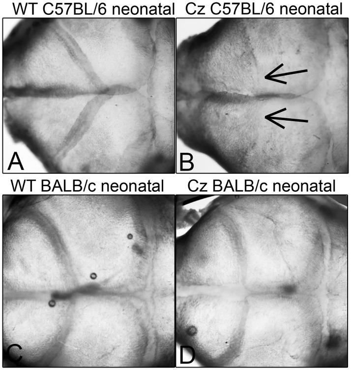
Because severity of craniosynostosis and associated craniofacial shape defects are variable on the mixed genetic background, FGFR2C342Y/+mice were backcrossed with BALB/c and C57BL/6 mice (obtained from Charles River Laboratories) for at least fifteen generations prior to experiments. BALB/c FGFR2C342Y/+mice have a more moderate form of Crouzon syndrome with craniosynostosis in the form of point fusions across the coronal suture first apparent between three and four weeks after birth [25]. C57BL/6 mice have a more severe form of Crouzon syndrome with craniosynostosis in the form of suture obliteration first apparent in neonatal mice (Fig 1). Both mice also exhibit abnormalities in the cranial base and deficient cranial base growth. Genotyping was performed as previously described [11, 25]. Briefly, DNA from tail digests was amplified by polymerase chain reaction using 5'-gagtaccatgctgactgcatgc-3' and 5'-ggagaggcatctctgtttcaagacc-3’ primers to yield a 200 base pair band for FGFR2 and a 300 base pair band for mutant FGFR2C342Y. Mice were fed ad libitum and housed under standard 12 hour dark/light cycles. Litters were randomly assigned to treatment/no treatment groups. Treated mice were injected with 1.0 x 10⁷ transforming units lentivirus or an equivalent volume of phosphate buffered saline via the jugular vein after birth. BALB/c mice (n = 12 FGFR2+/+ vehicle treated mice, n = 14 FGFR2C342Y/+ vehicle treated mice, n = 16 FGFR2C342Y/+ TNAP lentivirus treated mice) were euthanized by CO2 overdose at four weeks post-natal and C57BL/6 mice (n = 14 FGFR2+/+ vehicle treated mice, n = 14 FGFR2C342Y/+ vehicle treated mice, n = 17 FGFR2C342Y/+ TNAP lentivirus treated mice) were euthanized by CO2 overdose at three weeks post-natal for analyses. BALB/c mice were sacrificed at a later age than C57BL/6 mice because craniosynostosis onset occurs later in BALB/C than in C57BL/6 FGFR2C342Y/+ mice. Blood was collected by aortic puncture under surgical anesthesia. Mice were weighed, and body length was measured for each animal. All animal procedures were prospectively approved of by the University of Michigan’s University Committee on Use and Care of Animals (UCUCA, protocol PRO00006815). All samples were de-identified as to genotype and treatment group for analyses, then unblinded for statistical comparison. The primary outcome assessment was craniosynostosis incidence. Secondary outcome assessments included malocclusion incidence, cranial bone micro CT measurements, craniofacial shape measurements, and cranial base bone lengths.

Fig 1. Neonatal coronal suture fusion in C57BL/6 but not BALB/c FGFR2C342Y/+ mice.

Fig 1

Representative images of dissected C57BL/6 FGFR2+/+ (A), C57BL/6 FGFR2C342Y/+ (B), BALB/c FGFR2+/+ (C) and BALB/c FGFR2C342Y/+ (D) post-natal day 3 mouse skulls are shown. Parietal with frontal bone overlap is evident in all shown mice. Coronal suture fusion is only present in C57BL/6 FGFR2C342Y/+ mice. Arrows point to loss of the coronal suture in C57BL/6 FGFR2C342Y/+ (Cz) mice.

Serum analyses

Mice were fasted for six hours prior to blood collection. Alkaline phosphatase activity (AP) in serum was quantified using the colorimetric reagent 4-nitrophenyl-phosphate disodium hexahydrate (Sigma-Aldrich), as compared to a standard curve using commercially available alkaline phosphatase enzyme (Sigma-Aldrich). One unit of AP was defined as the amount of enzyme needed to generate 1 μmol of p-nitrophenol per minute. Inorganic phosphate quantifications were performed using commercially available kits (Pointe Scientific), also as compared to standard curves. The levels of alanine amino transferase (ALT) and Aspartate amino transferase (AST) were determined using commercially available colorimetric assay kits according to manufacturer instructions (Sigma-Aldrich) using serum from control and treated BALB/c mice, as these mice yielded more serum per mouse. One unit of ALT was defined as the amount of enzyme that would generate 1 nmol of pyruvate per minute. One unit of AST was defined as the amount of enzyme that would generate 1 nmol of glutamate per minute.

Micro Computed Tomography (micro CT)

Whole skulls were scanned at an 18 μm isotropic voxel resolution using the eXplore Locus SP micro CT imaging system (GE Healthcare Pre-Clinical Imaging, London, ON, Canada). Regions of interest (ROI’s) for parietal and frontal bones were established as 0.5 mm in length, 0.5 mm in width, and depth equivalent to thickness of bone, as previously described [17, 25]. Density, volume and mineral content of cranial bones from mice were measured using previously established methods using Microview version 2.2 software (GE Healthcare Pre-Clinical Imaging, London, ON) and established algorithms [34, 35]. Linear measurements of cranial base bones were measured on micro CT scans using Dolphin imaging software (Dolphin Imaging & Management Solutions).

Cranial suture and cranial base synchondrosis assessment

Fusions between frontal and parietal cranial bones (fusion of the coronal suture), fusion between parietal bones (fusion of the sagittal suture) and fusion of the intersphenoidal synchondrosis (ISS) and spheno-occipital synchondrosis (SOS) were identified on micro CT scans of mouse skulls. Cranial sutures and synchondroses were viewed using the two-dimensional micro CT slices in an orthogonal view across the entire length of the suture or synchondrosis, as previously described [17, 25]. Synchondroses were identified as fused or not fused. To distinguish between more vs. less severe forms of craniosynostosis, fusion of cranial suture was scored in the following categories: 0) normal open suture, 1) diminished suture width with no fusion 2) diminished suture width with point fusions, and 3) obliteration of the suture.

Reliability of suture fusion assessment was verified by both intra-operator and inter-operator reliability statistics by calculating intraclass correlation coefficients. Intra-operator reliability statistics was carried out by assessing suture fusion status of the coronal and sagittal sutures as well as the ISS and SOS synchondroses on fifteen micro CT scans by one investigator two times separated by a two-month period. Inter-operator reliability was carried out by analyzing fifteen micro CT scans by a second investigator. The intraclass correlation coefficient for intraoperator reliability for fusion assessment is .970 (p≤.0001) and the intraclass correlation coefficient for interoperator reliability is .972 (p≤.0001). Thus, there is high intraoperator and interoperator reliability for fusion assessment.

Linear measurements

Craniofacial linear skeletal measurements were taken using digital calipers on dissected skulls. Linear measurements were calculated using previously reported craniofacial skeletal landmarks [25, 36, 37], including standard measurements currently in use by the Craniofacial Mutant Mouse Resource of Jackson Laboratory (Bar Harbor, ME). Linear measurements were normalized to total skull length (measured from nasale to opisthion) to account for size differences between FGFR2+/+ and FGFR2C342Y/+ mice. Measurements were performed twice and an average of the two measurements was utilized for statistical comparison by genotype and treatment. Cranial base anterior-posterior bone lengths were measured on micro CT scans using Dolphin Imaging 11.0 software (Dolphin Imaging and Management Solutions, Chatsworth, CA), as previously described [26].

Statistics

Results from previous studies and this study demonstrate no sex difference for craniosynostosis in FGFR2C342Y/+ mice, therefore sexes were combined for analyses [25]. Normality of data was evaluated by D’Agostino & Pearson Tests. Students t test was used to compare groups that were of normal distribution and the Mann Whitney test was utilized to compare groups that were of non-normal distribution. Because serum AP levels varied in mice injected with the lentivirus, linear regressions were performed to determine if, and to what extent serum AP levels associated with changes body weight and serum liver enzyme levels. Incidences of craniosynostosis severity category, synchondrosis fusion and malocclusion were analyzed by the Fisher’s exact test.

Results

Injection with the TNAP expression lentivirus significantly increased serum AP levels in all of the treated mice (Table 1). BALB/c FGFR2C342Y/+ mice injected with the lentivirus increased serum AP levels by 1.2 U/mL when compared to BALB/c vehicle treated FGFR2C342Y/+ mice (p<0.0001) when measured at four weeks old. C57BL/6 FGFR2C342Y/+ mice injected with the TNAP expression lentivirus increased serum AP levels by 1.8 U/mL when compared to vehicle treated C57BL/6 FGFR2C342Y/+ mice (p<0.0001) when measured at three weeks old. No significant difference in serum AP levels were seen between untreated FGFR2C342Y/+ and FGFR2+/+ mice on the BALB/c or C57BL/6 backgrounds. Injection with the lentivirus did not alter serum inorganic phosphate (Pi) levels.

Table 1. Serum alkaline phosphatase and inorganic phosphate measurements in vehicle vs. TNAP treated BALB/c and C57BL/6 mice.

Strain Genotype Treatment Serum AP Level (units/ml) Serum Pi Level (mg/dl)
BALB/c FGFR2+/+ vehicle 0.03 +/- 0.01 10.9 +/- 0.8
BALB/c FGFR2C342Y/+ vehicle 0.03 +/- 0.78 9.9 +/- 1.4
BALB/c FGFR2C342Y/+ TNAP 1.30 +/- 0.54# 9.8 +/- 1.1
C57BL/6 FGFR2+/+ vehicle 0.01 +/- 0.01 9.5 +/- 1.0
C57BL/6 FGFR2C342Y/+ vehicle 0.02 +/- 0.01 8.7 +/- 1.1
C57BL/6 FGFR2C342Y/+ TNAP 1.93 +/- 0.77# 9.0 +/- 0.6

# p value < 0.01 between treatment groups.

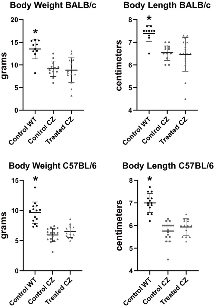
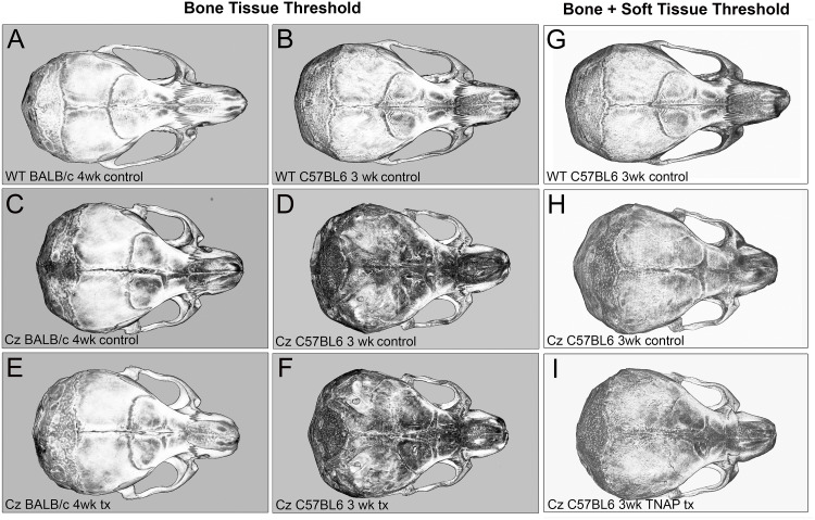
Statistical comparison of groups by Mann Whitney showed that FGFR2C342Y/+ mice weigh less and are shorter in body length than their FGFR2+/+ littermates, regardless of genetic background (Fig 2). Qualitative analysis of craniofacial skeletal shape suggested that FGFR2C342Y/+ mice differ in morphology from their FGFR2+/+ counterparts, and that delivery of mineral-targeted TNAP via lentivirus did not impact skull morphology (Fig 3).

Fig 2. Body weight and length of BALB/c and C57BL/6 mice.

Fig 2

Body weight and length of vehicle vs. TNAP treated mice are shown. FGFR2C342Y/+ mice are lighter and smaller than FGFR2+/+ mice, regardless of strain. Treatment does not alter body weight or body length. *p<0.01 between genotypes.

Fig 3. Isosurface micro CT images of control and TNAP treated FGFR2C342Y/+ mice.

Fig 3

Micro CT isosurface axial images of representative day 28 BALB/c and day 21 C57BL/6 mouse skulls are shown. Denser bone tissues are lighter in color. Control indicates no lentiviral delivery of TNAP. Tx indicates lentiviral delivery of TNAP. Comparison of BALB/c FGFR2+/+ (WT) mice (A), BALB/c FGFR2C342Y/+ (CZ) vehicle treated mice (C), and BALB/c FGFR2C342Y/+ (CZ) TNAP treated mice (E) indicates morphologic differences between genotypes but not treatment groups. Comparison of C57BL/6 FGFR2+/+ (WT) mice (B), C57BL/6 /c FGFR2C342Y/+ (CZ) control mice (D), and C57BL/6 FGFR2C342Y/+ (CZ) TNAP treated mice (F) indicates morphologic differences between genotypes but not treatment groups. (A-F) Isosurface images taken at a bone threshold. Skull images of control (D) and TNAP treated (F) C57BL/6 FGFR2C342Y/+ mice show both cranial vault and underlying cranial base due translucent poorly mineralized cranial bones in these mice. (G-I) Isosurface images taken at a threshold that includes both bone and soft tissue are provided for C57BL/6 FGFR2+/+ (WT) mice (G), C57BL/6 FGFR2C342Y/+ control mice (H), and C57BL/6 FGFR2C342Y/+ TNAP treated mice (I) are provided for improved visualization of these skulls.

Consistent with images shown in Fig 3, craniofacial skeletal linear measurements normalized to total skull length revealed many differences between FGFR2342Y/+ and FGFR2+/+ mice on both congenic backgrounds (Table 2). BALB/c FGFR2C342Y/+ mice had increased cranial height, cranial width, inner canthal distance, parietal bone length and cranial height to width ratios, with decreased nasal bone length. C57BL/6 FGFR2C342Y/+ mice had increased cranial height, cranial width, inner canthal distance, frontal bone length, parietal bone length and cranial height to width ratios, with decreased nose and nasal bone lengths. Treatment with the TNAP lentivirus did not alter craniofacial skeletal measurements in FGFR2C342Y/+ mice on either genetic background.

Table 2. Linear craniofacial skeletal measurements reveal.

Strain Measurement FGFR2+/+ vehicle FGFR2C342Y/+ vehicle FGFR2C342Y/+ TNAP
BALB/c Cranial Height 0.36 +/- 0.01* 0.45 +/- 0.01 0.45 +/- 0.01
BALB/c Cranial Width 0.55 +/- 0.01* 0.63 +/- 0.01 0.62 +/- 0.01
BALB/c Inner Canthal Distance 0.20 +/- 0.01* 0.25 +/- 0.01 0.26 +/- 0.01
BALB/c Nose Length 0.65+/- 0.01 0.65 +/- 0.01 0.65 +/- 0.01
BALB/c Nasal Bone Length 0.33 +/- 0.02* 0.32 +/- 0.01 0.32 +/- 0.03
BALB/c Frontal Bone length 0.33 +/- 0.02 0.33 +/- 0.01 0.34 +/- 0.03
BALB/c Parietal Bone Length 0.20 +/- 0.01* 0.25 +/- 0.02 0.26 +/- 0.01
BALB/c Ratio Height to Width 0.66 +/- 0.02* 0.72 +/- 0.02 0.72 +/- 0.02
C57BL/6 Cranial Height 0.38 +/- 0.01* 0.52 +/- 0.01 0.52 +/- 0.03
C57BL/6 Cranial Width 0.55 +/- 0.01* 0.64 +/- 0.01 0.64 +/- 0.02
C57BL/6 Inner Canthal Distance 0.23 +/- 0.01* 0.29 +/- 0.01 0.29 +/- 0.01
C57BL/6 Nose Length 0.65 +/- 0.01* 0.62 +/- 0.01 0.63 +/- .03
C57BL/6 Nasal Bone Length 0.32 +/- 0.01* 0.23 +/- 0.02 0.23 +/- 0.01
C57BL/6 Frontal Bone length 0.35 +/- 0.01* 0.40 +/- 0.02 0.41 +/- 0.03
C57BL/6 Parietal Bone Length 0.22 +/- 0.01* 0.26 +/- 0.02 0.27 +/- 0.02
C57BL/6 Ratio Height to Width 0.38 +/- 0.01* 0.52 +/- 0.01 0.52 +/- 0.03

No Rescue of Craniofacial Shape by TNAP Treatment in FGFR2C342Y/+ Mice.

Measurements are reported as normalized to total skull length.

* p value < 0.01 between genotypes

No statistical differences between treatment groups were found.

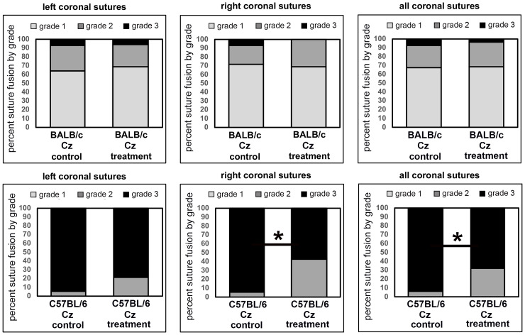
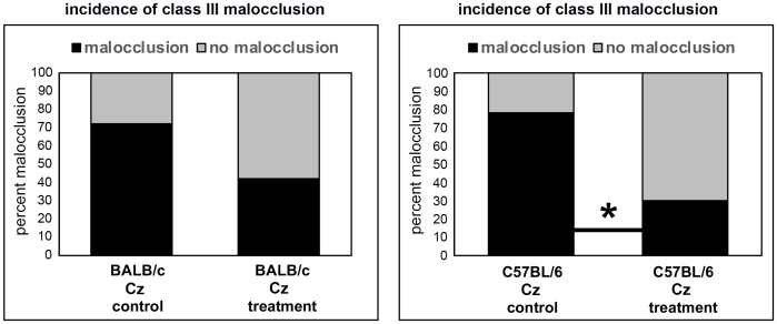
Qualitative micro CT based analysis of coronal suture fusions revealed diminished suture width (grade 1) in the majority of BALB/c FGFR2C342Y/+ vehicle treated mice with point fusions (grade 2) evident in approximately 30% of the mice. In the more severe C57BL/6 FGFR2C342Y/+ vehicle treated mice, the majority of the mice had obliteration of the suture (grade 3) with point fusions (grade 2) evident in the rest of the mice (Fig 4). The sagittal suture was not fused in any of the mice, and no cranial suture fusions were evident in FGFR2+/+ mice on either background. Treatment with recombinant TNAP had no significant impact on the incidence of grade 1, 2 or 3 of the coronal suture in the BALB/c FGFR2C342Y/+ mice. While there was only a trend for decreased fusion in the left coronal suture fusion upon treatment (control 94% suture obliteration vs. treated 78% suture obliteration), there was a significant decrease in coronal suture fusion upon treatment in the right coronal suture (control 94% suture obliteration vs. treated 57% suture obliteration, p<0.03). There was also a significant decrease in coronal suture fusion when combining both right and left coronal sutures (control 94% suture obliteration vs. treated 67% suture obliteration, p<0.02). Consistent with rescue of coronal suture fusion in C57BL/6 but not BALB/c mice, the incidence of malocclusion was also significantly decreased by treatment from 82.4% to 34.7% in C57BL/6 but not BALB/c FGFR2C342Y/+ mice (Fig 5, p<0.03).

Fig 4. Coronal suture fusion in vehicle and TNAP treated FGFR2C342Y/+ mice.

Fig 4

Percentage of mice with fusion of the coronal suture are shown. Fusion was scored in the following categories: 0) normal open suture, 1) diminished suture width with no fusion, 2) diminished suture width with point fusions across the suture, and 3) obliteration of the suture. Results show diminished suture obliteration in C57BL/6 FGFR2C342Y/+ (Cz) mice with no changes noted in BALB/c FGFR2C342Y/+ (Cz) mice. *p<0.03 between treatment groups.

Fig 5. Incidence of malocclusion in vehicle and TNAP treated FGFR2C342Y/+ mice.

Fig 5

Percentage of FGFR2C342Y/+ (Cz) mice with a class III malocclusion are shown. Treatment with TNAP lentivirus significantly diminished malocclusion in C57BL/6 FGFR2C342Y/+ (Cz) but not BALB/c FGFR2C342Y/+ (Cz) mice. *p<0.03.

Analysis of cranial base synchondrosis fusions revealed a 100% incidence of fusion of the inter-sphenoidal synchondrosis (ISS) in both strains of FGFR2C342Y/+ mice, with no fusions evident in FGFR2+/+ mice. Comparison of vehicle vs. treated mice revealed no significant change in the incidence of ISS fusion in FGFR2C342Y/+ mice on either genetic background. Spheno-occipital synchondronsis (SOS) fusion was seen in approximately half of C57BL/6 FGFR2C342Y/+ mice but rarely in BALB/c FGFR2C342Y/+ mice. Comparison of vehicle vs. treated mice revealed no significant change in the incidence of SOS fusion in FGFR2C342Y/+ mice on either genetic background. Measurements of cranial base bone lengths demonstrated decreased length of the basis-sphenoid and pre-sphenoid bones in both BALB/c and C57BL/6 vehicle treated FGFR2C342Y/+ as compared to FGFR2+/+ mice (Table 3). Treatment increased length of the pre-sphenoid bone in BALB/c and C57BL/6 FGFR2C342Y/+ mice, but not to the equivalent of pre-sphenoid bone length seen in FGFR2+/+ mice.

Table 3. Cranial base bone measurements in vehicle vs. TNAP treated BALB/C and C57Bl/6 mice.

Strain Genotype Treatment Basis Occipitus (mm) Basis Sphenoid (mm) Pre-Sphenoid (mm)
Balb/C FGFR2+/+ vehicle 3.1 +/- 0.1 2.9 +/- 0.1* 2.5 +/- 0.1*
Balb/C FGFR2C342Y/+ vehicle 2.9 +/- 0.3 2.4 +/- 0.3 1.8 +/- 0.1
Balb/C FGFR2C342Y/+ TNAP 2.9 +/- 0.2 2.5 +/- 0.3 1.9 +/- 0.1#
C57BL/6 FGFR2+/+ vehicle 2.8 +/- 0.2 2.9 +/- 0.2* 2.2 +/- 0.1*
C57BL/6 FGFR2C342Y/+ vehicle 2.8 +/- 0.1 2.7 +/- 0.1 1.7 +/- 0.1
C57BL/6 FGFR2C342Y/+ TNAP 3.0 +/- 0.2 2.6 +/-0.1 1.9 +/- 0.1#

* p value < 0.01 between genotypes.

# p value < 0.01 between treatment groups.

Micro CT based analyses of cranial bones demonstrated significantly diminished bone mineral density, tissue mineral content, tissue mineral density and bone volume fraction in frontal bones, plus significantly diminished tissue mineral density and bone volume fraction in parietal bones of vehicle treated FGFR2C342Y/+ mice when compared to FGFR2+/+ littermates on both BALB/c and C57BL/6 backgrounds (Table 4). Injection with the TNAP lentivirus significantly increased frontal bone mineral density, tissue mineral content and bone volume fraction, plus parietal bone volume fraction in FGFR2C342Y/+ mice on the BALB/c background. Injection with the TNAP lentivirus did not significantly impact any of the cranial bone parameters in FGFR2C342Y/+ mice on the C57BL/6 background.

Table 4. Cranial bone volume, density and mineral content in control and TNAP treated BALB/c and C57BL/6 mice.

Genotype Treatment Cranial Bone Bone Mineral Content (mg) Bone Mineral Density (mg/cc) Tissue Mineral Content (mg) Tissue Mineral Density (mg/cc) Bone Volume Fraction
BALB/c FGFR2+/+ vehicle Frontal 0.035 +/- 0.004 405 +/- 14* 0.028 +/- 0.007* 692 +/- 14* 0.41 +/- 0.03*
BALB/c FGFR2C342Y/+ vehicle Frontal 0.031 +/- 0.008 361 +/- 63 0.020 +/- 0.006# 671 +/- 18 0.36 +/- 0.00
BALB/c FGFR2C342Y/+ TNAP Frontal 0.035 +/- 0.005 401 +/- 26# 0.026 +/- 0.015# 683 +/- 29 0.42 +/- 0.06#
BALB/c FGFR2+/+ vehicle Parietal 0.034 +/- 0.004 405 +/- 12 0.023 +/- 0.005 693 +/- 15* 0.43 +/- 0.03*
BALB/c FGFR2C342Y/+ vehicle Parietal 0.031 +/- 0.007 396 +/- 39 0.020 +/- 0.005 669 +/- 24 0.36 +/- 0.06
BALB/c FGFR2C342Y/+ TNAP Parietal 0.034 +/- 0.006 403 +/- 27 0.025 +/- 0.012 691 +/- 36 0.42 +/- 0.07#
C57BL/6 FGFR2+/+ vehicle Frontal 0.017 +/- 0.003 245 +/- 25* 0.006 +/- 0.001* 570 +/- 18* 0.12 +/- 0.01*
C57BL/6 FGFR2C342Y/+ vehicle Frontal 0.013 +/- 0.002 209 +/- 25 0.004 +/- 0.001 519 +/- 26 0.10 +/- 0.01
C57BL/6 FGFR2C342Y/+ TNAP Frontal 0.016 +/- 0.004 225 +/- 30 0.004 +/- 0.001 553 +/- 40 0.11 +/- 0.01
C57BL/6 FGFR2+/+ vehicle Parietal 0.015 +/- 0.001 237 +/- 19 0.005 +/- 0.001 590 +/- 13* 0.12 +/- 0.02*
C57BL/6 FGFR2C342Y/+ vehicle Parietal 0.012 +/- 0.003 217 +/- 18 0.004 +/- 0.001 558 +/- 36 0.10 +/- 0.01
C57BL/6 FGFR2C342Y/+ TNAP Parietal 0.013 +/- 0.004 232 +/- 42 0.004 +/- 0.001 575 +/- 32 1.11 +/- 0.01

* p value < 0.01 between genotypes.

# p value < 0.01 between treatment groups.

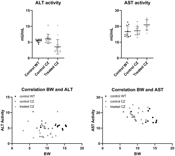
Biodistribution of lentivirus containing the recombinant mineral-targeted form of TNAP was previously shown to result in highest viral expression levels in the liver [30]. Because the treated mice in this study received recombinant TNAP via lentivirus, while the control group received no virus, we measured serum liver enzymes levels to determine if liver toxicity due to lentivirus could account for a change in phenotype in the treated mice, and/or the increase in serum AP levels seen in the treated mice. Alanine amino transferase (ALT) and Aspartate amino transferase (AST) are liver enzymes that, when seen at high levels in serum, are indicative of liver toxicity [38, 39]. Therefore, we tested serum ALT and AST levels in the mice. When compared by genotypes, no differences were noted between FGFR2+/+ and control FGFR2C342Y/+ mice for either ALT or AST enzymes. When compared by treatment groups, FGFR2C342Y/+ mice that were treated had significantly lower ALT levels than their control counterparts, and significantly higher AST levels than their control counterparts (Fig 6). We next performed correlation studies between ALT and AST serum levels with serum AP levels and body weight to better understand if lentiviral delivery of TNAP led to liver toxicity. No significant correlations were found between serum AP and AST levels for any of the groups. A significant reverse correlation was found between serum AP and ALT levels only for the treated FGFR2C342Y/+ group (r = -.73; 95% confidence interval of -.898 to -.361, p < .005). No significant correlations were found between serum AST or ALT levels and body weight for any of the groups. Together, these data show that the effect of lentiviral TNAP treatment decreased ALT and increased AST enzymes in serum, but had no effect on body weight of the mice and was not likely the cause of the observed increase in serum AP levels in treated mice.

Fig 6. Liver toxicity enzyme tests in vehicle and TNAP treated FGFR2C342Y/+ mice.

Fig 6

Alanine amino transferase (ALT) and aspartate amino transferase (AST) liver enzymes were measured in serum of vehicle and treated C57BL/6 mice. When compared by groups, no differences were seen between FGFR2+/+ and FGFR2C342Y/+ vehicle treated mice. Treatment decreased serum ALT (A) and increased serum AST (B) in FGFR2C342Y/+ mice. Linear regression analyses showed a significant reverse correlation (r = -.73; 95% CI of -.898 to -.361, p < .005) between AP and ALT enzyme levels in FGFR2C342Y/+ mice (C). No correlation was found between AP and AST enzyme levels in FGFR2C342Y/+ mice (D). No correlations were found between ALT (E) or AST (F) enzyme levels and body weight.

Discussion

Here we found that neonatal lentiviral delivery of recombinant TNAP increased cranial bone density, mineral content and volume fraction in the milder BALB/c FGFR2C342Y/+ model of Crouzon syndrome but not in the more severe C57BL/6 FGFR2C342Y/+ model of Crouzon syndrome. Increases in cranial bone density, mineral content and volume fraction by TNAP treatment in BALB/c Crouzon mice is consistent with results showing that recombinant mineral targeted TNAP treatment rescues mineralization of craniofacial and long bones in in the Alpl-/- mouse model of infantile hypophosphatasia (HPP) and humans with infantile and childhood HPP [30, 31, 33, 40]. It is curious that delivery of mineral-targeted TNAP did not increase cranial bone parameters in the more severe C57BL/6 FGFR2C342Y/+ mice. FGFR2+/+ C57BL/6 mice have significantly less cranial bone density, mineral content and volume fraction than BALB/c FGFR2+/+ mice, which is diminished to an even greater extent in FGFR2C342Y/+ C57BL/6 mice. While not quantified here, it is possible that the lack of rescue by TNAP is due to diminished bone matrix available to mineralize and/or a lack of cranial bone progenitor cells available to generate additional bone in the C57BL/6 mice, as TNAP is known to be essential for matrix mineralization [41] and for the formation of cranial bone progenitor cells [42].

Craniosynostosis severity was significantly diminished by treatment in the C57BL/6 FGFR2C342Y/+ mice. The vast majority of these mice typically exhibit obliteration of the coronal suture within a few days after birth (Fig 1). Suture obliteration went from 94% to 67% in the C57BL/6 FGFR2C342Y/+ mice upon treatment with exogenous TNAP by three weeks after birth. This result is consistent with the rescue of craniosynostosis seen in Alpl-/- mice treated with mineral targeted recombinant TNAP protein [33]. While far from a complete rescue, the data provided here demonstrate the potential efficacy of TNAP for diminishing severity of craniosynostosis in Crouzon syndrome. The data also support the idea that convergence exists between changes downstream of TNAP activity and FGFR2 signaling leading to coronal suture fusion. We recently showed that TNAP regulates expression of FGFR2 and Erk1,2 activity [42]. While the current study was originally based upon the hypothesis that FGF signaling regulates expression of TNAP, our more recent data suggest the alternative hypothesis that exogenous delivery of TNAP rescues Crouzon craniosynostosis because TNAP diminishes the overactive FGF and Erk1,2 signaling in FGFR2C342Y/+ cells. We are currently working to confirm this latter hypothesis and delineate how TNAP may mediate these changes.

This study was designed to include two mouse models of Crouzon syndrome: a severe form (C57BL/6 FGFR2C342Y/+ mice) that exhibited neonatal onset of coronal suture obliteration and a more moderate form (BALB/c FGFR2C342Y/+ mice) that exhibited small point fusions across the coronal suture evident by four weeks after birth. We anticipated that neonatal delivery of TNAP via lentivirus would have greater effects on the later onset BALB/c FGFR2C342Y/+ mouse model, as the drug would be delivered prior to onset of craniosynostosis. Results showed instead that the treatment significantly diminished severity of coronal suture fusion in the neonatal onset C5BL/6 FGFR2C342Y/+ mouse with no effect on coronal suture fusion in the postnatal onset BALB/c FGFR2C342Y/+ mouse. This data suggest that exogenous TNAP may be able to prevent cranial suture obliteration but not point fusions across the suture. The data also indicate that cellular mechanisms leading to craniosynostosis in FGFR2C342Y/+ mice are both independent and dependent upon TNAP.

While lentiviral TNAP treatment did not rescue cranial base synchondrosis fusion in the FGFR2C342Y/+ mice, length of the pre-sphenoid bone was mildly increased in the treated mice. This result is consistent with our previous study using archival lots of the lentivirus [27] which suggested changes in inferior skull morphology and may indicate that TNAP can promote cranial base growth in Crouzon syndrome. Delivery of recombinant TNAP also decreased the incidence of class III malocclusion in the C57BL/6 but not BALB/c FGFR2C342Y/+ mice. The malocclusion rescue is likely due to changes in coronal suture fusion as opposed to changes in the cranial base, as treatment with TNAP had minimal and similar changes on cranial base synchondroses and cranial base bone lengths in both C57BL/6 and BALB/c FGFR2C342Y/+ mice, with an effect on coronal suture fusion only in C57BL/6 FGFR2C342Y/+ mice.

A major limitation of this study is the method and timing of TNAP delivery. Lentiviral delivery of TNAP was not ideal, as the control group did not receive lentivirus which can lead to liver toxicity and increase serum AP levels. While we found that delivery of the TNAP lentivirus decreased serum ALT and increased serum AST levels in the treated mice, there was no impact on body weight of the mice. We interpret these findings to indicate that the treatment had minimal significant impact on overall health of the mice. In addition, serum AP levels did not correlate with serum AST levels and had a reverse correlation with serum ALT levels in treated FGFR2C342Y/+ mice. We interpret this latter data to indicate that while changes in the liver may have occurred, liver toxicity was not the cause for increased serum AP levels in the treated mice. Yet, we cannot be certain that the lentiviral vector itself had no impact on the craniofacial skeletal phenotype of the treated FGFR2C342Y/+ mice. A more ideal study design would incorporate empty lentiviral vector delivery to the control mice, or delivery of a recombinant TNAP protein to the mice. It would also be worthwhile to attempt prenatal delivery of TNAP, to determine if earlier treatment would have a greater impact on rescue of coronal suture fusion in C57BL/6 FGFR2C342Y/+ mice.

It is also worth noting that earlier time points to assess changes in the coronal suture were not performed. The decreased incidence of coronal suture obliteration in C57BL/6 Crouzon mice at 3 weeks postnatal suggests that onset occurred later in these mice. Earlier time points assessing the coronal suture after treatment could have confirmed that this was indeed the case. Histologic assessments of the suture at incremental time points during treatment would enable us to determine if the treatment delayed vs. reversed suture fusion.

Earlier onset of craniosynostosis increases morbidity, as brain growth is limited earlier and for a longer duration. In addition, the extent of cranial bone fusion can influence the type of surgical intervention needed for correction. Point fusions across the suture may only require an endoscopic suturectomy procedure, while suture obliteration requires more invasive full cranial vault remodeling procedures that carry increased risk of blood loss and long operation duration [43]. If TNAP postpones or reverses fusion of the coronal suture and/or allows for less invasive surgical procedures in Crouzon syndrome, treatment with TNAP could potentially diminish morbidity. Because the C57BL/6 FGFR2C342Y/+ Crouzon mice show neonatal onset of coronal suture fusion such that earlier intervention might be more efficacious, in future studies it might be appropriate to consider fetal delivery of TNAP. Most likely this would involve TNAP delivery using an adeno-associated virus (AAV), as opposed to the lentiviral virus utilized here, because AAV based gene delivery is considered safer due to the fact that there exists no known AAV based human diseases and because AAV rarely inserts into the genome, thereby diminishing risk for insertional mutagenesis [44]. Yet, fetal gene therapy comes with inherent risks including potential viral incorporation into germ cells leading to offspring transmission, as well as unintended disruption of developmental processes due to inappropriate timing and/or location of viral protein expression [45]. Our results overall indicate that use of an AAV based method for immediate post-birth TNAP delivery would likely diminish severity of craniosynostosis in the FGFR2C342Y/+ mice, suggesting potential utility in human neonates with severe Crouzon syndrome.

Acknowledgments

We thank Prof. José Luis Millán (La Jolla, CA) and Prof. Takashi Shimada (Tokyo, Japan) for providing us with the lentiviral vector expressing mineral targeted TNAP, as reported in Yamamoto et al., 2011 (30).

Data Availability

The data underlying the results presented in the study are available from the University of Michigan's Deep Blue Database; https://deepblue.lib.umich.edu/data/collections/7m01bk78r?locale=en.

Funding Statement

This work was supported by grant R01DE02582701. to N.E.H. from the National Institute of Dental and Craniofacial Research (NIDCR). The funders played no role in the study design, data collection, data analysis, decision to publish, or preparation of the manuscript.

References

  • 1.Flapper WJ, Anderson PJ, Roberts RM, et al. Intellectual outcomes following protocol management in Crouzon, Pfeiffer, and Muenke syndromes. J Craniofac Surg. 2009;20(4):1252–5. Epub 2009/07/25. 10.1097/SCS.0b013e3181acdf9a . [DOI] [PubMed] [Google Scholar]
  • 2.McCarthy JG, Warren SM, Bernstein J, et al. Parameters of care for craniosynostosis. Cleft Palate Craniofac J. 2012;49 Suppl:1S–24S. Epub 2011/08/19. 10.1597/11-138 . [DOI] [PubMed] [Google Scholar]
  • 3.Morriss-Kay GM, Wilkie AO. Growth of the normal skull vault and its alteration in craniosynostosis: insights from human genetics and experimental studies. J Anat. 2005;207(5):637–53. Epub 2005/11/30. 10.1111/j.1469-7580.2005.00475.x . [DOI] [PMC free article] [PubMed] [Google Scholar]
  • 4.Okajima K, Robinson LK, Hart MA, et al. Ocular anterior chamber dysgenesis in craniosynostosis syndromes with a fibroblast growth factor receptor 2 mutation. Am J Med Genet. 1999;85(2):160–70. Epub 1999/07/16. . [DOI] [PubMed] [Google Scholar]
  • 5.Rasmussen SA, Yazdy MM, Frias JL, et al. Priorities for public health research on craniosynostosis: summary and recommendations from a Centers for Disease Control and Prevention-sponsored meeting. Am J Med Genet A. 2008;146A(2):149–58. Epub 2007/12/15. 10.1002/ajmg.a.32106 . [DOI] [PubMed] [Google Scholar]
  • 6.Seruya M, Oh AK, Boyajian MJ, et al. Treatment for delayed presentation of sagittal synostosis: challenges pertaining to occult intracranial hypertension. J Neurosurg Pediatr. 2011;8(1):40–8. Epub 2011/07/05. 10.3171/2011.4.PEDS1160 . [DOI] [PubMed] [Google Scholar]
  • 7.Abe H, Ikota T, Akino M, et al. Functional prognosis of surgical treatment of craniosynostosis. Childs Nerv Syst. 1985;1(1):53–61. Epub 1985/01/01. 10.1007/BF00706732 . [DOI] [PubMed] [Google Scholar]
  • 8.Baird LC, Gonda D, Cohen SR, et al. Craniofacial reconstruction as a treatment for elevated intracranial pressure. Childs Nerv Syst. 2012;28(3):411–8. Epub 2011/11/10. 10.1007/s00381-011-1615-6 . [DOI] [PubMed] [Google Scholar]
  • 9.Renier D, Lajeunie E, Arnaud E, et al. Management of craniosynostoses. Childs Nerv Syst. 2000;16(10–11):645–58. Epub 2001/01/11. 10.1007/s003810000320 . [DOI] [PubMed] [Google Scholar]
  • 10.Chen L, Li D, Li C, et al. A Ser252Trp [corrected] substitution in mouse fibroblast growth factor receptor 2 (Fgfr2) results in craniosynostosis. Bone. 2003;33(2):169–78. Epub 2003/09/23. 10.1016/s8756-3282(03)00222-9 . [DOI] [PubMed] [Google Scholar]
  • 11.Eswarakumar VP, Horowitz MC, Locklin R, et al. A gain-of-function mutation of Fgfr2c demonstrates the roles of this receptor variant in osteogenesis. Proc Natl Acad Sci U S A. 2004;101(34):12555–60. Epub 2004/08/19. 10.1073/pnas.0405031101 . [DOI] [PMC free article] [PubMed] [Google Scholar]
  • 12.Reardon W, Winter RM, Rutland P, et al. Mutations in the fibroblast growth factor receptor 2 gene cause Crouzon syndrome. Nat Genet. 1994;8(1):98–103. Epub 1994/09/01. 10.1038/ng0994-98 . [DOI] [PubMed] [Google Scholar]
  • 13.Wilkie AO, Slaney SF, Oldridge M, et al. Apert syndrome results from localized mutations of FGFR2 and is allelic with Crouzon syndrome. Nat Genet. 1995;9(2):165–72. Epub 1995/02/01. 10.1038/ng0295-165 . [DOI] [PubMed] [Google Scholar]
  • 14.Whyte MP, Greenberg CR, Salman NJ, et al. Enzyme-replacement therapy in life-threatening hypophosphatasia. N Engl J Med. 2012;366(10):904–13. Epub 2012/03/09. 10.1056/NEJMoa1106173 . [DOI] [PubMed] [Google Scholar]
  • 15.Collmann H, Mornet E, Gattenlohner S, et al. Neurosurgical aspects of childhood hypophosphatasia. Childs Nerv Syst. 2009;25(2):217–23. Epub 2008/09/05. 10.1007/s00381-008-0708-3 . [DOI] [PubMed] [Google Scholar]
  • 16.Fraser D. Hypophosphatasia. Am J Med. 1957;22(5):730–46. Epub 1957/05/01. 10.1016/0002-9343(57)90124-9 . [DOI] [PubMed] [Google Scholar]
  • 17.Liu J, Nam HK, Campbell C, et al. Tissue-nonspecific alkaline phosphatase deficiency causes abnormal craniofacial bone development in the Alpl(-/-) mouse model of infantile hypophosphatasia. Bone. 2014;67:81–94. Epub 2014/07/12. 10.1016/j.bone.2014.06.040 . [DOI] [PMC free article] [PubMed] [Google Scholar]
  • 18.Galvin BD, Hart KC, Meyer AN, et al. Constitutive receptor activation by Crouzon syndrome mutations in fibroblast growth factor receptor (FGFR)2 and FGFR2/Neu chimeras. Proc Natl Acad Sci U S A. 1996;93(15):7894–9. Epub 1996/07/23. 10.1073/pnas.93.15.7894 . [DOI] [PMC free article] [PubMed] [Google Scholar]
  • 19.Neilson KM, Friesel RE. Constitutive activation of fibroblast growth factor receptor-2 by a point mutation associated with Crouzon syndrome. J Biol Chem. 1995;270(44):26037–40. Epub 1995/11/03. 10.1074/jbc.270.44.26037 . [DOI] [PubMed] [Google Scholar]
  • 20.Robertson SC, Meyer AN, Hart KC, et al. Activating mutations in the extracellular domain of the fibroblast growth factor receptor 2 function by disruption of the disulfide bond in the third immunoglobulin-like domain. Proc Natl Acad Sci U S A. 1998;95(8):4567–72. Epub 1998/05/16. 10.1073/pnas.95.8.4567 . [DOI] [PMC free article] [PubMed] [Google Scholar]
  • 21.Pfaff MJ, Xue K, Li L, et al. FGFR2c-mediated ERK-MAPK activity regulates coronal suture development. Dev Biol. 2016;415(2):242–50. Epub 2016/04/02. 10.1016/j.ydbio.2016.03.026 . [DOI] [PMC free article] [PubMed] [Google Scholar]
  • 22.Hatch NE, Li Y, Franceschi RT. FGF2 stimulation of the pyrophosphate-generating enzyme, PC-1, in pre-osteoblast cells is mediated by RUNX2. J Bone Miner Res. 2009;24(4):652–62. Epub 2008/12/04. 10.1359/jbmr.081213 . [DOI] [PMC free article] [PubMed] [Google Scholar]
  • 23.Hatch NE, Nociti F, Swanson E, et al. FGF2 alters expression of the pyrophosphate/phosphate regulating proteins, PC-1, ANK and TNAP, in the calvarial osteoblastic cell line, MC3T3E1(C4). Connect Tissue Res. 2005;46(4–5):184–92. Epub 2006/03/21. 10.1080/03008200500237203 . [DOI] [PubMed] [Google Scholar]
  • 24.Liu J, Kwon TG, Nam HK, et al. Craniosynostosis-associated Fgfr2(C342Y) mutant bone marrow stromal cells exhibit cell autonomous abnormalities in osteoblast differentiation and bone formation. Biomed Res Int. 2013;2013:292506 Epub 2013/06/14. . [DOI] [PMC free article] [PubMed] [Google Scholar]
  • 25.Liu J, Nam HK, Wang E, et al. Further analysis of the Crouzon mouse: effects of the FGFR2(C342Y) mutation are cranial bone-dependent. Calcif Tissue Int. 2013;92(5):451–66. Epub 2013/01/30. 10.1007/s00223-013-9701-2 . [DOI] [PMC free article] [PubMed] [Google Scholar]
  • 26.Nam HK, Sharma M, Liu J, et al. Tissue Nonspecific Alkaline Phosphatase (TNAP) Regulates Cranial Base Growth and Synchondrosis Maturation. Front Physiol. 2017;8:161 Epub 2017/04/06. 10.3389/fphys.2017.00161 . [DOI] [PMC free article] [PubMed] [Google Scholar]
  • 27.Wang E, Nam HK, Liu J, et al. The effects of tissue-non-specific alkaline phosphatase gene therapy on craniosynostosis and craniofacial morphology in the FGFR2C342Y/+ mouse model of Crouzon craniosynostosis. Orthod Craniofac Res. 2015;18 Suppl 1:196–206. Epub 2015/04/14. 10.1111/ocr.12080 . [DOI] [PMC free article] [PubMed] [Google Scholar]
  • 28.Hanawa H, Yamamoto M, Zhao H, et al. Optimized lentiviral vector design improves titer and transgene expression of vectors containing the chicken beta-globin locus HS4 insulator element. Mol Ther. 2009;17(4):667–74. Epub 2009/02/19. 10.1038/mt.2009.1 . [DOI] [PMC free article] [PubMed] [Google Scholar]
  • 29.Nishioka T, Tomatsu S, Gutierrez MA, et al. Enhancement of drug delivery to bone: characterization of human tissue-nonspecific alkaline phosphatase tagged with an acidic oligopeptide. Mol Genet Metab. 2006;88(3):244–55. Epub 2006/04/18. 10.1016/j.ymgme.2006.02.012 . [DOI] [PMC free article] [PubMed] [Google Scholar]
  • 30.Yamamoto S, Orimo H, Matsumoto T, et al. Prolonged survival and phenotypic correction of Akp2(-/-) hypophosphatasia mice by lentiviral gene therapy. J Bone Miner Res. 2011;26(1):135–42. Epub 2010/08/06. 10.1002/jbmr.201 . [DOI] [PMC free article] [PubMed] [Google Scholar]
  • 31.Millan JL, Narisawa S, Lemire I, et al. Enzyme replacement therapy for murine hypophosphatasia. J Bone Miner Res. 2008;23(6):777–87. Epub 2007/12/19. 10.1359/jbmr.071213 . [DOI] [PMC free article] [PubMed] [Google Scholar]
  • 32.Yadav MC, Lemire I, Leonard P, et al. Dose response of bone-targeted enzyme replacement for murine hypophosphatasia. Bone. 2011;49(2):250–6. Epub 2011/04/05. 10.1016/j.bone.2011.03.770 . [DOI] [PMC free article] [PubMed] [Google Scholar]
  • 33.Liu J, Campbell C, Nam HK, et al. Enzyme replacement for craniofacial skeletal defects and craniosynostosis in murine hypophosphatasia. Bone. 2015;78:203–11. Epub 2015/05/12. 10.1016/j.bone.2015.05.005 . [DOI] [PMC free article] [PubMed] [Google Scholar]
  • 34.Meganck JA, Kozloff KM, Thornton MM, et al. Beam hardening artifacts in micro-computed tomography scanning can be reduced by X-ray beam filtration and the resulting images can be used to accurately measure BMD. Bone. 2009;45(6):1104–16. Epub 2009/08/05. 10.1016/j.bone.2009.07.078 . [DOI] [PMC free article] [PubMed] [Google Scholar]
  • 35.Umoh JU, Sampaio AV, Welch I, et al. In vivo micro-CT analysis of bone remodeling in a rat calvarial defect model. Phys Med Biol. 2009;54(7):2147–61. Epub 2009/03/17. 10.1088/0031-9155/54/7/020 . [DOI] [PubMed] [Google Scholar]
  • 36.Perlyn CA, DeLeon VB, Babbs C, et al. The craniofacial phenotype of the Crouzon mouse: analysis of a model for syndromic craniosynostosis using three-dimensional MicroCT. Cleft Palate Craniofac J. 2006;43(6):740–8. Epub 2006/11/16. 10.1597/05-212 . [DOI] [PubMed] [Google Scholar]
  • 37.Richtsmeier JT, Baxter LL, Reeves RH. Parallels of craniofacial maldevelopment in Down syndrome and Ts65Dn mice. Dev Dyn. 2000;217(2):137–45. Epub 2000/03/08. . [DOI] [PubMed] [Google Scholar]
  • 38.Franca RF, Zucoloto S, da Fonseca BA. A BALB/c mouse model shows that liver involvement in dengue disease is immune-mediated. Exp Mol Pathol. 2010;89(3):321–6. Epub 2010/08/03. 10.1016/j.yexmp.2010.07.007 . [DOI] [PubMed] [Google Scholar]
  • 39.Huang F, Zhang W, Gong G, et al. Experimental infection of Balb/c nude mice with Hepatitis E virus. BMC Infect Dis. 2009;9:93 Epub 2009/06/16. 10.1186/1471-2334-9-93 . [DOI] [PMC free article] [PubMed] [Google Scholar]
  • 40.Whyte MP, Simmons JH, Moseley S, et al. Asfotase alfa for infants and young children with hypophosphatasia: 7 year outcomes of a single-arm, open-label, phase 2 extension trial. Lancet Diabetes Endocrinol. 2018. Epub 2018/12/19. 10.1016/S2213-8587(18)30307-3 . [DOI] [PubMed] [Google Scholar]
  • 41.Murshed M, Harmey D, Millan JL, et al. Unique coexpression in osteoblasts of broadly expressed genes accounts for the spatial restriction of ECM mineralization to bone. Genes Dev. 2005;19(9):1093–104. Epub 2005/04/19. 10.1101/gad.1276205 . [DOI] [PMC free article] [PubMed] [Google Scholar]
  • 42.Nam HK, Vesela I, Siismets E, et al. Tissue nonspecific alkaline phosphatase promotes calvarial progenitor cell cycle progression and cytokinesis via Erk1,2. Bone. 2018;120:125–36. Epub 2018/10/21. 10.1016/j.bone.2018.10.013 . [DOI] [PMC free article] [PubMed] [Google Scholar]
  • 43.Warren SM, Proctor MR, Bartlett SP, et al. Parameters of care for craniosynostosis: craniofacial and neurologic surgery perspectives. Plast Reconstr Surg. 2012;129(3):731–7. Epub 2012/03/01. 10.1097/PRS.0b013e3182412a50 . [DOI] [PubMed] [Google Scholar]
  • 44.Zheng CX, Wang SM, Bai YH, et al. Lentiviral Vectors and Adeno-Associated Virus Vectors: Useful Tools for Gene Transfer in Pain Research. Anat Rec (Hoboken). 2018;301(5):825–36. Epub 2017/11/18. 10.1002/ar.23723 . [DOI] [PMC free article] [PubMed] [Google Scholar]
  • 45.DeWeerdt S. Prenatal gene therapy offers the earliest possible cure. Nature. 2018;564(7735):S6–S8. Epub 2018/12/14. 10.1038/d41586-018-07643-z . [DOI] [PubMed] [Google Scholar]

Decision Letter 0

JJ Cray Jr

6 Mar 2020

PONE-D-20-01614

Viral delivery of tissue nonspecific alkaline phosphatase diminishes craniosynostosis in one of two FGFR2C342Y/+ mouse models of Crouzon syndrome.

PLOS ONE

Dear Dr Hatch,

Thank you for submitting your manuscript to PLOS ONE. After careful consideration, we feel that it has merit but does not fully meet PLOS ONE’s publication criteria as it currently stands. Therefore, we invite you to submit a revised version of the manuscript that addresses the points raised during the review process.

There are some minor issues to address. Also please consider the addition of the histological data requested by the reviewer, and an explanation as to my caliper measures were used instead of quantitating the micro-ct.

We would appreciate receiving your revised manuscript by Apr 20 2020 11:59PM. When you are ready to submit your revision, log on to https://www.editorialmanager.com/pone/ and select the 'Submissions Needing Revision' folder to locate your manuscript file.

If you would like to make changes to your financial disclosure, please include your updated statement in your cover letter.

To enhance the reproducibility of your results, we recommend that if applicable you deposit your laboratory protocols in protocols.io, where a protocol can be assigned its own identifier (DOI) such that it can be cited independently in the future. For instructions see: http://journals.plos.org/plosone/s/submission-guidelines#loc-laboratory-protocols

Please include the following items when submitting your revised manuscript:

  • A rebuttal letter that responds to each point raised by the academic editor and reviewer(s). This letter should be uploaded as separate file and labeled 'Response to Reviewers'.

  • A marked-up copy of your manuscript that highlights changes made to the original version. This file should be uploaded as separate file and labeled 'Revised Manuscript with Track Changes'.

  • An unmarked version of your revised paper without tracked changes. This file should be uploaded as separate file and labeled 'Manuscript'.

Please note while forming your response, if your article is accepted, you may have the opportunity to make the peer review history publicly available. The record will include editor decision letters (with reviews) and your responses to reviewer comments. If eligible, we will contact you to opt in or out.

We look forward to receiving your revised manuscript.

Kind regards,

JJ Cray Jr., Ph.D.

Academic Editor

PLOS ONE

Journal Requirements:

When submitting your revision, we need you to address these additional requirements:

1. Please ensure that your manuscript meets PLOS ONE's style requirements, including those for file naming. The PLOS ONE style templates can be found at http://www.plosone.org/attachments/PLOSOne_formatting_sample_main_body.pdf and http://www.plosone.org/attachments/PLOSOne_formatting_sample_title_authors_affiliations.pd

2. We note that you have stated that you will provide repository information for your data at acceptance. Should your manuscript be accepted for publication, we will hold it until you provide the relevant accession numbers or DOIs necessary to access your data. If you wish to make changes to your Data Availability statement, please describe these changes in your cover letter and we will update your Data Availability statement to reflect the information you provide.

3. Your ethics statement must appear in the Methods section of your manuscript. If your ethics statement is written in any section besides the Methods, please move it to the Methods section and delete it from any other section. Please also ensure that your ethics statement is included in your manuscript, as the ethics section of your online submission will not be published alongside your manuscript.

[Note: HTML markup is below. Please do not edit.]

Reviewers' comments:

Reviewer's Responses to Questions

Comments to the Author

1. Is the manuscript technically sound, and do the data support the conclusions?

The manuscript must describe a technically sound piece of scientific research with data that supports the conclusions. Experiments must have been conducted rigorously, with appropriate controls, replication, and sample sizes. The conclusions must be drawn appropriately based on the data presented.

Reviewer #1: Yes

Reviewer #2: Yes

**********

2. Has the statistical analysis been performed appropriately and rigorously?

Reviewer #1: Yes

Reviewer #2: Yes

**********

3. Have the authors made all data underlying the findings in their manuscript fully available?

The PLOS Data policy requires authors to make all data underlying the findings described in their manuscript fully available without restriction, with rare exception (please refer to the Data Availability Statement in the manuscript PDF file). The data should be provided as part of the manuscript or its supporting information, or deposited to a public repository. For example, in addition to summary statistics, the data points behind means, medians and variance measures should be available. If there are restrictions on publicly sharing data—e.g. participant privacy or use of data from a third party—those must be specified.

Reviewer #1: Yes

Reviewer #2: Yes

**********

4. Is the manuscript presented in an intelligible fashion and written in standard English?

PLOS ONE does not copyedit accepted manuscripts, so the language in submitted articles must be clear, correct, and unambiguous. Any typographical or grammatical errors should be corrected at revision, so please note any specific errors here.

Reviewer #1: Yes

Reviewer #2: Yes

**********

5. Review Comments to the Author

Please use the space provided to explain your answers to the questions above. You may also include additional comments for the author, including concerns about dual publication, research ethics, or publication ethics. (Please upload your review as an attachment if it exceeds 20,000 characters)

Reviewer #1: The manuscript “Tissue nonspecific alkaline phosphatase improves bone quality but does not alleviate craniosynostosis in the FGFR2C342Y/+ mouse model of Crouzon syndrome” has been revised well. In particular, the changes made to the introduction and discussion have improved the manuscript significantly. The study that aims to determine if delivery of TNAP prevents or diminishes FGFR2C342Y driven craniosynostosis. This is an interesting and worthwhile investigation seeking to identify a pharmacological treatment for craniosynostosis. Though the authors have addressed the major concerns from the previous review, a number of minor issues still need to be addressed.

Minor Revisions:

1. Abstract Line 33: The use of the word improved is vague. As you have not defined the quality/ described normal/ unaffected bone, improved is confusing. Consider changing this language to be directional, as in “increased bone volume as compared to control.”

2. Introduction line 65: “… we found statistical differences” – What does this mean? Perhaps significant would be a better word in place of statistical?

3. Introduction lines 74-76: Consider rephrasing. The way that this is written indicates one model of Crouzon syndrome, and though only one genetic target was tested here there are in fact two different models of the human disease being studied. Please rephrase the second half of this sentence “ a model of Crouzon craniosynostosis syndrome”

4. Methods section Micro Computed Tomography, Line 149: Please be consistent with referring to Micro Computed Tomography. Either always write it out or abbreviate it at the first instance. It is in different forms (written out, micro CT, micro-CT) throughout the methods, figure legends, and results.

5. Methods section Statistics, Line 186, 187: There are extra words in this sentence. Please revise carefully.

6. Results Table 1, Lines 204-205: Please be consistent with capitalization in the table title. If measurements is capitalized, then vehicle, treatment, and mice should also be capitalized. Also, be consistent with table format. Have the heading bolded all the time or none of the time.

7. Results Table 2, Lines 231-232: Please be consistent with capitalization in the table title. Also, be consistent with table format. Have the heading bolded all the time or none of the time.

8. Results, Line 249: This seems to be the first instance of the term “wild type” please use the gene instead as I believe you mean unaffected and not a true wild-type.

9. Results Table 3, Lines 272-273: Please be consistent with capitalization in the table title. Also, be consistent with table format. Have the heading bolded all the time or none of the time.

10. Results, Line 296: The term “wild type” is used again and I again believe you mean unaffected, which would make using the gene notation more appropriate.

11. Results Table 4, Lines 302-303: Please be consistent with capitalization in the table title. Also, be consistent with table format. Have the heading bolded all the time or none of the time.

12. Results Line 324-327: Please revise this sentence to the following as data is always plural, and the sentence in currently in multiple tenses: “Together, these fata show that the effect of lentiviral TNAP treatment decreased ALT and increased AST enzymes in serum, but had not effect on body weight of the mice and was not likely the cause of the observed increase in serum AP levels in treated mice.”

13. Discussion Line 330: The use of the word improved here is also vague. Consider changing this language as suggested above.

14. Discussion Line 351: delete “does”

15. Discussion Line 352: As data is always a plural (datum is singular) supports should be changes to support. Please check for agreement of this throughout your discussion, Lines 356, 368,369 need to be revised.

16: Discussion line 369: is there a functional difference between point fusions across the suture and complete obliteration? Perhaps this could factor into the discussion to some degree as a means of better situating this study within a clinical context in the treatment of craniosynostosis.

17. Figure Legends Figure 1, Line 571: As you have indicated that the fusion of the coronal suture occurs early (pre-natally) in the C57BL/6 model please indicate that the images here are from post-natal day 3 mice. Also, within Figure 1 the notation should be consistent. Either always CZ or always Crouzon, and always C57BL6 or C57Bl6. Please check that you are consistent with these notations throughout the manuscript, tables, legends, and figures.

18. Figure Legends Figure 4, Line 592: Please be consistent throughout the manuscript, particularly within figures and legends. Either use Crouzon or CZ, not both.

19. Figure 3: what does the notation no tx vs. tx stand for? This is not clear in the legend.

20. Figure 4. Please revise this figure to have on legend that includes all of the colors and display if possible all colors on each graph. Also, please use proper and consistent capitalization on titles throughout this figure and all other figures.

21. Figure 5: Why are there not titles on these graphs?

22. Figure 6: Please indicate on the graphs and or in the legend which strain was used for these experiments.

Reviewer #2: Overall this is an interesting study, but it feels unfinished, even though the work presented here is a continuation -with significant overlap- from previous work. The study can easily be improved by the addition of histology data of the coronal suture for example. If the treatment with TNAP does indeed delay or reverse the process of synostosis this could have been shown by a longitudinal study of the suture, analysing the morphology of the suture and the histology of the sutural mesenchyme.

Material and methods – Linear measurements: I don’t understand why the authors decided to perform skull measurements using a caliper when they have microCT scans available. Quantitative data using microCT scans is far superior. It would be helpful if the authors could explain why they have decided to not do this.

Material and methods – Statistics: While the authors mention a previous study that found no differences between male and female Fgfr2-C342Y mice regarding the craniosynostosis phenotype, it is currently considered bad practise to use a mixed sex cohort, especially when testing a pharmacological intervention. I recommend the authors change this in the future.

Discussion: It would be interesting if the authors could comment on the feasibility of administering TNAP in utero.

The term perinatal is not used correctly. It refers to the period shortly before to shortly after birth. In the Abstract (line 35) perinatal is used where it should be postnatal. Also, in line 67, the addition of the term perinatal to prenatal and postnatal is superfluous.

In Figure 3, it would be helpful if the authors commented on if the images are representative for their cohorts. The quality of the images in D and F (arguably the most interesting) is poor due to a likely issue with contrast. As a result the coronal sutures -or what’s left of them- are impossible to see.

In Table 3, the # referring to the non-significant differences is missing.

**********

6. PLOS authors have the option to publish the peer review history of their article (what does this mean?). If published, this will include your full peer review and any attached files.

If you choose “no”, your identity will remain anonymous but your review may still be made public.

Do you want your identity to be public for this peer review? For information about this choice, including consent withdrawal, please see our Privacy Policy.

Reviewer #1: No

Reviewer #2: No

[NOTE: If reviewer comments were submitted as an attachment file, they will be attached to this email and accessible via the submission site. Please log into your account, locate the manuscript record, and check for the action link "View Attachments". If this link does not appear, there are no attachment files to be viewed.]

While revising your submission, please upload your figure files to the Preflight Analysis and Conversion Engine (PACE) digital diagnostic tool, https://pacev2.apexcovantage.com/. PACE helps ensure that figures meet PLOS requirements. To use PACE, you must first register as a user. Registration is free. Then, login and navigate to the UPLOAD tab, where you will find detailed instructions on how to use the tool. If you encounter any issues or have any questions when using PACE, please email us at figures@plos.org. Please note that Supporting Information files do not need this step.

PLoS One. 2020 May 29;15(5):e0234073. doi: 10.1371/journal.pone.0234073.r003

Author response to Decision Letter 0


20 Apr 2020

Response to Journal:

1. Please ensure that your manuscript meets PLOS ONE's style requirements, including those for file naming.

References are now in PLOS ONE style.

2. We note that you have stated that you will provide repository information for your data at acceptance. Should your manuscript be accepted for publication, we will hold it until you provide the relevant accession numbers or DOIs necessary to access your data. If you wish to make changes to your Data Availability statement, please describe these changes in your cover letter and we will update your Data Availability statement to reflect the information you provide.

Online data repository information is now provided with the submission.

3. Your ethics statement must appear in the Methods section of your manuscript. If your ethics statement is written in any section besides the Methods, please move it to the Methods section and delete it from any other section. Please also ensure that your ethics statement is included in your manuscript, as the ethics section of your online submission will not be published alongside your manuscript.

Animal use ethics statement including prior approval by university committee, animal protocol number and type of euthanasia performed is now included in the methods section.

Response to Reviewers:

We would like to thank the reviewers again for their thorough manuscript review and thoughtful critiques. We appreciate that the reviewers agreed that we: 1) provide a technically sound manuscript in which the data support the conclusions, 2) performed an appropriate and rigorous statistical analysis, 3) made all data underlying the findings in the manuscript fully available and 4) presented the manuscript in an intelligible fashion written in standard English.

We agree with and have made relevant changes in response to both Reviewer #1 and Reviewer #2 critiques and suggestions. The only exception to this is our inability to provide histologic data at this time (due to COVID-19 based university research activity ramp down). We were fully prepared to provide histology at final time points until this ramp down. In an attempt to provide some additional data given the lack of histology, in this revision we have an additional figure showing images of dissected skulls of the mice (Fig. 4). We hope that the editor and reviewers agree that this manuscript submission is worthy of publication.

Reviewer #1: The manuscript “Tissue nonspecific alkaline phosphatase improves bone quality but does not alleviate craniosynostosis in the FGFR2C342Y/+ mouse model of Crouzon syndrome” has been revised well. In particular, the changes made to the introduction and discussion have improved the manuscript significantly. The study that aims to determine if delivery of TNAP prevents or diminishes FGFR2C342Y driven craniosynostosis. This is an interesting and worthwhile investigation seeking to identify a pharmacological treatment for craniosynostosis. Though the authors have addressed the major concerns from the previous review, a number of minor issues still need to be addressed.

Minor Revisions:

1. Abstract Line 33: The use of the word improved is vague. As you have not defined the quality/ described normal/ unaffected bone, improved is confusing. Consider changing this language to be directional, as in “increased bone volume as compared to control.”

We agree that “improved” is vague. The word has been replaced by “increased” and “as compared to controls” has been added in the abstract.

2. Introduction line 65: “… we found statistical differences” – What does this mean? Perhaps significant would be a better word in place of statistical?

The word “statistical” has been replaced with “significant” in this sentence.

3. Introduction lines 74-76: Consider rephrasing. The way that this is written indicates one model of Crouzon syndrome, and though only one genetic target was tested here there are in fact two different models of the human disease being studied. Please rephrase the second half of this sentence “ a model of Crouzon craniosynostosis syndrome”

This phrase has been replaced with, “in BALB/c FGFR2C342Y/+ mice, a model of less severe Crouzon craniosynostosis syndrome and/or in C67BL/6 FGFR2C342Y/+ mice, a model of more severe Crouzon craniosynostosis syndrome” in the introduction section to the manuscript (lines 75-77).

4. Methods section Micro Computed Tomography, Line 149: Please be consistent with referring to Micro Computed Tomography. Either always write it out or abbreviate it at the first instance. It is in different forms (written out, micro CT, micro-CT) throughout the methods, figure legends, and results.

To eliminate inconsistencies with regard to terminology for micro computed tomography, the methods section is now titled: Micro Computed Tomography (micro CT) and all references within the text of the manuscript have been changed to “micro CT).

5. Methods section Statistics, Line 186, 187: There are extra words in this sentence. Please revise carefully.

We apologize for this confusing sentence and thank the reviewer for pointing this out. The sentence has been revised with elimination of extra words, which should clarify interpretation of text (lines 187-189).

6. Results Table 1, Lines 204-205: Please be consistent with capitalization in the table title. If measurements is capitalized, then vehicle, treatment, and mice should also be capitalized. Also, be consistent with table format. Have the heading bolded all the time or none of the time.

Thank you for catching these errors. All tables now have bolded headings. Capitalization of sentences should also now be consistent.

7. Results Table 2, Lines 231-232: Please be consistent with capitalization in the table title. Also, be consistent with table format. Have the heading bolded all the time or none of the time.

All tables now have bolded headings. Capitalization of sentences should also now be consistent.

8. Results, Line 249: This seems to be the first instance of the term “wild type” please use the gene instead as I believe you mean unaffected and not a true wild-type.

For improved clarity, “wild type” is now referred to as FGFR2+/+ throughout the manuscript.

9. Results Table 3, Lines 272-273: Please be consistent with capitalization in the table title. Also, be consistent with table format. Have the heading bolded all the time or none of the time.

All tables now have bolded headings. Capitalization of sentences should also now be consistent.

10. Results, Line 296: The term “wild type” is used again and I again believe you mean unaffected, which would make using the gene notation more appropriate.

“wild type” is now referred to as FGFR2+/+ throughout the manuscript.

11. Results Table 4, Lines 302-303: Please be consistent with capitalization in the table title. Also, be consistent with table format. Have the heading bolded all the time or none of the time.

All tables now have bolded headings. Capitalization of sentences should also now be consistent.

12. Results Line 324-327: Please revise this sentence to the following as data is always plural, and the sentence in currently in multiple tenses: “Together, these data show that the effect of lentiviral TNAP treatment decreased ALT and increased AST enzymes in serum, but had not effect on body weight of the mice and was not likely the cause of the observed increase in serum AP levels in treated mice.”

The final sentence of results sections on ALT/AST levels (lines 346-349) has been changed to the reviewer suggested sentence (lines 395-398).

13. Discussion Line 330: The use of the word improved here is also vague. Consider changing this language as suggested above.

Thank for this reminder to avoid ambiguous terminology. The word “improved” has been changed to “increased” here. Use of the word “improved” has also been eliminated from the manuscript text (line 352).

14. Discussion Line 351: delete “does”

This word has been deleted (line 452).

15. Discussion Line 352: As data is always a plural (datum is singular) supports should be changes to support. Please check for agreement of this throughout your discussion, Lines 356, 368,369 need to be revised.

The word “supports” has been changed to “support” here. Grammar has also been corrected to consistently refer to data as plural in the rest of the discussion text.

16: Discussion line 369: is there a functional difference between point fusions across the suture and complete obliteration? Perhaps this could factor into the discussion to some degree as a means of better situating this study within a clinical context in the treatment of craniosynostosis.

This is an excellent point. Discussion of surgical risk for correction of point fusions vs. suture obliteration are now included at the end of the discussion section, including an additional reference (lines 516-522).

17. Figure Legends Figure 1, Line 571: As you have indicated that the fusion of the coronal suture occurs early (pre-natally) in the C57BL/6 model please indicate that the images here are from post-natal day 3 mice. Also, within Figure 1 the notation should be consistent. Either always CZ or always Crouzon, and always C57BL6 or C57Bl6. Please check that you are consistent with these notations throughout the manuscript, tables, legends, and figures.

The phrase “day three” has been replaced with “post-natal day 3” mice in the legend for Figure 1.

C57BL/6 is now used consistently throughout text, tables and figure legends.

18. Figure Legends Figure 4, Line 592: Please be consistent throughout the manuscript, particularly within figures and legends. Either use Crouzon or CZ, not both.

Figure legends for figures 4 and 5 now refer to Cz as opposed to Crouzon, to be consistent with other figure legends. The figures have also been changed so as to refer to Cz, not Crouzon.

19. Figure 3: what does the notation no tx vs. tx stand for? This is not clear in the legend.

The legend for figure 3 is now labeled “control” as opposed to no tx. The legend also now includes reference to the fact that “tx” refers to lentiviral TNAP delivery and control refers to no lentiviral delivery of TNAP.

20. Figure 4. Please revise this figure to have on legend that includes all of the colors and display if possible all colors on each graph. Also, please use proper and consistent capitalization on titles throughout this figure and all other figures.

Figure 5 (previously figure 4) is revised to include all colors for all grades of suture fusion. Revision of figure title also now has consistent title capitalization.

21. Figure 5: Why are there not titles on these graphs?

Figure 6 (previously figure 5) now has titles above graphs and on the vertical axis.

22. Figure 6: Please indicate on the graphs and or in the legend which strain was used for these experiments.

The strain is now indicated in the legend for figure 7 (previously figure 6).

Reviewer #2: Overall this is an interesting study, but it feels unfinished, even though the work presented here is a continuation -with significant overlap- from previous work. The study can easily be improved by the addition of histology data of the coronal suture for example. If the treatment with TNAP does indeed delay or reverse the process of synostosis this could have been shown by a longitudinal study of the suture, analysing the morphology of the suture and the histology of the sutural mesenchyme.

We agree that it would have been ideal to do analyses at earlier time points of the treatment to determine if treatment delayed or reversed fusion. Had this reviewer suggestion been provided in the critique of the 1st manuscript submission, we would have euthanized mice at internal time points to perform histology while we were generating additional control and treated mice to obtain power for the 2nd submission. At this point, we hope that expanded discussion of this limitation is adequate for publication (lines 427-428).

Additionally, we had planned to provide histologic sections of animals from groups at the final time points for this submission. Unfortunately, in this time of COVID-19, we have been unable to pursue these studies in a timely manner to meet the resubmission deadline. We are fully prepared to provide histology of bone and suture, but will not be able to do so until our university again allows for research activity. If histology is deemed necessary, we will proceed but cannot now say when that can be completed.

In an attempt to address this critique under current circumstances in which we cannot pursue histology, this revision includes an additional figure (Figure 4) showing the coronal suture in dissected C57BL/6 mouse skulls. We recognize that this is not the same as histology but, given the poor bone mineralization and therefore difficulty visualizing the micro CT images in Figure 3, we thought inclusion of Figure 4 would help with visualization of the rescued coronal suture in a TNAP treated mouse.

Material and methods – Linear measurements: I don’t understand why the authors decided to perform skull measurements using a caliper when they have microCT scans available. Quantitative data using microCT scans is far superior. It would be helpful if the authors could explain why they have decided to not do this.

We did not include micro CT based linear methods because we have done both micro CT based linear measurements plus digital caliper based measurements for numerous mouse models and find that measurements from the two methods yield similar results. So now we have taken to commonly providing only digital caliper based measurements because this method produces reproducible, high quality, quantifiable data that can provide phenotype information efficiently and prior to micro CT.

Material and methods – Statistics: While the authors mention a previous study that found no differences between male and female Fgfr2-C342Y mice regarding the craniosynostosis phenotype, it is currently considered bad practice to use a mixed sex cohort, especially when testing a pharmacological intervention. I recommend the authors change this in the future.

Thank you for this suggestion. We will proceed in the future by not using a mixed sex cohort.

Discussion: It would be interesting if the authors could comment on the feasibility of administering TNAP in utero.

Discussion of in utero TNAP delivery is now included (lines 432-444).

The term perinatal is not used correctly. It refers to the period shortly before to shortly after birth. In the

Abstract (line 35) perinatal is used where it should be postnatal. Also, in line 67, the addition of the term perinatal to prenatal and postnatal is superfluous.

Perinatal has been replaced with the term postnatal in the abstract (line 35) and perinatal has been removed from the introduction section describing craniosynostosis onset (line 67).

In Figure 3, it would be helpful if the authors commented on if the images are representative for their cohorts.

Figure 3 legend now includes reference to the fact that mice in these images are representative of their cohorts.

In addition, introduction to the craniofacial linear measurement data now includes the phrase, “Consistent with images shown in Fig. 3” (line 243).

The quality of the images in D and F (arguably the most interesting) is poor due to a likely issue with contrast. As a result the coronal sutures -or what’s left of them- are impossible to see.

It is true that isosurface micro CT images of the C57BL/6 FGFR2C342Y/+ mice in figure 3 (D,F) are difficult to interpret for coronal suture fusion. This is because the cranial bones of these mice are so poorly mineralized that you can see through them to the cranial base, such that the cranial base and cranial vault appear superimposed when the image is taken at a bone limited threshold. To address this concern, we now also include micro CT isosurface images of the C57BL/6 FGFR2C342Y/+ mice at a lower threshold that includes both bone and soft tissue. Please note that we did not include comments on coronal suture fusion assessment with these images, because it is not appropriate to do so. These images provide overall skull morphology.

In Table 3, the # referring to the non-significant differences is missing.

Thank you for noting this. “# p value < 0.01 between treatment groups” is now written under Table 3.

Attachment

Submitted filename: Response to Reviewers.docx

Decision Letter 1

JJ Cray Jr

12 May 2020

PONE-D-20-01614R1

Viral delivery of tissue nonspecific alkaline phosphatase diminishes craniosynostosis in one of two FGFR2C342Y/+ mouse models of Crouzon syndrome.

PLOS ONE

Dear Dr Hatch,

Thank you for submitting your manuscript to PLOS ONE. After careful consideration, we feel that it has merit but does not fully meet PLOS ONE’s publication criteria as it currently stands. Therefore, we invite you to submit a revised version of the manuscript that addresses the points raised during the review process.

There are a couple of outstanding point with the figures that should be addressed prior to final approval.

We would appreciate receiving your revised manuscript by Jun 26 2020 11:59PM. When you are ready to submit your revision, log on to https://www.editorialmanager.com/pone/ and select the 'Submissions Needing Revision' folder to locate your manuscript file.

If you would like to make changes to your financial disclosure, please include your updated statement in your cover letter.

To enhance the reproducibility of your results, we recommend that if applicable you deposit your laboratory protocols in protocols.io, where a protocol can be assigned its own identifier (DOI) such that it can be cited independently in the future. For instructions see: http://journals.plos.org/plosone/s/submission-guidelines#loc-laboratory-protocols

Please include the following items when submitting your revised manuscript:

  • A rebuttal letter that responds to each point raised by the academic editor and reviewer(s). This letter should be uploaded as separate file and labeled 'Response to Reviewers'.

  • A marked-up copy of your manuscript that highlights changes made to the original version. This file should be uploaded as separate file and labeled 'Revised Manuscript with Track Changes'.

  • An unmarked version of your revised paper without tracked changes. This file should be uploaded as separate file and labeled 'Manuscript'.

Please note while forming your response, if your article is accepted, you may have the opportunity to make the peer review history publicly available. The record will include editor decision letters (with reviews) and your responses to reviewer comments. If eligible, we will contact you to opt in or out.

We look forward to receiving your revised manuscript.

Kind regards,

JJ Cray Jr., Ph.D.

Academic Editor

PLOS ONE

[Note: HTML markup is below. Please do not edit.]

Reviewers' comments:

Reviewer's Responses to Questions

Comments to the Author

1. If the authors have adequately addressed your comments raised in a previous round of review and you feel that this manuscript is now acceptable for publication, you may indicate that here to bypass the “Comments to the Author” section, enter your conflict of interest statement in the “Confidential to Editor” section, and submit your "Accept" recommendation.

Reviewer #1: All comments have been addressed

Reviewer #2: (No Response)

**********

2. Is the manuscript technically sound, and do the data support the conclusions?

The manuscript must describe a technically sound piece of scientific research with data that supports the conclusions. Experiments must have been conducted rigorously, with appropriate controls, replication, and sample sizes. The conclusions must be drawn appropriately based on the data presented.

Reviewer #1: Yes

Reviewer #2: Yes

**********

3. Has the statistical analysis been performed appropriately and rigorously?

Reviewer #1: Yes

Reviewer #2: Yes

**********

4. Have the authors made all data underlying the findings in their manuscript fully available?

The PLOS Data policy requires authors to make all data underlying the findings described in their manuscript fully available without restriction, with rare exception (please refer to the Data Availability Statement in the manuscript PDF file). The data should be provided as part of the manuscript or its supporting information, or deposited to a public repository. For example, in addition to summary statistics, the data points behind means, medians and variance measures should be available. If there are restrictions on publicly sharing data—e.g. participant privacy or use of data from a third party—those must be specified.

Reviewer #1: Yes

Reviewer #2: Yes

**********

5. Is the manuscript presented in an intelligible fashion and written in standard English?

PLOS ONE does not copyedit accepted manuscripts, so the language in submitted articles must be clear, correct, and unambiguous. Any typographical or grammatical errors should be corrected at revision, so please note any specific errors here.

Reviewer #1: Yes

Reviewer #2: Yes

**********

6. Review Comments to the Author

Please use the space provided to explain your answers to the questions above. You may also include additional comments for the author, including concerns about dual publication, research ethics, or publication ethics. (Please upload your review as an attachment if it exceeds 20,000 characters)

Reviewer #1: The authors of this manuscript have done a very careful revision. I would like to thank them for their careful attention to consistency of language and their revision of word choice away from vague terms. I have very few minor comments as follow:

Line 138-139: There are extra words in this sentence. I suggest deleting “as the.”

Line 168-169: Please spell out ICC

Table 2: If possible please add the ones place digit for all values (0.12 instead of .12)

Table 4: If possible please add the ones place digit for all values (0.12 instead of .12). If not possible, please be consistent and never have the ones place digit included when it is 0.

Line 640: Missing word, revise to “underlying cranial base due to translucent poorly mineralized”…

Figure 4 legend: Please add “Tx indicates delivery of TNAP” to the legend.

Reviewer #2: The authors have addresses all of the minor and the paper -especially the Discussion- has improved.

However, the two main concerns are still left somewhat unresolved.

• Regarding caliper versus microCT measurements of skull dimensions, the authors claim that they “…find that measurements from the two methods yield similar results.” If this is the case I would have expected the authors to either show this as part of this paper or refer to previously published work. Mainly because this is counter-intuitive and because this will be very useful information for colleagues performing similar analyses in other laboratories. However, I will take the authors on their word that this is indeed the case.

• The paper’s main finding is that “Viral delivery of tissue nonspecific alkaline phosphatase diminishes craniosynostosis…” (from the title). This is supported by data in Figure 3 and 5, which highlights the effects of TNAP treatment on C57BL/6 Crouzon mice. I hope the authors agree that scoring the patency of the suture by looking at the external surface only tells part of the story, hence my request of an histological analysis of the underlying suture pathology. The new data in Figure 4 does* not add anything to support the effect of TNAP on the coronal suture and I suggest the authors remove this figure. The new images in Figure 3 are a useful addition, but I would like to see the same image (using the soft tissue threshold) for the WT to make it possible to compare the ‘rescued’ C57BL/6 suture with the WT one.

Under the circumstances I am prepared to accept the paper without the histological data (partly because of the improved Figure 3), but I am very much looking forward to a follow-up study that analyses the impact of TNAP treatment on the coronal suture bone and mesenchyme at the cellular and molecular level. This study represent some extremely interesting findings and it deserves a more thorough investigation.

Minor points

• Figure 1 Please include an image of the BALB/c strain at the same time point if available. I didn’t spot this in the previous submission, but it would help to highlight the difference between the two Crouzon mouse strains here.

• Table 2, line 255 “Measures are reported as normalized to total skull length.” Measures should be measurements.

• Table 3, line 312 “No significant differences between treatment groups were found.” I suspect this should be deleted.

*in my opinion (and the Oxford English Dictionary’s) the word data should be treated as a mass noun and thus takes a singular verb.

**********

7. PLOS authors have the option to publish the peer review history of their article (what does this mean?). If published, this will include your full peer review and any attached files.

If you choose “no”, your identity will remain anonymous but your review may still be made public.

Do you want your identity to be public for this peer review? For information about this choice, including consent withdrawal, please see our Privacy Policy.

Reviewer #1: No

Reviewer #2: No

[NOTE: If reviewer comments were submitted as an attachment file, they will be attached to this email and accessible via the submission site. Please log into your account, locate the manuscript record, and check for the action link "View Attachments". If this link does not appear, there are no attachment files to be viewed.]

While revising your submission, please upload your figure files to the Preflight Analysis and Conversion Engine (PACE) digital diagnostic tool, https://pacev2.apexcovantage.com/. PACE helps ensure that figures meet PLOS requirements. To use PACE, you must first register as a user. Registration is free. Then, login and navigate to the UPLOAD tab, where you will find detailed instructions on how to use the tool. If you encounter any issues or have any questions when using PACE, please email us at figures@plos.org. Please note that Supporting Information files do not need this step.

PLoS One. 2020 May 29;15(5):e0234073. doi: 10.1371/journal.pone.0234073.r005

Author response to Decision Letter 1


16 May 2020

Response to Reviewer comments:

Reviewer #1: The authors of this manuscript have done a very careful revision. I would like to thank them for their careful attention to consistency of language and their revision of word choice away from vague terms. I have very few minor comments as follow:

Line 138-139: There are extra words in this sentence. I suggest deleting “as the.”

This word was deleted.

Line 168-169: Please spell out ICC.

ICC is now spelled out as intraclass correlation coefficient.

Table 2: If possible please add the ones place digit for all values (0.12 instead of .12)

Thank you for pointing this out. A digit was added at the ones place for all values.

Table 4: If possible please add the ones place digit for all values (0.12 instead of .12). If not possible, please be consistent and never have the ones place digit included when it is 0.

Thank you for pointing this out. A digit was added at the ones place for all values.

Line 640: Missing word, revise to “underlying cranial base due to translucent poorly mineralized”…

Thank you for catching this. The phrase was revised for this submission.

Figure 4 legend: Please add “Tx indicates delivery of TNAP” to the legend.

This figure was removed per Reviewer #2 request.

Reviewer #2: The authors have addressed all of the minor and the paper -especially the Discussion- has improved. However, the two main concerns are still left somewhat unresolved.

• Regarding caliper versus microCT measurements of skull dimensions, the authors claim that they “…find that measurements from the two methods yield similar results.” If this is the case I would have expected the authors to either show this as part of this paper or refer to previously published work. Mainly because this is counter-intuitive and because this will be very useful information for colleagues performing similar analyses in other laboratories. However, I will take the authors on their word that this is indeed the case.

We agree with reviewer #2 that there are situations in which a micro CT based morphologic analysis is appropriate, particularly (for example) if there is a need for performing shape analyses that require 3D coordinate data of landmarks, or analyses of specific regions of the skull that cannot be assessed using caliper measurements on dissected skulls. For example, in this manuscript we measured cranial base bones on micro CT images because these measurements cannot be performed well on dissected skulls.

That said, we have found that digital caliper measurements of the overall skull, if done accurately, can relatively quickly convey differences in craniofacial shape, and that data from digital caliper measurements reflects data from micro CT based measurements. We first discovered this working with the Alpl-/- mouse model of infantile hypophosphatasia. The craniofacial bones of Alpl-/- mice are present but severely under-mineralized, making visualization of some skeletal landmarks difficult on micro computed tomographic images. Therefore, at that time we proceeded with digital caliper measurements using landmarks that included five standard measurements in use by the Craniofacial Mutant Mouse Resource of the Jackson Laboratory (Bar Harbor, ME). Results demonstrated that Alpl-/- mice are acrocephalic (taller) and brachycephalic (wider) relative to anterior-posterior length when compared to Alpl+/+ mice.1 We subsequently performed a more comprehensive morphologic analysis of the same set of mice using micro CT. Results again showed that Alpl-/- mice are acrocephalic (taller) and brachycephalic (wider) relative to anterior-posterior length when compared to Alpl+/+ mice, and that only those Alpl-/- mice with a severe bone hypomineralization defect develop the abnormal craniofacial shape.2 We have now performed digital caliper and micro CT based measurements on additional mouse models and find that conclusions of results from both methods are similar. We find that the most important factor in reporting accurate skull morphologic differences is appropriate normalization for mouse/skull size differences (regardless of method used to create craniofacial linear measurements). As primary author, I am extremely confident that use of the digital calipers for linear measurements is appropriate in this context, and that the skull morphology data reported in this manuscript accurately reflects what is seen in the mice.

1. Liu J, Nam HK, Campbell C, Gasque KC, Millan JL, Hatch NE. Tissue-nonspecific alkaline phosphatase deficiency causes abnormal craniofacial bone development in the Alpl(-/-) mouse model of infantile hypophosphatasia. Bone 2014;67:81-94.

2. Durussel J, Liu J, Campbell C, Nam HK, Hatch NE. Bone mineralization-dependent craniosynostosis and craniofacial shape abnormalities in the mouse model of infantile hypophosphatasia. Dev Dyn 2016;245:175-182.

• The paper’s main finding is that “Viral delivery of tissue nonspecific alkaline phosphatase diminishes craniosynostosis…” (from the title). This is supported by data in Figure 3 and 5, which highlights the effects of TNAP treatment on C57BL/6 Crouzon mice. I hope the authors agree that scoring the patency of the suture by looking at the external surface only tells part of the story, hence my request of a histological analysis of the underlying suture pathology.

The new data in Figure 4 does* not add anything to support the effect of TNAP on the coronal suture and I suggest the authors remove this figure.

We agree that Figure 4 does not add additional supportive data. We provided it because the images were available during this time in which we cannot return to the laboratory to generate additional data. For this revision, Figure 4 was removed from the manuscript.

The new images in Figure 3 are a useful addition, but I would like to see the same image (using the soft tissue threshold) for the WT to make it possible to compare the ‘rescued’ C57BL/6 suture with the WT one.

In response to this request, an image of a representative WT C57BL/6 mouse skull using the soft tissue threshold is now included in Figure 3.

Under the circumstances I am prepared to accept the paper without the histological data (partly because of the improved Figure 3), but I am very much looking forward to a follow-up study that analyses the impact of TNAP treatment on the coronal suture bone and mesenchyme at the cellular and molecular level. This study represent some extremely interesting findings and it deserves a more thorough investigation.

This author wants to thank Reviewer #2 for their appreciation both of the significance of results shown here, and of how the current laboratory closures are negatively impacting our ability to generate additional data. We are very interested in understanding how TNAP and FGF signaling interact to control craniofacial development and will most certainly report more mechanistic cell and tissue data in future manuscripts.

Minor points

• Figure 1 Please include an image of the BALB/c strain at the same time point if available. I didn’t spot this in the previous submission, but it would help to highlight the difference between the two Crouzon mouse strains here.

In response to this request, Figure 1 now includes images of the BALB/c strain at the same time point.

• Table 2, line 255 “Measures are reported as normalized to total skull length.” Measures should be measurements.

Thank you for catching this typo. It was corrected for this resubmission.

• Table 3, line 312 “No significant differences between treatment groups were found.” I suspect this should be deleted.

This line was deleted.

*in my opinion (and the Oxford English Dictionary’s) the word data should be treated as a mass noun and thus takes a singular verb.

Controversy regarding use of data as singular or plural has been ongoing for the past century. All verbs for data were previously revised based upon reviewer #1’s critique of a previous resubmission (“Discussion Line 352: As data is always a plural (datum is singular) supports should be changes to support. Please check for agreement of this throughout your discussion, Lines 356, 368,369 need to be revised”).

This author is happy to treat the term data as singular or plural, if the reviewers can agree on which it should be. For now, in this 3rd resubmission, data remains treated as plural, based upon earlier reviewer #1 feedback.

Attachment

Submitted filename: Response to Reviewers.docx

Decision Letter 2

JJ Cray Jr

19 May 2020

Viral delivery of tissue nonspecific alkaline phosphatase diminishes craniosynostosis in one of two FGFR2C342Y/+ mouse models of Crouzon syndrome.

PONE-D-20-01614R2

Dear Dr. Hatch,

We are pleased to inform you that your manuscript has been judged scientifically suitable for publication and will be formally accepted for publication once it complies with all outstanding technical requirements.

Within one week, you will receive an e-mail containing information on the amendments required prior to publication. When all required modifications have been addressed, you will receive a formal acceptance letter and your manuscript will proceed to our production department and be scheduled for publication.

Shortly after the formal acceptance letter is sent, an invoice for payment will follow. To ensure an efficient production and billing process, please log into Editorial Manager at https://www.editorialmanager.com/pone/, click the "Update My Information" link at the top of the page, and update your user information. If you have any billing related questions, please contact our Author Billing department directly at authorbilling@plos.org.

If your institution or institutions have a press office, please notify them about your upcoming paper to enable them to help maximize its impact. If they will be preparing press materials for this manuscript, you must inform our press team as soon as possible and no later than 48 hours after receiving the formal acceptance. Your manuscript will remain under strict press embargo until 2 pm Eastern Time on the date of publication. For more information, please contact onepress@plos.org.

With kind regards,

JJ Cray Jr., Ph.D.

Academic Editor

PLOS ONE

Additional Editor Comments (optional):

Reviewers' comments:

Acceptance letter

JJ Cray Jr

21 May 2020

PONE-D-20-01614R2

Viral delivery of tissue nonspecific alkaline phosphatase diminishes craniosynostosis in one of two FGFR2C342Y/+ mouse models of Crouzon syndrome.

Dear Dr. Hatch:

I am pleased to inform you that your manuscript has been deemed suitable for publication in PLOS ONE. Congratulations! Your manuscript is now with our production department.

If your institution or institutions have a press office, please notify them about your upcoming paper at this point, to enable them to help maximize its impact. If they will be preparing press materials for this manuscript, please inform our press team within the next 48 hours. Your manuscript will remain under strict press embargo until 2 pm Eastern Time on the date of publication. For more information please contact onepress@plos.org.

For any other questions or concerns, please email plosone@plos.org.

Thank you for submitting your work to PLOS ONE.

With kind regards,

PLOS ONE Editorial Office Staff

on behalf of

Dr. JJ Cray Jr.

Academic Editor

PLOS ONE

Associated Data

    This section collects any data citations, data availability statements, or supplementary materials included in this article.

    Supplementary Materials

    Attachment

    Submitted filename: 122719 response to critiques.pdf

    Attachment

    Submitted filename: Response to Reviewers.docx

    Attachment

    Submitted filename: Response to Reviewers.docx

    Data Availability Statement

    The data underlying the results presented in the study are available from the University of Michigan's Deep Blue Database; https://deepblue.lib.umich.edu/data/collections/7m01bk78r?locale=en.


    Articles from PLoS ONE are provided here courtesy of PLOS

    RESOURCES