KEY POINTS
Clinicians should be aware of the risk of Lyme carditis in patients presenting with atrioventricular (AV) block, especially those with a history of outdoor exposure in Lyme endemic areas, even if they do not endorse tick exposure or history of erythema migrans.
Urgent electrocardiography shoud be obtained and antibiotics started early if there is suspicion of Lyme carditis, without waiting for serologic confirmation.
In patients with suspected Lyme carditis, monitoring with telemetry for at least 24–48 hours should be considered and availability of transcutaneous pacing ensured in case of deterioration.
Clinicians should recognize the potential for rapid progression of AV nodal block and symptomatic bradycardia, as well as sudden cardiac death in patients with Lyme carditis.
In early July, a previously healthy 37-year-old man presented to a primary care clinic with fever, sore throat, nasal congestion and migratory arthralgia. He also reported circular, erythematous lesions having developed on his back and chest in late June, but he thought he had sustained these lesions while picking raspberries. The lesions had faded within 1 week. He had a history of tick exposure earlier in June, in southern Manitoba, but did not note having removed an engorged tick. A viral infection was suspected by the primary care physician; the patient’s symptoms resolved spontaneously and he felt well.
In late July, he presented to a walk-in clinic with a 3-day history of dyspnea, palpitations and chest discomfort on exertion. He was then referred to the emergency department and admitted to hospital with a heart rate of 42 beats/min. He had no previous cardiac history. Electrocardiography (ECG) showed complete heart block. The patient’s blood pressure was 141/93 mm Hg, he was afebrile and oxygen saturation was normal. Cardiac examination was normal, and no rashes or joint swelling were noted. Cardiac rhythm during continuous monitoring showed complete heart block alternating with sinus rhythm with first-degree and 2:1 second-degree heart block.
Lyme disease was suspected owing to the presence of atrioventricular (AV) block and prior symptoms. Lyme serology was ordered, and ceftriaxone was started empirically. Echocardiography was planned for the following day.
About 12 hours after admission, the patient became diaphoretic and hypotensive. He was bradycardic, with a heart rate of about 30 beats/min in 2:1 second-degree heart block. The resuscitation team was called to the bedside. Bradycardia was managed with atropine and dopamine infusion as a bridge to transvenous pacing. However, before pacing could be initiated, he transitioned into unstable ventricular tachycardia requiring cardiopulmonary resuscitation and defibrillation. Despite efforts to stabilize the arrythmia, the patient’s condition deteriorated into pulseless electrical activity with junctional rhythm and third-degree AV block. Ongoing resuscitation failed to achieve return of spontaneous circulation. He was intubated, and extracorporeal membrane oxygenation was initiated. A ventricular assist device was placed for cardiovascular support.
Arterial blood gases showed an elevated lactate level and profound metabolic acidosis. There was evidence of severe anoxic brain injury on imaging. The chance for meaningful neurologic recovery was felt to be low. After discussion with his family, his care strategy was transitioned to a comfort-focused approach, and invasive supportive measures were withdrawn. The patient died about 48 hours after admission.
Lyme serology showed a positive immunoglobulin M immunoblot with 3/3 bands present, a high-positive C6 enzyme-linked immunosorbent assay, a weakly positive immunoglobulin G (IgG) immunoblot with 4/10 bands present, and weak p58 and strong VlsE (variable major protein–like sequence, expressed), consistent with acute infection. Testing for IgG antibodies to Anaplasma phagocytophilum was positive with a titre of 1:512, stable when tested against a stored serum sample and consistent with nonacute exposure. Investigations for other causes of heart block were negative, including screening for electrolyte, metabolic and structural abnormalities, autoimmune conditions and other infections.
An autopsy showed hemorrhage into cardiac tissues and interstitial lymphocytic infiltration consistent with Lyme myocarditis. Polymerase chain reaction (PCR) testing on cardiac tissue was positive for Borrelia burgdorferi.
Discussion
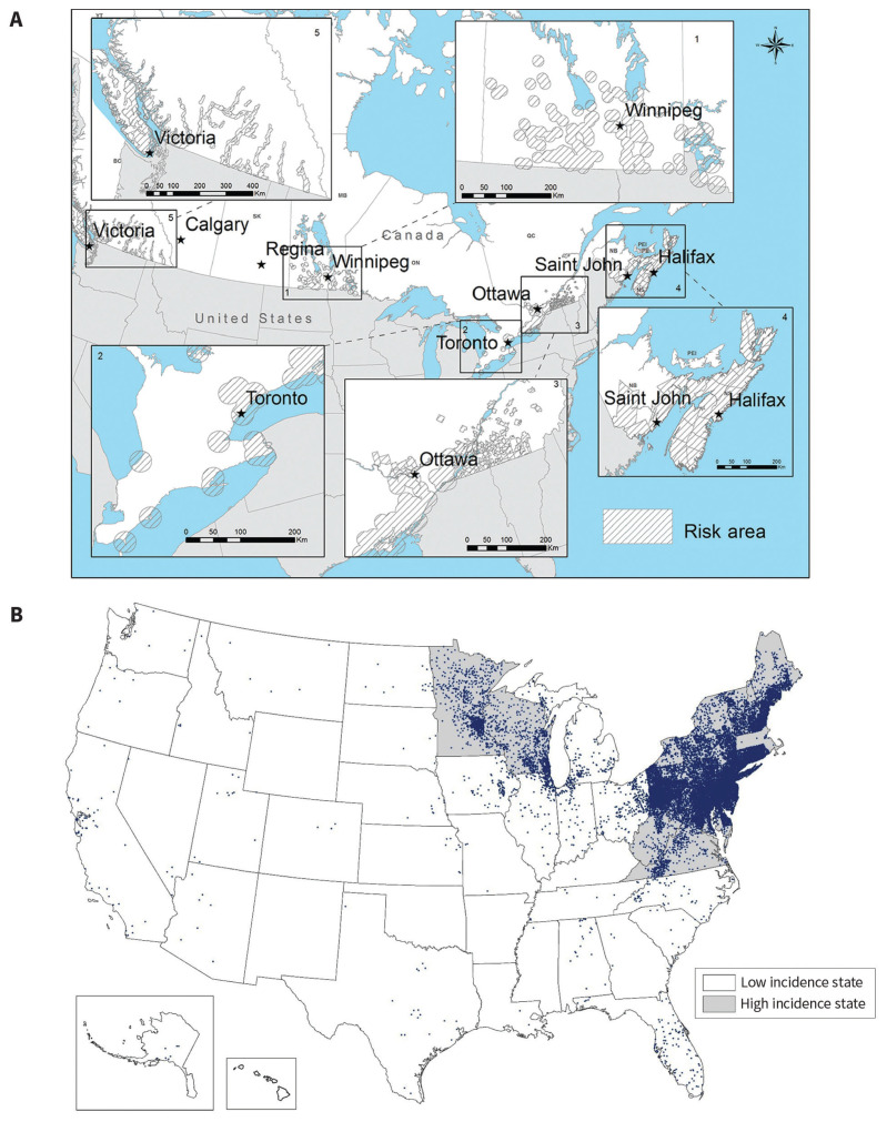
Lyme disease is caused by the spirochete B. burgdorferi, which is transmitted in North America by ticks in the Ixodes genus. Although early cases of Lyme disease were concentrated in the northeastern United States, the distribution in North America has expanded and shifted over time. Cases are increasingly recognized in more northern latitudes, including in Canada (Figure 1).1 Climate change, an increase in human exposure to ticks, and changes in distribution of both ticks and host animals are all thought to contribute to the changing epidemiology.2
Figure 1:
(A) Risk areas for Lyme disease as determined by the presence of ticks capable of Borrelia burgdorferi transmission (cross-hatched areas). Based on surveillance data collected by the Public Health Agency of Canada (2009–2016). © All rights reserved. Risk of Lyme disease to Canadians. Public Health Agency of Canada, 2018. Adapted and reproduced with permission from the Minister of Health, 2019. “Lyme disease risk areas map” is available at www.canada.ca/en/public-health/services/diseases/lyme-disease/risk-lyme-disease.html#map. (B) Reported cases of Lyme disease in the United States, 2018. Map from the Centers for Disease Control and Prevention (CDC). Each dot represents 1 case of Lyme disease and is placed randomly in the patient’s county of residence. The presence of a dot in a state does not necessarily mean that Lyme disease was acquired in that state. Grey shading indicates states with high incidence. CDC materials used are in the public domain. “Reported cases of Lyme disease — United States, 2018” is available at www.cdc.gov/lyme/datasurveillance/maps-recent.html.
Lyme carditis refers to the range of cardiac abnormalities associated with B. burgdorferi infection. From surveillance reports, it is estimated that cardiac complications will develop in about 3%–4% of patients with Lyme disease.1 However, the diagnosis of Lyme carditis is often difficult to confirm, and incidence estimates range from 1% to 10% in the literature.3,4 One of the difficulties in diagnosing Lyme carditis can be a lack of classic features of Lyme disease on assessment. In a systematic review, erythema migrans was reported in only 50% of patients with Lyme carditis, recognized history of tick bites in 30.7%, and history of outdoor activity or exposure in about 40%.5 A risk score intended to be used to screen patients with possible Lyme carditis in endemic areas has been proposed but not yet validated.5
The most recognized form of cardiac involvement in Lyme disease is AV block, which can range from first-degree to complete heart block and may be fluctuating and intermittent in nature.3,5,6 Prior reviews of Lyme carditis presenting with high-degree (second- and third-degree) AV block have highlighted the frequency of AV nodal block reversibility; very few patients who receive appropriate treatment with antimicrobials require pacing long-term.5–7 Fluctuations in heart rhythm may occur rapidly, and both complete heart block and unstable ventricular arrhythmias can develop, necessitating urgent intervention.6
Sudden cardiac death can occur in a small subset of patients with Lyme carditis. A review by the Centers for Disease Control and Prevention of 1696 cases of Lyme carditis in the US from 1995 to 2013 found 5 cases of sudden cardiac death.4 Three cases were confirmed based on autopsy findings, and clinical presentation was felt to be consistent in the other 2 cases.4 Two additional cases, diagnosed on autopsy, had been described before that time in the US. Subsequently, 3 more cases have been published, most recently in 2019 (Appendix 1, available at www.cmaj.ca/lookup/suppl/doi:10.1503/cmaj.191194/-/DC1).
Eight of the 10 described North American patients were male, and ages ranged from 17 to 66 years. Cases occurred from June through November, consistent with the usual window of activity of tick vectors. Available clinical information is limited owing to the retrospective diagnosis in several cases; however, only 4 patients were reported to have had erythema migrans. In those cases with available information, symptoms ranged from nonspecific manifestations, such as fever and malaise, to cardiac symptoms, including dyspnea and chest pain.
Borrelia burgdorferi appears to show affinity for cardiac tissue. Spirochete infiltration, extensive inflammatory changes, and interstitial lymphocytic pancarditis have been shown in autopsy studies.8 In this case, the diagnosis was confirmed based on cardiac autopsy specimens in addition to serologic test results consistent with acute Lyme disease. Confirmatory PCR testing of cardiac tissue using a 2-step process targeting B. burgdorferi 23S ribosomal RNA and the ospA gene target was carried out by Canada’s National Microbiology Laboratory. The assays can detect the equivalent of less than 50 Borrelia spirochetes, and specificity approaches 100%.9,10 Polymerase chain reaction testing for Lyme disease has previously been used on cardiac tissue to confirm the diagnosis of Lyme carditis in fatal cases (Appendix 1).
The diagnosis of Lyme carditis is based on clinical suspicion and serology consistent with acute Lyme disease. Unfortunately, diagnosis can be delayed while serology is being processed, and clinical suspicion should guide empiric treatment.6,11 Given that the early diagnosis is primarily clinical, cases may be overlooked by clinicians, especially as Lyme disease moves into new geographic areas. A review of 5 cases in Kingston, Ontario, found that Lyme disease was considered in only a small proportion of patients presenting with AV block, highlighting that Lyme carditis can be underrecognized even in relatively high-prevalence areas.7
Patients presenting with AV block should be questioned about a preceding history of rash or constitutional symptoms, exposure to ticks and time spent in Lyme-endemic areas. Clinicians should be aware of the distribution of Lyme disease and its vector in their practice area. Conversely, patients with suspected or confirmed Lyme disease should be questioned regarding cardiac symptoms, such as dyspnea, syncope, chest pain and decreased exercise tolerance. Those who are asymptomatic from a cardiac perspective on presentation should be educated regarding the possibility of cardiac involvement and advised to seek care immediately should they develop any cardiac symptoms. Current guidelines do not comment on the utility of screening ECG in patients with Lyme disease who do not have cardiac symptoms.11
If there is suspicion of Lyme carditis, patients should undergo urgent ECG and be considered for admission to a monitored setting equipped with cardiac telemetry.11 Empiric treatment with appropriate antibiotics should be started immediately, before serologic confirmation of the diagnosis.6,11 Temporary pacing may be required in some cases, and timely access to transcutaneous or transvenous pacing should be ensured, especially if AV block of any degree is noted on ECG. Permanent pacing is generally not needed, provided the patient receives appropriate treatment with antibiotics.6,7,11 The Infectious Diseases Society of America is in the process of updating guidelines for the treatment of Lyme disease, including Lyme carditis. Current guidelines support the use of either oral doxycycline or intravenous ceftriaxone for treatment depending on clinical presentation, with intravenous ceftriaxone recommended for patients requiring hospital admission.11
For a video testimonial by the patient’s family, see www.cmaj.ca/lookup/suppl/doi:10.1503/cmaj.191194/-/DC3.
Acknowledgement
The authors are grateful to the patient’s family for allowing his story to be shared, and for the essay (Box 1) they have written, which highlights the importance of recognizing Lyme disease in all its guises.
Box 1:
Family’s perspective
At a family gathering in the beginning of July 2018, Samuel told us that he didn’t feel well. Samuel was normally so strong and energetic — the life of our gatherings; it seemed unusual to see fatigue in his eyes.
“It’s the flu,” he said. “No big deal.”
We went swimming later that day and when he took off his shirt, I noticed circular rashes on his back and chest. He looked surprised as well. He hadn’t seen them before.
“I was picking raspberries,” he said. “I must have gotten scratched.”
I mentioned Lyme disease then. “It’s tick season. You shouldn’t ignore a rash plus the flu, Samuel.”
“But I haven’t been bitten by a tick!” he said, and with that, he totally dismissed the idea.
A worrier, I felt relieved to hear that he went to visit the doctor the following week after his flu symptoms worsened. He took some tests and was sent home with the conclusion that he was experiencing a bad flu. Samuel wasn’t the kind of guy to rest and recuperate. He kept working long-hour workdays, exercising and trying all the natural remedies he could think of to build his immune system and fight this sickness. He was strong and in charge of his health.
Or so we thought.
The next time I saw him, we were celebrating his 37th birthday. I asked him how he was feeling. He said he felt better, and that it had been the worst flu he’d ever experienced.
Then, a week and a half later, he messaged us to say he had gone back to the doctor because he was experiencing some minor heart symptoms. He texted as if it was no big deal, just a small hiccup in his work week. They wanted to keep him in overnight, he said. But he’d get out soon. They’d figure it out and the symptoms wouldn’t last.
I wondered if it really was as insignificant as he made it sound. “Should we worry?” I asked my husband that day. “I wonder if it’s more serious than he thinks it is.”
I did some reading that night and I sent him an article on Lyme carditis. “I think you should ask for antibiotics,” I texted. “Just in case.”
I’m not sure if he ever read the text.
We got a call at 4 in the morning that his heart had stopped. We rushed into the city and sat by his bedside. He was on life support. We half dared to hope that he’d wake up and we’d laugh about his near-death experience. It was just like him to play a prank on us and get out of it laughing and unscathed.
“This isn’t funny, Samuel,” I told him. “This is too real.”
Over the next day or so, it became very clear that he wasn’t waking up. We said our goodbyes. It was heartbreaking. Although he was hanging on with machines, we knew he was gone. Not only was I losing my brother, I felt heart-wrenched for his new wife and 4-month-old son. The reality of their loss seemed insurmountable.
His death and funeral were a blur. “What happened?” so many people asked. But we couldn’t answer. We didn’t know. His death was as much of a shock to us as it was to them.
His tests came back positive for Lyme disease several weeks after he passed. His autopsy was also clear — he had died from complications of Lyme carditis. Finally, we had some answers.
But with the results came more questions than comfort. How had he contracted the disease? What could have been done differently and how could it have been prevented?
With all the speculation and the unknown, it can be difficult to continue our lives without fear. Since Samuel’s death, we have met multiple others who have been affected by this mysterious disease. Perhaps it is only our awareness of the disease that is growing; however, it also seems like the ravages of Lyme disease are becoming more and more prevalent in our world. With all its hurt and devastation, we hope and pray that answers may also become more widespread, and that we can begin to rest peacefully at night knowing strides are being made to understand the disease, and we can keep ourselves and our loved ones safe from its harm in the future.
Editor’s note: This essay was authored by the patient’s sister. We have not included the author’s name to protect the privacy of the family.
See related articles at www.cmaj.ca/lookup/doi/10.1503/cmaj.191279 and www.cmaj.ca/lookup/doi/10.1503/cmaj.191660
Footnotes
Competing interests: None declared.
This article has been peer reviewed.
The authors have obtained consent from the patient’s next of kin.
Contributors: All authors conceived of the article. Milena Semproni drafted the manuscript, which all authors revised. All authors gave final approval of the version to be published and agree to be accountable for all aspects of the work.
References
- 1.Gasmi S, Ogden NH, Lindsay LR, et al. Surveillance for Lyme disease in Canada: 2009–2015. Can Commun Dis Rep 2017;43:194–9. [DOI] [PMC free article] [PubMed] [Google Scholar]
- 2.Bouchard C, Dibernardo A, Koffi J, et al. Increased risk of tick-borne diseases with climate and environmental changes. Can Commun Dis Rep 2019;45:83–9. [DOI] [PMC free article] [PubMed] [Google Scholar]
- 3.Robinson ML, Kobayashi T, Higgins Y, et al. Lyme carditis. Infect Dis Clin North Am 2015;29:255–68. [DOI] [PubMed] [Google Scholar]
- 4.Forrester JD, Meiman J, Mullins J, et al. Notes from the field: update on Lyme carditis, groups at high risk, and frequency of associated sudden cardiac death — United States. MMWR Morb Mortal Wkly Rep 2014;63:982–3. [PMC free article] [PubMed] [Google Scholar]
- 5.Besant G, Wan D, Yeung C, et al. Suspicious index in Lyme carditis: systematic review and proposed new risk score. Clin Cardiol 2018;41:1611–6. [DOI] [PMC free article] [PubMed] [Google Scholar]
- 6.Yeung C, Baranchuk A. Diagnosis and treatment of Lyme carditis: JACC review topic of the week. J Am Coll Cardiol 2019;73:717–26. [DOI] [PubMed] [Google Scholar]
- 7.Wan D, Blakely C, Branscombe P, et al. Lyme carditis and high-degree atrioventricular block. Am J Cardiol 2018;121:1102–4. [DOI] [PubMed] [Google Scholar]
- 8.Muehlenbachs A, Bollweg BC, Schulz TJ, et al. Cardiac tropism of Borrelia burgdorferi: an autopsy study of sudden cardiac death associated with Lyme carditis. Am J Pathol 2016;186:1195–205. [DOI] [PubMed] [Google Scholar]
- 9.Courtney JW, Kostelnik LM, Zeidner NS, et al. Multiplex real-time PCR for detection of Anaplasma phagocytophilum and Borrelia burgdorferi. J Clin Microbiol 2004;42:3164–8. [DOI] [PMC free article] [PubMed] [Google Scholar]
- 10.Tokarz R, Tagliafierro T, Cucura DM, et al. Detection of Anaplasma phagocytophilum, Babesia microti, Borrelia burgdorferi, Borrelia miyamotoi, and Powassan virus in ticks by a multiplex real-time reverse transcription-PCR assay. MSphere 2017;2:pii: e00151-17. [DOI] [PMC free article] [PubMed] [Google Scholar]
- 11.Wormser GP, Dattwyler RJ, Shapiro ED, et al. The clinical assessment, treatment, and prevention of Lyme disease, human granulocytic anaplasmosis, and babesiosis: clinical practice guidelines by the Infectious Diseases Society of America. Clin Infect Dis 2006;43:1089–134. [DOI] [PubMed] [Google Scholar]


