1. Introduction
Apocrine glands of the external auditory canal (EAC) are found within the dermis contiguous to the cartilaginous region and their secretory cerumen is thought to help prevent EAC infection [1]. The inappropriate proliferation of apocrine sweat glands can lead to the development of apocrine hidrocystomas, also known as cystadenoma, generally occurring in the head and neck. Though rare cases of EAC apocrine hidrocystomas have been published in the literature, they have been singular events with no reports of post-excisional recurrence [2, 3]. Here, we present a unique case of recurrent apocrine hidrocystoma of the EAC in a 49-year old male presenting on three occasions leading to a current state of remission.
2. Case Report
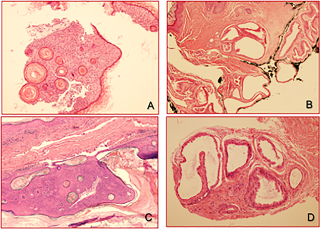
A 49-year-old male with a history of recurrent lesions within the right EAC presented complaining of bloody ear drainage. Past medical history consisted of a right EAC lesion that was initially biopsied twice in 2003, demonstrating seborrheic keratosis (Fig. 1A). The lesion recurred in 2007, at which point it was excised including part of the underlying cartilage and diagnosed as an apocrine hidrocystoma (Fig. 1B). The patient presented again 7 years later with bloody ear drainage after cleaning his ear with a Q-tip. He was otherwise asymptomatic and denied otorrhea, otalgia, hearing loss, or tinnitus. Examination demonstrated a papillomatous lesion along the superior cartilaginous EAC with a clear tympanic membrane negative for effusion or retraction (Figs. 1C & 1D), judged to be a recurrence of his apocrine hidrocystoma. The lesion was again excised with margins which were negative. In 2016, for the third time, a papillomatous lesion recurred along the posterior cartilaginous EAC with overlying bloody crusting blocking 40–50% of the canal. Re-excision was discussed, but the patient opted for observation. Patient was administered nystatin and triamcinolone ointment for pruritus and asked to follow up one year later. Upon follow ups in 2017 and 2018, the lesion was no longer observed within the EAC. Patient was instructed to continue nystatin and triamcinolone ointment as needed.
Figure 1.
Right auditory canal biopsy showing an irritated seborrheic keratosis in 2003 (A); at the same location, an apocrine hidrocystoma was excised in 2007, extending to the inked margin (B); after 7 years, recurrent pigmented seborrheic keratosis (C) and adjacent apocrine hidrocystoma (D) at the same right auditory canal.
3. Discussion
Apocrine hidrocystoma of the EAC are extremely rare cases that have been reported as singular events [1, 2], making this presentation, to our knowledge, the first published case of recurrence. Developing from an abnormal proliferation of apocrine sweat glands, apocrine hidrocystoma has been characterized as a solitary, adenomatous, translucent nodule 3–15 mm in diameter, with mobility upon palpitation and idiopathic etiology in the majority of cases [4, 5]. Histological studies are important tools to assure correct diagnosis and differentiation from other possibilities including but not limited to eccrine hidrocystomas, epidermal inclusion cysts, mucoid cysts, seborrheic keratosis, melanoma, and haemangiomas.
Histologically, apocrine hidrocystomas are well-circumscribed cystic lesions containing cytokine or decapitation secretion and are enclosed by fibrotic tissue [6]. Cytologically, they have low mitotic activity with little to no nuclear atypia and do not typically contain papillary or pseudopapillary growth into the cystic cavity [6]. The presence of apocrine lesions with true papillary growth is more reserved for apocrine cyst adenomas. Once diagnosed, treatment via excision, incision and drainage, carbon dioxide laser vaporization, or topical management can be discussed and pursued.
It is suggested that the occurrence of multiple hidrocystomas can be associated with two rare genetic disorders of Schopf-Schulz-Passarge and Goltz-Gorlin syndrome [7], however; we would like to make the distinction of presence of lesions in different locations with our case of recurrence in the same region. This paper intends to present a unique case of recurrence in EAC apocrine hidrocystoma with two main teaching points. First, it is not only important to correctly diagnose this rare entity in order to ensure the most appropriate discussion and treatment plan, but that it is also beneficial to follow-up with the excisional site to monitor for the newly-illustrated potential recurrence. We show that our patient’s lesion has been in remission 2 years post recurrence via conservative management and the use of nystatin- triamcinolone ointment, making surgical and medical managements both possibly viable treatment routs in the case of EAC apocrine hidrocystoma recurrence.
Footnotes
Conflict of Interest: None
Financial Disclosure: None
References
- 1.Stoeckelhuber M, Matthias C, Andratschke M, Stoeckelhuber BM, Koehler C, Herzmann S, et al. Human ceruminous gland: ultrastructure and histochemical analysis of antimicrobial and cytoskeletal components. Anat Rec A Discov Mol Cell Evol Biol 2006; 288(8): 877–84. [DOI] [PubMed] [Google Scholar]
- 2.Ioannidis DG, Drivas EI, Papadakis CE, Feritsian A, Bizakis JG, Skoulakis CE. Hidrocystoma of the external auditory canal: a case report. Cases J 2009; 2(1): 79. [DOI] [PMC free article] [PubMed] [Google Scholar]
- 3.Wu KC, Lin HC, Chang KM. External auditory canal apocrine hidrocystoma. Otol Neurotol 2011; 32(7): e54–55. [DOI] [PubMed] [Google Scholar]
- 4.Smith JD, Chernosky ME. Apocrine hidrocystoma (cystadenoma). Arch Dermatol 1974; 109(5): 700–2. [PubMed] [Google Scholar]
- 5.Obaidat NA, Ghazarian DM. Bilateral multiple axillary apocrine hidrocystomas associated with benign apocrine hyperplasia. J Clin Pathol 2006; 59(7): 779. [DOI] [PMC free article] [PubMed] [Google Scholar]
- 6.Sugiyama A, Sugiura M, Piris A, Tomita Y, Mihm MC. Apocrine cystadenoma and apocrine hidrocystoma: examination of 21 cases with emphasis on nomenclature according to proliferative features. J Cutan Pathol 2007; 34(12): 912–7. [DOI] [PubMed] [Google Scholar]
- 7.Vani D,TRD,HBS,MB, Kumar HR, Ravikumar V. Multiple apocrine hidrocystomas: a case report. J Clin Diagn Res 2013; 7(1): 171–2. [DOI] [PMC free article] [PubMed] [Google Scholar]


