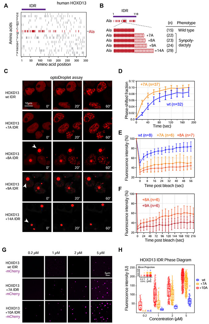
Figure 2. Synpolydactyly-associated repeat expansions enhance HOXD13 IDR phase separation.
(A) Amino acid composition of human HOXD13. Ticks represent amino acids indicated on the y-axis at the positions indicated on the x-axis. The IDR cloned for subsequent experiments is highlighted with a purple bar.
(B) Alanines within the HOXD13 IDR sequence are indicated as red ticks. The central alanine repeat consists of 15As in the wild type protein.
(C) Representative images of live HEK-293T nuclei expressing wt and repeat-expanded HOXD13 IDR-mCherry-CRY2 fusion proteins. Cells were stimulated with 488nm laser every 20s for 3 minutes. Arrowheads highlight spontaneously forming IDR condensates present without 488nm laser stimulation.
(D) Quantification of the fraction of the nuclear area occupied by HOXD13 wt IDR-mCherry-CRY2 and HOXD13 +7A IDR-mCherry-CRY2 droplets in HEK-293T cells over time. Data displayed as mean+/−SEM.
(E) Fluorescence intensity of light-induced wt, +7A and +8A HOXD13 IDR droplets before, during and after photobleaching. Data displayed as mean+/−SD.
(F) Fluorescence intensity of +8A and +9A spontaneously formed HOXD13 IDR condensates before, during and after photobleaching. Data displayed as mean+/−SD.
(G) Representative images of droplet formation by purified HOXD13 IDR-mCherry fusion proteins in droplet formation buffer.
(H) Phase diagram of HOXD13 IDR-mCherry fusion proteins. Every dot represents a detected droplet. The inset depicts the projected average size of the droplets as mean+/− SD (middle circle: mean, inner and outer circle: SD). n.d.: not detected
See also Figure S2.

