Abbreviations
- COVID‐19
coronavirus disease 2019
- HFU
high‐frequency ultrasound
- RCM
reflectance confocal microscopy
- SARS‐CoV‐2
severe acute respiratory syndrome coronavirus 2
Dear Editor,
1.
Dermoscopy, reflectance confocal microscopy (RCM), and high‐frequency ultrasound (HFU) are noninvasive imaging tools and widely applied in diagnosis and follow‐up with the advantages of real‐time and dynamic features. 1 Recently, Jakhar et al described how to perform dermoscopy during the pandemic of COVID‐19. 2 After peak period of the epidemic, how to resume noninvasive imaging detectings safely is a concerning issue for both dermatologists and patients.
Wuhan is the hardest‐hit city by the COVID‐19. After 3 months of circuit policy, large backlog patients made the resumption of noninvasive imaging tests urgent. However, the existence of asymptomatic and extended incubation period COVID‐19 patients made difficulties in screening out all patients. Besides, the contact of scan probe with the patient's skin is unavoidable, and the SARS‐CoV‐2 was found capable of staying alive on such plastic or steel surfaces for 48‐72 hours with the possible danger of cross‐transmission of COVID‐19. 3 Consequently, the infection‐control measures while resuming routine dermatologic care are still of great necessity. We take some practical safety measures in the stabilized epidemic period as the followings:
1. Weigh the potential risk of delaying detection against the COVID‐19 infection to the patient according to the overall local epidemic situation and then make the timeframe of resume the noninvasive imaging detection.
On 23 January, Wuhan city was lockdown, and our dermatology outpatient clinic was closed on the same day. From 11 March, the number of new confirmed COVID‐19 infections was less than 10/day, and dermatological clinic was partially opened for severe patients. From 18 March, almost none of new confirmed infections. Three weeks later, we completely resumed outpatients visits and noninvasive imaging detection on 8 April There are three‐time points (23 January, 11 March, and 8 April) from shutdown to reopen, and the setting of each time point depends on the epidemic situation of the local city.
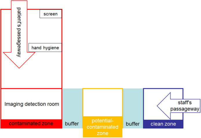
2. We recommend dividing the outpatient department into three zones, including clean zone, potential‐contaminated zone, and contaminated zone. Also, there is a buffer area between each zone. Patients and staffs have different passageways. The noninvasive imaging detection room located in the contaminated zone (Figure 1). Staffs enter through the staff's passageway, wear protective equipment and then enter the detection room. After the shift, staffs remove protective equipment in the potential‐contaminated zone and then they return back
FIGURE 1.

Diagram of the “three zones and two passageways.” The red indicates contaminated zone; the yellow indicates potential‐contaminated zone, and the blue indicates clean zone. The patients and staff get into the noninvasion imagine detection room by their, respectively, passageway
Patients should be screened using a rapid questionnaire and body temperature at the entrance of a patient's passageway, conduct hand hygiene, and then enter the detection room.
3. During procedures
Dermoscopy: It is recommended to use a disposable glass slide to isolate skin lesions and the lens of dermoscopy. After disinfecting, medium applied (such as medical alcohol), cover the skin lesions with a disposable glass slide, and then conduct dermoscopy detection; otherwise, using disposable isolation lens cover contact the skin lesion.
RCM: Skin lesion was disinfected, medium applied (medical alcohol), then wrap the disposable cling film on scan head for detection.
HFU: Skin lesion was disinfected, ultrasound gel applied, and then perform HFU. Remove the gel after the procedure and wiped the scanner or water cyst with an alcohol wipe.
Using the disposable glass slide and cling film isolated equipment and skin lesion avoiding direct contact. It is a simple and effective method to avoid cross‐infection.
4. Special site detection
Facial skin that needs to take off mask should be avoided detection; otherwise, the patient should provide negative test results of COVID‐19 in the latest 1 week. The periocular, perioral, vulvar, and mucosa should be avoided as far as possible.
5. Disinfection and cleaning the surfaces of the devices after finished all the detection are warranted.
CONFLICT OF INTEREST
None declared.
ACKNOWLEDGMENTS
This work was supported by HUST COVID‐19 Rapid Response Call Program (2020kfyXGYJ056) and Hubei Provincial Emergency Science and Technology Program for COVID‐19 (2020FCA037).
Xiangjie An and Zexing Song are First equal author
Funding information Huazhong University of Science and Technology (HUST) COVID‐19 Rapid Response Call Program; Hubei Provincial Emergency Science and Technology Program for COVID‐19 Grant/Award Number: 2020kfyXGYJ056; 2020FCA037; HUST, Grant/Award Number: 2020FCA037
REFERENCES
- 1. Welzel J, Schuh S. Noninvasive diagnosis in dermatology. J Dtsch Dermatol Ges. 2017;15(10):999‐1016. [DOI] [PubMed] [Google Scholar]
- 2. Jakhar D, Kaur I, Kaul S. Art of performing dermoscopy during the times of Coronavirus Disease (COVID‐19): simple change in approach can save the day! J Eur Acad Dermatol Venereol. 2020. 10.1111/jdv.16412. (Early view). [DOI] [PubMed] [Google Scholar]
- 3. van Doremalen N, Bushmaker T, Morris DH, et al. Aerosol and surface stability of SARS‐CoV‐2 as compared with SARS‐CoV‐1. N Engl J Med. 2020;382(16):1564‐1567. [DOI] [PMC free article] [PubMed] [Google Scholar]


