Figure 2.
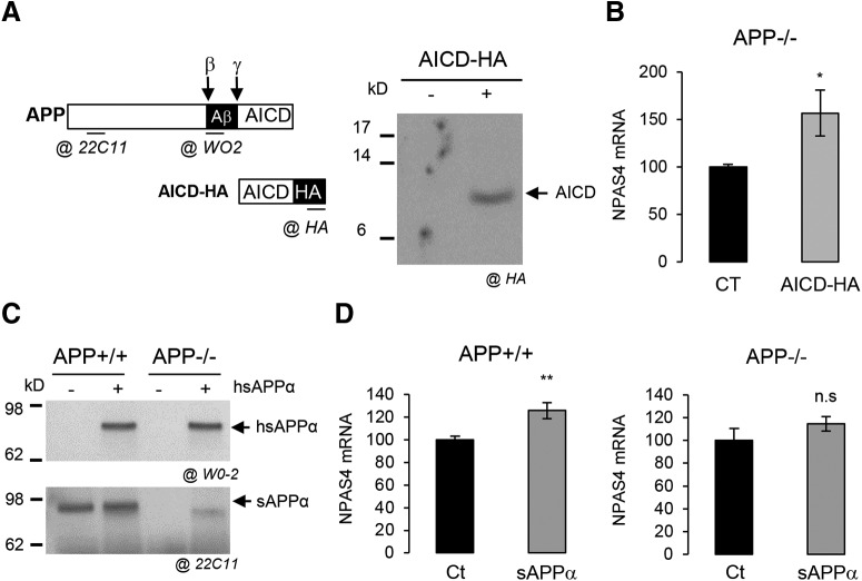
APP metabolites regulate NPAS4 expression. A, Schematic representation of APP, its fragments, the AICD-HA construct along with and the localization of the epitopes recognized by the different antibodies used. Western blot analysis of AICD-HA expression after 3 d of lentiviral infection in cells with control or AICD-HA-expressing vectors. Total cell lysate was analyzed with anti-HA antibody. B, Quantification by qPCR of NPAS4 mRNA in neurons APP−/− at DIV7 infected with lentiviral vector expressing AICD-HA (n = 6, N = 3). Results are expressed as percentage of control (Ct; mean± SEM); *p = 0.0291, Student’s t test. C, Medium of sAPPα-treated APP+/+ or APP−/− neurons was subjected to Western blot analysis using anti-human APP antibody (clone W0-2) to detect the exogenous human sAPPα (h sAPPα) and an anti-mouse APP antibody (clone 22C11) to detect both endogenous and exogenous sAPPα (h + m sAPPα). Medium was collected after 16 h of treatment. D, Quantification by qPCR of NPAS4 mRNA level in APP+/+ (n = 8, N = 4) or in APP−/− neurons at DIV7 treated with 20 nm sAPPα for 16 h (n = 6, N = 3). Results are expressed as percentage of control (Ct; mean ± SEM); **p = 0.0055, n.s. = non-significant, Student’s t test. Given that primary cultures of cortical neurons at DIV7 also contain astrocytes, the astrocytic pattern of NPAS4 expression is described in Extended Data Figure 2-1.

