To the Editor,
We read with great interest the study authored by Angeletti et al 1 regarding the role of nonstructural proteins nsp2 and nsp3 in the pathogenesis of coronavirus disease 2019 (COVID‐19). COVID‐19 emerged in December 2019 in Wuhan City, Hubei Province, China and is caused by a novel viral variant belonging to the viral variant belonging to the severe acute respiratory syndrome‐related coronavirus (SARS‐rCoV) species of subgenus Sarbecovirus, genus Betacoronavirus, family Coronaviridae and named SARS‐CoV‐2. 2 The novel SARS‐CoV‐2 virus shows the typical genome organization of the betacoronaviruses, which consists of a 5′‐untranslated region (5′‐UTR), replicase complex (orf1ab), S gene, E gene, M gene, N gene, 3′‐UTR, and other open reading frames (ORFs) yet to be characterized. In addition to SARS‐CoV‐2, this genus has other two highly pathogenic CoVs: the SARS‐CoV and the Middle East respiratory syndrome CoV (MERS‐CoV). All three viruses are originated from bats and have emerged in the 21st century causing outbreaks with high mortality rates. SARS‐CoV and MERS‐CoV infection results in higher mortality rates compared to SARS‐CoV‐2, but only the latter was capable to establishing sustained human‐to human‐transmission.
In their article, the authors used several computational tools to analyze the ORF1a and ORF1b of SARS‐CoV‐2. The ORF1a/b comprise about two‐thirds of the viral genome and codes for 16 nonstructural proteins (nsp1‐16). There is a −1 frameshift between ORF1a and ORF1b, leading to production of two polypeptides (pp1a and pp1ab), which are further processed by viral‐encoded proteases into 16 nsp. They found a segment within the nsp2 and the nsp3 regions that has no homology with other CoVs and suggested that “the stabilizing mutation falling in the endosome‐associated‐protein‐like domain of the nsp2 protein could account for COVID‐2019 high ability of contagious, while the destabilizing mutation in nsp3 proteins could suggest a potential mechanism differentiating COVID‐2019 from SARS.” The work is of unquestionable value, but we would like to make some important considerations from a molecular virology point of view.
First, according to the International Committee on Taxonomy of Viruses the correct nomenclature for the novel coronavirus is SARS‐CoV‐2 and not COVID‐2019 as written by the authors. The virus is the etiological agent of the disease named by WHO as “coronavirus disease 2019” (abbreviated “COVID‐19”). 3
Second, phenotypic inference from sequence information alone can lead to wrong conclusions. Viral pathogenesis is usually a polygenic trait, even though some specific mutations can have a great impact by themselves. As an example, a highly pathogenic porcine reproductive and respiratory syndrome virus (PRRSV) emerged in China in 2006 causing a widespread outbreak of disease in pigs that was very atypical. Initially, the unusual high pathogenicity of this novel PRRSV strain was associated with a unique 30‐amino‐acid deletion in its Nsp2 protein. However, subsequent studies using reverse genetics found that this large genomic deletion was not responsible to PRRSV virulence. 4 Actually, the increased virulence of this PRRSV strain was mapped to the Nsp9 and Nsp10 proteins. 5
Mutations are also context‐dependent. The PB1‐F2 protein of influenza A viruses (IAV) has been reported as an important virulence factor and the N66S polymorphism in this protein played a role in the high lethality of the 1918 and other IAV. Pena and coworkers used reverse genetics to study the role of PB1‐F2 (and the N66S mutation) in the virulence of different IAV and demonstrated that their effects and were host and strain‐dependent and ranged from higher virulence, no phenotype, and even lower virulence. 6 , 7 , 8
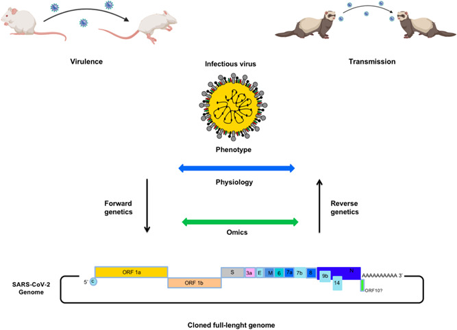
Identification of molecular markers of SARS‐CoV‐2 virulence, host range, and transmissibility can be accomplished unequivocally by reverse genetics (Figure 1). This powerful technique involves the generation of an infectious virus from a cloned full‐length complementary DNA and evaluation of the role of specific genes (and mutations) in adequate cell culture systems and animal models. Although the large genome of CoVs and its instability in bacterial plasmid vectors creates difficulties in the construction of reverse genetics systems, many technological advances have allowed the successful engineering of several CoVs, including SARS‐CoV and MERS‐CoV. 9 The development of reverse genetics systems for SARS‐CoV‐2 will shed light on many aspects of its biology and will pave the way to the development of vaccines and antivirals against this lethal pathogen.
Figure 1.

Schematic representation of forward and reverse genetic approaches. Forward genetics aims to identify the viral genotype that is responsible for a specific phenotype, whereas reverse genetic techniques enable the generation of an infectious virus from a cloned full‐length genome and the subsequent studies of the phenotypic effects of specific gene sequences in a biological system. Relevant animal models can used to study viral phenotypes, including virulence and transmissibility. ORF, open reading frame; SAR‐CoV‐2, Severe acute respiratory syndrome‐coronavirus 2
CONFLICT OF INTERESTS
The author declares that there are no conflict of interests.
REFERENCES
- 1. Angeletti S, Benvenuto D, Bianchi M, Giovanetti M, Pascarella S, Ciccozzi M. COVID‐2019: the role of the nsp2 and nsp3 in its pathogenesis. J Med Virol. 2020;92(6):584‐588. [DOI] [PMC free article] [PubMed] [Google Scholar]
- 2. Lorusso A, Calistri P, Petrini A, Savini G, Decaro N. Novel coronavirus (SARS‐CoV‐2) epidemic: a veterinary perspective. Vet Ital. 2020. [DOI] [PubMed] [Google Scholar]
- 3. Coronaviridae Study Group of the International Committee on Taxonomy of viruses . The species Severe acute respiratory syndrome‐related coronavirus: classifying 2019‐nCoV and naming it SARS‐CoV‐2. Nat Microbiol. 2020;5:536‐544. [DOI] [PMC free article] [PubMed] [Google Scholar]
- 4. Zhou L, Zhang J, Zeng J, et al. The 30‐amino‐acid deletion in the Nsp2 of highly pathogenic porcine reproductive and respiratory syndrome virus emerging in China is not related to its virulence. J Virol. 2009;83(10):5156‐5167. [DOI] [PMC free article] [PubMed] [Google Scholar]
- 5. Li Y, Zhou L, Zhang J, et al. Nsp9 and Nsp10 contribute to the fatal virulence of highly pathogenic porcine reproductive and respiratory syndrome virus emerging in China. PLoS Pathog. 2014;10(7):e1004216. [DOI] [PMC free article] [PubMed] [Google Scholar]
- 6. Pena L, Vincent AL, Loving CL, et al. Strain‐dependent effects of PB1‐F2 of triple‐reassortant H3N2 influenza viruses in swine. J Gen Virol. 2012;93(Pt 10):2204‐2214. [DOI] [PMC free article] [PubMed] [Google Scholar]
- 7. Pena L, Vincent AL, Loving CL, et al. Restored PB1‐F2 in the 2009 pandemic H1N1 influenza virus has minimal effects in swine. J Virol. 2012;86(10):5523‐5532. [DOI] [PMC free article] [PubMed] [Google Scholar]
- 8. Schmolke M, Manicassamy B, Pena L, et al. Differential contribution of PB1‐F2 to the virulence of highly pathogenic H5N1 influenza A virus in mammalian and Avian species. PLoS Pathog. 2011;7(8):e1002186. [DOI] [PMC free article] [PubMed] [Google Scholar]
- 9. Almazán F, Sola I, Zuñiga S, et al. Coronavirus reverse genetic systems: infectious clones and replicons. Virus Res. 2014;189:262‐270. [DOI] [PMC free article] [PubMed] [Google Scholar]


