Abstract
Johne’s disease (JD) is an infectious wasting condition of ruminants caused by Mycobacterium avium subsp. paratuberculosis (MAP) in domestic livestock of every country that has been investigated. Controlling JD is problematic due to the lack of sensitive, specific, efficient, and cost-effective diagnostic tests. A major challenge in the development of diagnostics like ELISA is the selection of an ideal antigen/(s) that is pathogen-specific and allows sensitive recognition. Therefore, the purpose of this study was to identify and use Mce-truncated protein-based ELISA assay for the diagnosis of MAP infection with high sensitivity and specificity. In silico epitope prediction by epitope mapping throughout the whole length of MAP2191 protein revealed that C-terminal portion of this protein presented potential T- and B-cell epitopes. Therefore, a novel Mce-truncated protein encoded by the selected region of MAP2191 gene was expressed, purified with Ni-NTA gel matrix and confirmed by SDS PAGE and western blot. A profiling ELISA assay was developed to evaluate sera from MAP infected and non-infected ruminant species for antibodies against Mce-truncated protein to infer the immunogenicity of this protein in the host. Using this Mce protein-based ELISA, 251 goats, 53 sheep, 117 buffaloes, and 33 cattle serum samples were screened and 49.4, 51.0, 69.2, and 54.6% animals, respectively, were found positive. Comparing with i-ELISA, the new Mce-based ELISA kit showed a relatively higher specificity but suffered from slightly reduced sensitivity. Mce-based ELISA excluded apparently false positive results of i-ELISA. Mce protein was found to be antigenic and Mce-ELISA test could be employed as a diagnostic test for JD in domestic livestock in view of the a relatively higher specificity and accuracy. The antigenic potential of Mce antigen can also be exploited for the development of a new vaccine for the control of MAP infection.
Introduction
Mycobacterium avium subsp. paratuberculosis (MAP) is the cause of chronic wasting disease in domestic livestock species commonly known as paratuberculosis or Johne’s disease (JD) [1, 2]. JD has the widest host range and has been isolated from domestic livestock, wild ruminants, and other animal species, including primates and human beings [3, 4–6]. The disease inflicts major economic losses to livestock production system and dairy industry by lowering productivity, both in terms of quality and quantity, of milk, meat, fiber and skin, by way of increased morbidity, mortality and early cullings [1, 6]. Large number of studies reported high bio-load of MAP in domestic livestock and in their milk [7, 8] and other secretions. MAP has also been associated with a number of incurable human illnesses such as Crohn’s disease / Inflammatory Bowel Disease, diabetes, rheumatoid arnthritis, thyroids, and other auto-immune type disorders [1, 2, 9]. Control and eradication of JD are challenging due to the insidious nature, long incubation period, and lack of rapid and sensitive diagnostic tests [10]. With the scale and complexity of the expanding problem of MAP, pressure is increasing to find a way to control MAP infections in animals in order to rescue high milk yielding breeds of domestic livestock, and reduce production and economic losses in the livestock, dairy industry and animal farming, and impending threat for large scale human infection by consumpton of milk and milk products contaminated with MAP bacilli [6, 11, 12].
Recent immuno-informatics tools have made in silico analysis possible; such analysis has been used successfully to predict the immune epitopes in the pathogens and identify immunogenic proteins [13]. Presently, ideal major antigen candidates for MAP for efficient immuno-diagnosis and immunization are not available [11, 13]. There is need for the identification of immunogenic candidate antigens, which can be assessed for the development of efficient diagnostic tests and also as effective vaccine candidates against MAP infection [11]. Mammalian cell entry (Mce) proteins are among the virulence-related proteins which are functionally analogous to ABC transporters and are thought to function in lipid uptake system [14–18]. It was shown that Mce proteins have possible role in virulence of Mycobacterium tuberculosis (MTB) [15, 19–21]. Information is limited to the individual Mce protein expression in terms of their contribution to virulence in other Mycobacteria such as MAP. In MAP K-10 reference genome, there are eight separate mce genes [14, 22, 23]. Mce5 operon of MAP possesses a cluster of six homologs of the mce-family (MAP2189 to MAP2194) genes predicted to encode proteins with potentially useful metabolic functions [24–26]. Mce5 cluster has previously been identified as an important player for invasion and survival within macrophages, and possibly for the virulence of MAP [25]. Expression studies revealed that the purified Mce family of proteins has the ability to elicit antibody responses during Mycobacterium tuberculosis infection in human [27, 28].
Present study identified immuno-dominant candidate antigens using immune-informatics analysis (T- and B-cell epitopes analysis) for the development of improved diagnostic tests. Mce-truncated protein is a partial part of Mce protein (MAP2191) which has been proposed to be expressed from the truncated gene of MAP2191, and used as the coating antigen in the ELISA platform for the sero-diagnosis of JD using animal sera from naturally infected and infection free controls.
Materials and methods
Ethics statement
Central Institute for Research on Goats (CIRG), Makhdoom, Mathura ethical committee chaired by Member Secretary, Institutional Animal Ethics Committee (IAEC) under the supervision of central Committee for the Purpose of Control and Supervision of Experiments on Animals (CPCSEA), New Delhi has approved the experiments which were performed under the project [grant number 5/8/5/28/TF/2013/ECD-I], awarded by Indian Council of Medical Research (ICMR), New Delhi, India. IAEC vide reference number IAEC/CIRG/16-17 dated: 12.05.2016, confirmed that this project do not have any ethical issue. Samples were collected only for laboratory analyses and avoided unnecessary pain and suffering to animals and were not from any endangered or protected species.
Serum samples
Serum samples were collected from four domestic livestock species (251 goats, 53 sheep, 117 buffaloes, and 33 cattle) suspected for MAP infection. Animals sampled were both from farm and farmer’s herds and flocks located in two states {(Uttar Pradesh (UP) and Madhya Pradesh (MP)} of country. A total of 251 goats (40-IGFRI- Indian Grassland and Fodder Research Institute, Jhansi, UP; 91-SADIL- State Anima Disease Investigation Laboratory, Bhopal, MP; 120-Veterinary college, Mhow, MP), 53 sheep (53-IGFRI- Indian Grassland and Fodder Research Institute, Jhansi, UP), 117 buffaloes (117-Veterinary college, Mhow, MP), and 33 cattle (33- SADIL- State Anima Disease Investigation Laboratory, Bhopal, MP) were included in this study. Animals were in variable age mainly from females and they have mixed physical conditions and 30–35% of the animals were suffering from clinical to advance clinical JD. All these animals were ear-tagged for identification.For the collection of blood samples goats and sheep by restraining manually by side of chest and gently holding the the head towards one side. Cattle and buffaloes were properly restrained in cattle crush and blood (2–3 ml) was collected in vacutainers, after properly sterilizing the jugular vein with 70.0% ethyl alcohol. Institute Animal Ethics committee (IAEC) of the CIRG, Mathura, UP, India has approved the technical program of the project. Samples were stored at -20°C until screened. Status of the animals with respect to MAP infection was determined using ‘Gold Standard’fecal culture, fecal microscopy and fecal IS900-PCR before screening through i-ELISA and Mce-ELISA.
Morphological and molecular characterization
Reference strain of MAP ‘S 5’ Indian Bison Type was provided by CIRG, Mathura, UP, India. Strain used in this study was recovered from an advance case of JD (terminally sick, extremely weak and recumbent) in a Jamunapari breed of goat [29], which later succumbed to disease [30]. The MAP strain was sub-cultured and colonies identified morphologically on Herrold’s egg yolk (HEY) agar medium, mycobactin dependency and acid fast (Ziehl-Neelsen) staining of smears. Method for fecal culture and decontamination methods explained in the text. Briefly, ingredients of HEYM weighed (Mineral salt solution—MSS) and pH was made 7.8 after adding glycerol. Agar powder added and MSS (870 mil) was autoclaved. Egg yolk (120 ml from 8 eggs) harvested after sterilizing in isopropanol. Antibiotic free eggs from local poultry farmers used. Harvested yolk and 5 ml of 2.0% malachite green were added to MSS at 560C and HEYM was stirred for 2 hr at 500C. Mycobactin J was added to HEYM and dispensed. Fecal smears were made from middle layer after triturating feces (2 gm) in 10 ml sterilized water and centrifuged at 5000 rpm for 60 min. Molecular characterization of the strain was done using IS900, IS1311 PCR and IS1311 PCR-REA [29].
Identification of antigenicity mce gene by in-silico analysis
In Silico analysis of the MAP2191 protein was carried out using CLC Genomics Workbench 7.5.1 program (CLC bio, QIAGEN, Germany) and Major histocompatibility complex (MHC) binding peptide prediction algorithms through Immune Epitope Database and Analysis Resource (IEDB-AR) site (http://www.iedb.org/). MAP2191 protein full-length coding sequence was subjected to a basic local alignment search (BLASTp) analysis at the National Center for Biotechnology Information (NCBI) GenBank. After that T cell MHC class I and II epitopes prediction were performed using the IEDB-AR analysis consensus tool, which combines predictions from the Artificial neural network (ANN), Stabilized matrix method (SMM) and combLib [13, 31–34]. T cell epitopes prediction were calculated on the basis of their binding affinity to MHC alleles of the mouse, using the half-maximal inhibitory concentration (IC50) values as follows: IC50s <50 nm (high-affinity binding); IC50s <500 nm (intermediate-affinity binding), and IC50s <5,000 nm (low-affinity binding). A lower IC50 for MAP2191 protein indicates a higher affinity of binding to host MHC alleles. Mouse MHC alleles H-2Db, H-2Dd, H-2Kb, H-2Kd, H-2Kk, and H-2Ld were used for peptide binding affinity to MHC-I epitopes and H-2IAb, H-2IAd, and H-2IEd were used for peptide binding affinity to MHC-II epitopes [31–33]. For the prediction of linear and conformational B cell epitopes, the MAP2191 protein sequence was subjected to an ElliPro suite [35]. We obtained a score for each predicted epitope defined by PI average over each amino acid residue present in the MAP2191 protein. Predicted B cell epitopes of MAP2191 protein had scores of between 0.5 and 0.9; the minimum prediction cutoff score was set at 0.80.
Amplification and cloning of mce-truncated gene in TA-cloning vector
Mce-truncated gene (Accession number: MG754208) was successfully amplified with the help of primers designed to have BamHI and HindIII restriction enzyme sites (Table 1). Thermocycling was done in Veriti 96 well thermal cycler (Applied Biosystem), with initial hot start denaturation step for 5 min at 94°C, followed by 37 cycles of 94°C for 1 min, 58°C for 30 sec, and 72°C for 1 min. Subsequently, a final extension at 72°C for 10 min was carried out. Purified mce-truncated was confirmed by nucleic acid sequencing and cloned with pTZ57R/T cloning vector. Ligation products were successfully transformed into E. coli DH5α/E. coli XL10-Gold ultra-competent cells followed by plating on Luria Bertani (LB) agar (Merck, Germany) containing X-gal, IPTG, Ampicillin, Tetracycline, and Chloramphenicol. Final confirmation of purified clones was done using restriction digestion followed by sequencing.
Table 1. Primers used for characterization of Mycobacterium avium subsp. paratuberculosis.
| Target | Primer | Primer sequence | Product size (bp) |
|---|---|---|---|
| MAP2191/ truncated | Forward | 5’ACGGGATTCACCCTCAATCAATCGCTGG 3’-BamH1 | 621 (MAP Mce) |
| Reverse | 5’CTAAAGCTTTCATCGCGAACCGCCCGGGATG 3’- HindIII |
Cloning and confirmation of mce-truncated gene in expression vector
Expression plasmid was generated by cloning mce-truncated gene of MAP using pET28a (+) expression vector as per Chaubey et al. (2018) [36] with some modifications. Prior to ligation reaction, pET28a vector was restricted with HindIII and BamHI endonuclease, de-phosphorylated by a fast alkaline phosphatase enzyme (Fermentas, USA) with 10X FastAP buffer, and gel purified. For ligation reaction, mce-truncated gene fragment was digested from a pTZ57R/T vector with HindIII and BamHI, gel purified, and ligated with de-phosphorylated and digested using pET28a plasmid. Ligation product was transformed into E. coli DH5α/XL-10 competent cells followed by plating on LB-agar containing 50 μg ml-1 Kanamycin. Six to ten colonies were picked for mce-truncated colony PCR for the confirmation of the insert. Plasmid DNA of concern clones was isolated and digested with HindIII and BamHI enzymes. In order to further confirm the insert, one of the confirmed pET28a-mce-truncated cloning vectors was sequenced using T7 universal primers.
Expression and purification of Mce-recombinant protein
To express Mce-truncated protein, cloned vector was further transformed into E. coli Rosetta and E. coli BL-21 (DE3) competent cells; a single colony was picked up and propagated in 10 ml fresh Luria Bertani broth supplemented with Kanamycin (50 μg ml-1) at 37°C with shaking (180 rpm) overnight. For expression, 500 ml LB-broth containing Kanamycin were inoculated with 1% overnight grown culture and propagated until the absorbance reached approximately 0.5 at 600 nm. In the next step, cells were induced with different concentrations of IPTG and cultures were grown at different temperatures for different time intervals. One ml of uninduced cells (as negative control) were collected just before the addition of IPTG and from the induced cells at different time intervals, i.e. 2, 4, 6, 8, 10, 20 and 24 hrs. post induction. Collected culture was centrifuged and the pellet was harvested.
Purification of His-tagged Mce-truncated protein was carried out by affinity chromatography on a nickel-nitrilo triacetic acid (Ni-NTA) gel matrix (Qiagen, USA) using batch method. In brief, Rosetta and BL21 cells, harboring mce-truncated pellets, were suspended by gentle stirring in 15 ml equilibration buffer (50 mm Tris. HCl, 200 mm NaCl, 5mm DTT, 1mm PMSF, pH 8.0). The 14 ml cell suspension was lysed on ice using sonication at 30% amplitude for 20 cycles (20 sec pulse on, 20 sec pulse off). Then, a 50 ml conical tube containing 12 ml crude lysate was equilibrated with 1ml of pre-washed Ni-NTA resin in a horizontal position for 8 hours at 4°C while mixing. After incubation, the equilibrated suspension was centrifuged (2000 rpm, 10 min, 4°C) and the pellet was washed three times with wash buffer. Captured Mce-truncated proteins were eluted by increasing the imidazole concentration to 250 mM in the elution buffer.
SDS-PAGE and western blotting
Expression of Mce-truncated protein was analyzed by 12% Sodium Dodecyl Sulfate Polyacrylamide Gel Electrophoresis (SDS-PAGE) to investigate the desired size of expressing proteins. For western blotting, Mce-truncated protein band was transferred to nitrocellulose membrane (GE water and process technologies) with a Transblot Turbo® (Biorad, USA) apparatus operated at 60 volts for 2 hrs. The unbound membrane sites were first blocked by incubation for 30 min in blocking buffer (3% skimmed milk powder) at room temperature (RT). Blocking buffer was removed, followed by three rounds of washing with phosphate buffer saline with tween 20 (PBST buffer). The membrane was dipped with anti His Tag monoclonal antibody (Sigma-Aldrich, Germany; 1:10000 dilutions) for 1 hr. at RT. After 3 final washing with PBS- Tween-20, the membrane was immersed in 0.02% substrate solution (diaminobenzidine (DAB) / Roche Applied Science) suspended in PBS containing 0.03 Hydrogen Peroxide. Color was developed by incubation in the dark at RT for 5–10 min until a deep brown band appeared. Concentration of the purified Mce-truncated protein was estimated by Bradford protein estimation assay. In Addition, another Western blotting was also performed on SDS-PAGE gel with sera from tuberculosis infected animals, to determine the cross reactivity of Mce-truncated protein. Serum samples used to check cross reactivity of protein, were obtained from six tuberculin positive cattle.
Recombinant Mce-truncated protein (antigen)
Recombinant protein Mce (MAP2191) was identified on the basis of it’s moleculara weight by SDS-PAGE and it’s immunoreactivity in western blot assay.
Mce-ELISA
Mce-ELISA was performed to quantify titer of the antibody in serum samples of naturally infected, suspected, and non-infected (Healthy) animals. Concentrations of serum (1:50), antigen and conjugate (1:8000 for goats and sheep; 1:6000 for cattle and buffaloes) were optimized by checker board analysis. Flat bottom 96-well Microtiter plates (Cat. No. 655061, Greiner bio-one, made in Germany) were coated with 100 μl of recombinant Mce-truncated protein containing 0.02 μg of antigen in coating buffer. Coated plates were incubated over-night at 4°C. After incubation, antigen-coated plates were washed one time with washing buffer (1X PBS containing 0.05% [v/v] Tween-20). Uncoated surfaces were blocked (100 μl/well) with blocking buffer (3.0% skimmed milk) for one hour at 37°C. Following three washes with washing buffer, 100 μl of diluted serum (1:50) in serum dilution buffer (1.0% BSA) were added in duplicate to each well. Plates were incubated at 37°C for one hour, emptied, and washed three times with washing buffer. Secondary antibodies used in this assay were peroxidase-labelled anti-species whole IgG antibody produced in rabbits (Cat. numbers A8919 for goats/sheep and A5295 for cattle/buffaloes, Sigma-Aldrich, Inc.), at 1:8000 dilution for goats and sheep and 1:6000 for cattle and buffaloes in 1xPBS. 100 μl of secondary anti-species antibody was added to each well and incubated for 50 minutes at 37°C. After washing four times, 100 μl of chromogenic substrate solution of O-Phenylenediamine dihydrochloride (OPD) (Cat. number P3804, Sigma-Aldrich, Inc.) was prepared as per manufacturer’s recommendation and added to each well. Plates were incubated for 10–15 minutes in the dark at 37°C. Extent of the color development (optical density) was measured at the absorbance of 450 nm using Bio-RAD i-mark ELISA plate reader. Positive and negative controls were selected from the animals that were fully characterized on cultural/ morphological characteristics (Ziehl-Neelsen staining), molecular characteristics (IS900 PCR, IS1311PCR and Real time PCR), serological screening (ELISA and Dot-ELISA) and other tests (LAT and FAT).
Indigenous ELISA kit (i-ELISA)
A robust and extensive validated indigenous ELISA kit based on semi-purified Protoplasmic antigen (sPPA) prepared from the characterized strain (S 5) 'Indian Bison Type' bio-type of MAP of goat origin (provided by CIRG) was used for the comparative studies. PPA used in this kit was harvested as whole cell sonicate. Lysate was centrifuged and supernatant was used as antigen after protein estimation. Initially, i-ELISA was developed for the screening of goats and sheep [30, 37, 38] and has since been standardized for screening cattle and buffaloes [39]. Serum samples were used in 1:50 dilution and anti-species horseradish peroxidase conjugates (Sigma Aldrich, USA) at the dilution of 1:5000 for goats and sheep and 1:4000 for cattle and buffaloes in 1xPBS. Serum samples from culture positive and negative animals were used as positive and negative controls, respectively.
S/P ratio for Mce: Optical densities (OD) were expressed as sample-to-positive (S/P) ratios as per Collins (2002) by the following formula (Table 2).
Table 2. S/P ratios (ELISA OD values and the status Johne’s disease in animals.
| S No. | Calculated value of S/P Ratio | Johne’s disease status |
|---|---|---|
| 1 | 0.00–0.09 | Negative (N) |
| 2 | 0.10–0.24 | Suspected or Borderline (S) |
| 3 | 0.25–0.39 | Low Positive (LP) |
| 4 | 0.4–0.99 | Positive (P) |
| 5 | 1.0–10.0 | Strong Positive (SP) |
Sensitivity and Specificity:
Sensitivity = True Positive X 100 / True Positive + False Negative
Specificity = True Negative X 100 / True Negative + False Positive
Statistical analyses
Statistical differences between the results of Mce-ELISA and i-ELISA were analyzed by McNemar’s and kappa agreement was applied to measure the statistical significance between the results of the two tests (GraphPad software, USA).
Results
Morphological and molecular characteristics of MAP
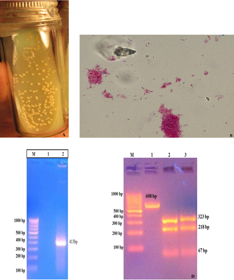
Colonies of the MAP reference strain (S 5) appeared on the slants of HEY medium. Colonies were small, straw color, translucent, hemispherical, raised with nipple, smooth, and glistening (Fig 1A). Staining of smears from MAP colonies by ZN-stain exhibited short pink staining cocco-bacillary nature (Fig 1B). Yield of specific PCR products in IS900 PCR, confirmed the presence of MAP (Fig 1C and 1D).
Fig 1. Characterization of sub-cultured of MAP reference strain (S5) on HEY medium.
(A) Characterization of sub-cultured MAP ‘S 5’ colonies (B) Microscopic view of acid fast bacilli (indistinguishable to MAP) under oil immersion (100X). Molecular Characterization of MAP ‘S5’ DNA: (C) IS900 PCR: Lane M- 100 base Marker (#SM0243, Fermentas), Lane 2: IS900 PCR product; (D) IS1311 PCR and IS1311-REA genotyping: Lane M: 100 base Marker (#SM0243, Fermentas), Lane 1: IS1311 PCR product, Lane 2: Digested PCR product of IS1311 from Positive control, Lane 3: Digested PCR product of IS1311 from sub-cultured colonies.
Bioinformatics analysis
Alignment and comparison of MAP K-10 and MAP (S 5) strain sequences illustrated highly conserved sequences, likely to be cross-reactive, as most epitopes were shared. Moreover, MAP2189-MAP2194 cluster organization was also conserved in both the strains. Based on Bioinformatics analysis, MAP2191 gene from MAP was of 1065bp and encodes for a typical linear Mce protein that has 354 amino acids with an estimated molecular weight of 37.510 kDa. Possible secondary structure of the MAP2191 protein was predicted using SOPMA website and analyzed results revealed that this protein consisted of 41.53% α-Helix, 16.95% extended strand, 11.58% β-turn and 29.94% random coils. C-terminal portion of MAP2191 protein was successfully expressed and purified in this study. Partial Mce-truncated protein sequence (amino acids 154 to 354) was chosen based on the predicted sequence to contain most dominant B- and T-cell epitopes. CLC software analysis also revealed that selected region of MAP2191 protein had high antigenic index and was more hydrophilic.
Amplification, cloning, and sub-cloning of mce-truncated gene
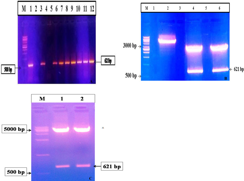
Whole DNA was isolated from MAP (S 5) strain, and target region of MAP2191 gene was successfully amplified by PCR. Agarose gel electrophoresis of PCR products showed a single band of 621bp amplicon (603bp mce gene and extra 18bp were used to restrict sites and bring the coding sequence in-frame) as positive for the truncated gene (Fig 2A). Mce-truncated amplicon was sequenced and the sequence results were aligned using BLASTp analysis; no mismatch was reported in sequence data. The amplified mce-truncated fragment was purified and ligated into pTZ57R/T TA cloning vector and transformed into E. coli competent cells. Transformants were confirmed for the presence of mce-truncated insert by specific colony PCR and restriction analysis (Fig 2B). Gene sequencing results confirmed the presence of mce-truncated gene segment in pTZ57R/T vector. Purified mce-truncated gene from TA-cloning vector was sub-cloned into pET28a (+) expression vector between the restriction sites BamHI and HindIII and transformed into XL10-Gold ultra-competent cells for the high percentage of recombinant plasmid yields. Presence of the insert was approved by colony PCR and double digestion using restriction enzymes (Fig 2C). Gene sequencing results confirmed the presence of mce-truncated gene in the pET28a vector.
Fig 2.
(A) Colony PCR for MAP mce-truncated gene, Lane M: 1kb DNA ladder (#SM0313, Fermentas), Lane 1: Positive control (MAP Indian Bison type DNA), Lane 2: Negative control (Nuclease Free Water), Lane 4: Negative in colony PCR for MAP Mce-truncated genes, Lanes 3 and 5–12 positive in colony PCR for MAP Mce-truncated genes; (B) Restriction digestion of confirmed colony PCR positive PTZ57R/T mce-truncated clones: Lane M: 1kb DNA ladder (#SM0313, Fermentas), Lane 2: Double digested pET28a plasmid, Lanes 4 and 6: Positive clones of PTZ57R/T mce-truncated plasmid; (C) Restriction digestion of confirmed colony PCR positive pET28a mce-truncated clones; Lane M: 1kb DNA ladder (RBC), lanes 1 and 2: Positive clones of PTZ57R/T mce-truncated plasmid.
Expression and purification of Mce-recombinant protein
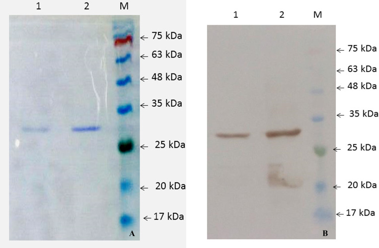
The pET28a-mce-truncated plasmid was transformed into Rosetta/BL-21 cells. Transformed cells were induced for expression with optimized concentration of 1.5 mM IPTG (Fig 3A). Samples were collected at different time intervals and were analyzed using 12.0% SDS-PAGE, along with the protein ladder (Precision plus make). The ~28–30 kDa Mce-truncated protein was expressed from 2 hrs post induction, while zero hour sample and mock controls showed no observable expression; distinct bands of intensity increased with time between 2 and 24 hrs as shown in Fig 3B. Optimized time interval for the best expression of Mce-truncated protein after induction with IPTG was 20 hrs. Expressed proteins were purified by nickel-nitrilotriacetic acid (Ni-NTA) gel matrix (Qiagen, USA) using batch method; purified Mce-truncated protein run on SDS-PAGE showed single band with an approximate expected molecular mass of ~28–30 kDa (Fig 4A). Mce-truncated proteins were confirmed using anti-His tagged mouse monoclonal antibody (Sigma Aldrich, Inc.) (Fig 4B). Final yield of pure Mce-truncated protein per liter for E. coli culture was approximately 0.8 gr/L using Bradford method.
Fig 3. Optimization condition for Mce-truncated protein expression.
(A) SDS-PAGE analysis of Mce-truncated protein expression level with different IPTG concentration at 200 C in E. coli strain BL21 (DE3) pLysS. (B) SDS-PAGE analysis of Mce-truncated protein production at different time intervals on the optimized concentration of IPTG (1.5mM IPTG). Lane M. Molecular weight marker (MBT092-100LN Hi-Media). Arrow indicates Mce-truncated protein.
Fig 4. Analysis of purified Mce-truncated protein expressed in E. coli strain BL21 (DE3) pLysS.
(A) SDS PAGE. (Left to Right), Lane 1. pET28a.Mce.truncated Elution 2; lane 2. pET28a.Mce.truncated Elution 1; Lane M. Protein molecular weight marker (SL7012, CinnaGen). (B) Western blot analysis using Anti-6X His-tagged monoclonal antibody (left to right) Lanes 1: and 2: Western blot from purified MAP Mce-truncated protein, Lane M: Protein molecular weight marker (SL7012, CinnaGen).
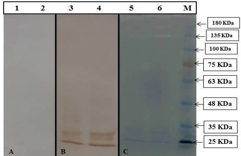
Cross reactions analysis by western blot also showed that the expressed Mce-truncated protein reacted with sera of MAP infected animals, but did not react with tuberculosis-infected ones (Fig 5). The result suggested that the recombinant Mce-truncated protein had no cross reactions with non-MAP mycobacterial infections, such as tuberculosis.
Fig 5. Analysis of cross reactivity and immunogenicity of purified Mce-truncated protein.
(A) Western blot analysis using HRP-conjugated anti-bovine immunoglobulin. (Left to Right), Lanes 1, 2. Western blot of Mce-truncated protein of different batch with tuberculin positive sera. (B) Western blot analysis using positive goat serum for MAP. (Left to Right), Lanes 3, 4. Western blot from Mce-truncated proteins of different batch reacted with MAP infected goat serum. (C) SDS PAGE of purified Mce-truncated proteins: Lanes 5, 6. Purified Mce-truncated proteins of different batch, Lane M: Protein molecular weight marker (SL7012, CinnaGen).
Antibody response with Mce-ELISA
Newly developed Mce-truncated protein based-ELISA was compared with i-ELISA (an in house ELISA using a semi-purified protoplasmic extract of the S5 'Indian Bison' MAP bio-type) (Table 3). Sample OD values (at 450 nm) were converted to sample-to-positive (S/P) ratios as per Collins (2002) [40]. Serum samples in the positive and strong positive categories were considered positive for MAP infection (Table 3). Serum samples from 251 goats, 53 sheep, 117 buffalos, and 33 cattle were screened by Mce-ELISA and i-ELISA tests. Antibody titers measured were 49.4, 51.0, 69.2 and 54.6%, by Mce-ELISA and 55.4, 58.5, 71.8 and 63.6%, by i-ELISA in goats, sheep, buffaloes, and cattle, respectively. Mce-truncated antigen had a higher specificity, but its sensitivity was lower than i-ELISA. However, absorbance values in Mce-ELISA were higher as compared to the values in i-ELISA.
Table 3. Comparison of species-wise status of Johne’s disease in ‘Indigenous ELISA’ and ‘Mce ELISA’ tests.
| Sera samples (n) | Status | Mce-ELISA (%) | Results (%) | i-ELISA (%) | Results (%) |
|---|---|---|---|---|---|
| Goats (251) | N | 81(32.3) | Negative 127 (50.6%) | 65 (25.9) | Negative 112 (44.6%) |
| S | 9 (3.5) | 12 (4.8) | |||
| LP | 37 (14.7) | 35 (13.9) | |||
| P | 102 (40.6) | Positive 124 (49.4%) | 124 (49.4) | Positive 139 (55.4%) | |
| SP | 22 (8.8) | 15 (6) | |||
| Sheep (53) | N | 19 (35.8) | Negative 26 (49%) | 14 (26.4) | Negative 22 (41.5%) |
| S | 5 (9.4) | 6 (11.3) | |||
| LP | 2 (3.8) | 2 (3.8) | |||
| P | 23 (43.4) | Positive 27 (51%) | 25 (47.2) | Positive 31 (58.5%) | |
| SP | 4 (7.5) | 6 (11.3) | |||
| Buffaloes (117) | N | 22 (18.8) | Negative 36 (30.8%) | 10 (8.6) | Negative 33 (28.2%) |
| S | 2 (1.7) | 8 (6.8) | |||
| LP | 12 (10.3) | 15 (12.8) | |||
| P | 61 (52.1) | Positive 81 (69.2%) | 65 (55.6) | Positive 84 (71.8%) | |
| SP | 20 (17.1) | 19 (16.2) | |||
| Cattle (33) | N | 12 (36.4) | Negative 15 (45.5%) | 7 (21.2) | Negative 12 (36.4%) |
| S | 0 (0) | 1 (3.1) | |||
| LP | 3 (9.1) | 4 (12.1) | |||
| P | 13 (39.4) | Positive 18 (54.5%) | 17 (51.5) | Positive 21 (63.6%) | |
| SP | 5 (15.1) | 4 (12.1) |
Goats
Of the 251 serum screened by Mce-ELISA and i-ELISA, 124 (49.4%) and 139 (55.4%) were positive, and 127 (50.6%) and 112 (44.6%) were negative, respectively (Table 3). Comparative sero-reactivity of Mce-ELISA with i-ELISA showed that results of positive and negative goats were comparable. A total of 21 (8.3%) positive samples in i-ELISA were missed by Mce-ELISA, while 6 (2.3%) samples-positive in Mce-ELISA were missed by i-ELISA (Table 4). Though i-ELISA appeared to be more sensitive of the two tests applied, but Mce-ELISA was more specific than i-ELISA (Table 5).
Table 4. Comparison of two ELISA test (Mce-ELISA and i-ELISA) combinations for the diagnosis of Johne’s disease in domestic livestock species.
| Tests | Combinations, n (%) | |||
|---|---|---|---|---|
| 1 | 2 | 3 | 4 | |
| Mce-ELISA | + | - | + | - |
| i-ELISA | + | - | - | + |
| Goats (251) | 118 (47.0) | 106 (42.2) | 6 (2.3) | 21 (8.3) |
| Sheep (53) | 27 (50.9) | 22 (41.5) | 0 (0.0) | 4 (7.5) |
| Buffaloes (117) | 78 (66.6) | 30 (25.6) | 3 (2.5) | 6 (5.0) |
| Cattle (33) | 18 (54.5) | 12 (36.3) | 0 (0.0) | 3 (9.0) |
*Value in parenthesis are percent.
Table 5. Comparative sensitivity and specificity of two ELISA tests (Mce-ELISA with i-ELISA) in four domestic livestock species.
| Species | Tests | TP | TN | FP | FN | Sen & Sp (%) |
|---|---|---|---|---|---|---|
| Goats (251) | Mce-ELISA | 118 | 106 | 6 | 21 | 84.8 & 94.6 |
| i-ELISA | 118 | 106 | 21 | 6 | 95.1 & 83.4 | |
| Sheep (53) | Mce-ELISA | 27 | 22 | 0 | 4 | 87.1 & 100.0 |
| i-ELISA | 27 | 22 | 4 | 0 | 100.0 & 84.6 | |
| Buffaloes (117) | Mce-ELISA | 78 | 30 | 3 | 6 | 92.8 & 90.0 |
| i-ELISA | 78 | 30 | 6 | 3 | 96.3 & 83.3 | |
| Cattle (33) | Mce-ELISA | 18 | 12 | 0 | 3 | 85.7 & 100.0 |
| i-ELISA | 18 | 12 | 3 | 0 | 100.0 & 80.0 |
TP- true positive; FP-false positive; TN- true negative; FN- false negative; Sen- sensitivity; Sp- specificity.
Sheep
Of the 53 serum screened by Mce-ELISA and i-ELISA, 27 (51.0%) and 31 (58.5%) were positive and 26 (49.0%) and 22 (41.5%) were negative, respectively (Table 3). I-ELISA showed higher number of serum samples positive and lower number as negatives than Mce-ELISA. Only 4 (7.5%) positive samples in i-ELISA were missed by Mce-ELISA (Table 4). Here too, i-ELISA was more sensitive, though specificity was lower than Mce-ELISA (Table 5).
Buffaloes
Of 117 buffaloes serum samples screened by Mce-ELISA and i-ELISA, 81 (69.2%) and 84 (71.8%) were positive and 36 (30.8) and 33 (28.2%) were negative, respectively (Table 3). Results were comparable; only 3 (2.5%) positive serum in i-ELISA were missed by Mce-ELISA (Table 4). These results showed higher specificity and lowered sensitivity in case of Mce-ELISA as compared to i-ELISA in buffaloes (Table 5).
Cattle
Of 33 cattle serum screened, 18 (54.5%) and 21 (63.6%) were positive, and 15 (45.5%) and 12 (36.4%) were negative by Mce-ELISA and i-ELISA, respectively (Table 3). The 3 (9.0%) serum positive in i-ELISA were missed by Mce-ELISA (Table 4). The results were similar in cattle and Mce-ELISA showed higher specificity and lowered sensitivity in comparison to i-ELISA (Table 5).
Sensitivity and specificity of Mce-ELISA
(a) Comparison of Mce-ELISA with i-ELISA. Sensitivity of Mce-ELISA was lower 84.8, 87.1, 92.8 and 85.7%, but specificity was higher 94.6%, 100.0%, 90.0 and 100.0% in goats, sheep, buffaloes, and cattle, respectively (Table 5).
(b) Comparison of Mce-ELISA with i-ELISA. Sensitivity of i-ELISA was higher 95.1, 100.0, 96.3 and 100.0%, but specificity was lower 83.4%, 84.6%, 83.3 and 80.0% in goats, sheep, buffaloes and cattle, respectively (Table 5).
Statistical analysis
Mc-Nemar test and kappa agreement were used for statistical analysis. P value and kappa agreement were calculated. P values were 0.0071, 0.1336, 0.5050 and 0.2482, and Kappa agreements were 0.785, 0.849, 0.815 and 0.814 in goats, sheep, cattle, and buffaloes, respectively. The strength of agreement was substantial in goats and perfect in another three livestock species (sheep, buffaloes, cattle), respectively (Table 6).
Table 6. P values (McNemar test) and Kappa agreement between Mce-ELISA and i-ELISA.
| Species | P values | Kappa | Strength of agreement | 95% Confidence interval | |
|---|---|---|---|---|---|
| Status | Value | ||||
| Goats | Very significantly different | 0.0071 | 0.785 | Substantial | 0.709 to 0.861 |
| Sheep | Not significantly different | 0.1336 | 0.849 | Perfect | 0.708 to 0.990 |
| Buffaloes | Not significantly different | 0.5050 | 0.815 | Perfect | 0.700 to 0.931 |
| Cattle | Not significantly different | 0.2482 | 0.814 | Perfect | 0.616 to 1.000 |
Kappa value (0.0–0.20, poor; 0.21–0.40, fair; 0.41–0.60, moderate; 0.61–0.80, substantial and 0.81–100, perfect).
Discussion
MAP, the cause of JD in domestic livestock [41], has also been associated with CD and other auto-immune disorders in human beings [2, 4, 5, 8]. Developing effective strategies for the diagnosis and control of MAP infection remains a major challenge for optimizing the animal health [42]. ELISA test being sensitive, quick and cost effective is preferred as ‘screening test’ in chronic infections like MAP [37]. Present study described genetic expression of a new Mce protein that could be possibly used as potential antigenic candidate for the development of a new ELISA test for the diagnosis of MAP infection.
Proteins of Mce-family of pathogenic mycobacteria have been associated with virulence by enhancing invasive power and survival in macrophages and various mammalian cell lines [15, 43–46]. Mce encoded products were found to conferring invasive attribute to otherwise a non-pathogenic strain of E. coli, invading non-phagocytic HeLa cells [16, 24, 47]. Kumar et al. [48] analyzed and compared the expression profiles of the mce operons of Mycobacterium tuberculosis, Mycobacterium bovis, and Mycobacterium smegmatis under different growth conditions and found the expression of mce1, mce3, and mce4 operons present in tissues collected from infected guinea pigs and rabbits. Hou et al. [49] found that genes similar to MTB mce genes were expressed by Mycobacterium avium during growth in macrophages. Another previous study also revealed presence of mce and PE/ PPE family genes in MAP as virulence factors [50]. In a study conducted by Wu et al. [51], showed that there were islands in both M. avium subsp. avium (MAC) and MAP which encode for different mce genes that were analogous in all the mycobacteria, indicating their functional importance.
MAP2191 gene is one of the mce5 operons (MAP2189 to MAP2194) of the MAP; mce5 cluster which has previously been identified as important for invasion and survival of bacilli within macrophages and also virulence of MAP [25, 26]. Eight putative Mce-encoding genes have been identified in the MAP strain K-10 genome. Some studies have been carried out to find the unique MAP-specific protein targets, which may elicit strong immune responses in the host [13, 52]. Targeting of the MAP2191 gene was based on the identification of putative B- and T-cell epitopes in MAP proteins encoded by the mce5 gene cluster and asses their antigenicity during infection. Using Bioinformatics analyses, we identified B- and T-cell epitopes in the C-terminal region of the MAP2191 putative Mce protein. The selected region of MAP2191 protein had a high antigenic index and was more hydrophilic as crevealed by the CLC software analysis. In this study, we generated and cloned a truncated MAP2121 DNA sequence, and subsequently generated the His-tagged recombinant protein in an E. coli host. Our purification method with Ni-NTA agarose resin was very effective and we obtained a large amount of soluble and purified Mce-truncated peptide.
Currently, antigen-based ELISA tests for detecting MAP with a mixture of proteins are available including avian or johnin, a MAP-PPD, whole-cell sonicated extract, protoplasmic antigens etc., [53]. Because analysis and sequencing of the entire MAP genome has been recorded [54], immune reactivity of several specific proteins of the MAP genome have been investigated [55]. In the present study also, serological ELISA assays were conducted and results have been reported for the new ELISA test developed using truncated MAP Mce protein and the traditional i-ELISA kit.
This in house i-ELISA kit has been validated with other commercial ELISA kits (ID-VET, Allied Monitor Inc., USA, antigen based indirect ELISA, Ethanol Vortex ELISA (EV-ELISA), recombinant secretory proteins based cocktail ELISA (c_ELISA) and natural secretory proteins-based ELISA test [10, 35, 37, 56, 57] and compared its efficacy in different livestock species (goats, sheep, cattle and buffaloes) [12, 58, 59]. In this series, Mce-ELISA is the most recently developed ELISA and used for the diagnosis of MAP infection. I-ELISA has been the most validated and robust test was used for evaluating newly developed Mce-ELISA in terms of specificity and sensitivity.
Our earlier studies reported enhanced sensitivity and specificity of the i-ELISA as compared to the other commercially available ELISA kits for the diagnosis of Johne’s disease in domestic livestock [38, 39]. For example, in a study, i-ELISA kit showed greater diagnostic efficiency than other commercial ELISA kits [38] and improved the detection rate of MAP in test samples [60]. We used S/P-ratios-based categorization of OD values as per Collin (2002), wherein serum samples in the positive and strong positives categories, were considered as positive for MAP infection. This type of categorization exhibited good correlation with fecal culture in some of our other studies [39, 61]. Chaubey et al. (2015) compared i-ELISA (Indigenous g-ELISA) with commercial antigen-based b-ELISA and commercial sr-ELISA and found 100% sensitivity. In another report, 91.4% sensitivity of i-ELISA was observed when compared with EV-ELISA kit [57]. These and other findings from other studies confirmed the enhanced sensitivity and specificity of i-ELISA in the detecting of MAP infection in domestic livestock in the country [35, 38].
In our study, Mce-truncated protein yielded higher absorbance values against the confirmed positive pooled serum samples, and low-level (but above background) antibody reactivity was also observed with confirmed negative pooled serum samples. The ELISA test using Mce-truncated protein was able to discriminate between the most positive and negative serum samples used in this study. However, the Mce-based ELISA showed a relatively higher specificity than i-ELISA but suffered from slight reduction in sensitivity. These results highlighted the potential capability of Mce-truncated ELISA in preventing false-positive reactions that occur when a milieu of antigens is used in i-ELISA tests. Mce-ELISA has shown perfect agreement with i-ELISA and there is no cross-reactivity with anti-M.bovis antibodies. Therefore, this test could be a better choice for screening of paratuberculosis especially in the region where bovine-TB is endemic. Overall the Mce-based ELISA was comparable to the the highly sensitive and robust ‘i-ELISA test’, indigenously developed for the screening of livestock population.
In conclusion, this is the first report of a Mce surface protein-based ELISA as new modified ELISA test, as diagnostic test against MAP infection. Affinity of purified Mce-truncated protein was detected by serum antibodies from animals infected with JD and supported the hypothesis that MAP Mce protein was immunogenic and highly expressed protein during MAP infection. True achievement of this study was the isolation and expreseeion of the Mce protein, ‘a new antigen’ with the huge potential to be further exploited not only as a diagnostic tool but also a new target antigen for the development of a highly effective vaccine for the control and eradication of the incurable Johne’s disease in domestic livestock.
Supporting information
(DOCX)
(DOCX)
Acknowledgments
The authors would like to thank Dr Rinkoo Devi Gupta, Assistant Professor from SAU, New Delhi, Dr. Pravin Kumar Singh, Assistnat Professor, GLA University, Mathura, U.P., India and from ICAR_CIRG (Director, Dr. K. Gururaj, Scientist, Mr. Saurabh Gupta, RA and Ms. Manju Singh, RA for their tremendous help and support during the studies.
Data Availability
All relevant data are within the paper and its Supporting Information files.
Funding Statement
This work was financially supported partially by a grant from Shiraz University (94GCU3M1304) and also Iran National Science Foundation (INSF) (No. 97004189), different institutions in India (ICAR_CIRG, Mathura, ICMR and DST_SERB New Delhi).
References
- 1.Faisal SM, Yan F, Chen TT, Useh NM, Guo S, Yan W, et al. Evaluation of a salmonella vectored vaccine expressing Mycobacterium avium subsp. paratuberculosis antigens against challenge in a goat model. PLoS ONE. 2013;8:70171 10.1371/journal.pone.0070171 [DOI] [PMC free article] [PubMed] [Google Scholar]
- 2.Gupta S, Singh SV, Gururaj K, Chaubey KK, Singh M, Lari B, et al. Comparison of IS900 PCR with ‘Taqman probe PCR’ and ‘SYBR green Real time PCR’ assays in patients suffering with thyroid disorder and sero-positive for Mycobacterium avium subspecies paratuberculosis. Indian J Biotechnol. 2017;228:234 URl: http://nopr.niscair.res.in/handle/123456789/42918 [Google Scholar]
- 3.Singh SV, Singh AV, Singh PK, Kumar A, Singh B. Molecular identification and characterization of Mycobacterium avium subspecies paratuberculosis in free living non-human primate (Rhesus macaques) from North India. Comp. Immunol Microbiol Infect Dis. 2011;34:267–71. 10.1016/j.cimid.2010.12.004 [DOI] [PubMed] [Google Scholar]
- 4.Kuenstner JT, Naser S, Chamberlin W, Borody T, Graham DY, McNees A, et al. The Consensus from the Mycobacterium avium ssp. paratuberculosis (MAP) Conference 2017. Front Public Health. 2017;5:208 10.3389/fpubh.2017.00208 [DOI] [PMC free article] [PubMed] [Google Scholar]
- 5.United States Department of Agriculture, "National Animal Health Reporting System—Reportable Diseases" Archived 2009-08-29 at the Wayback Machine.
- 6.Ahlstrom C, Barkema HW, Stevenson K, Zadoks RN, Biek R, Kao R, et al. Limitations of variable number of tandem repeat typing identified through whole genome sequencing of Mycobacterium avium subsp. paratuberculosis on a national and herd level. BMC. Genomics. 2015;16:161 10.1186/s12864-015-1387-6 [DOI] [PMC free article] [PubMed] [Google Scholar]
- 7.Singh SV, Stephen BJ, Singh M, Gupta S, Chaubey KK, Sahzad, et al. Comparison of newly standardized 'Latex milk agglutination test', with 'Indigenous milk ELISA' for 'on spot' screening of domestic livestock against Mycobacterium avium subsp. paratuberculosis infection. Indian J Biotechnol. 2016;15:511–17. URl: http://nopr.niscair.res.in/handle/123456789/41031 [Google Scholar]
- 8.Acharya KR, Dhand NK, Whittington RJ, Plain KM. Detection of Mycobacterium avium subspecies paratuberculosis in powdered infant formula using IS900 quantitative PCR and liquid culture media. Int J Food Microbiol. 2017;257:1–9. 10.1016/j.ijfoodmicro.2017.06.005 [DOI] [PubMed] [Google Scholar]
- 9.Sechi LA, Dow CT. Mycobacterium avium ss. paratuberculosis zoonosis, the hundred year war beyond Crohn’s disease. Front Microbiol. 2015;6:1–8. 10.3389/fimmu.2015.00096 [DOI] [PMC free article] [PubMed] [Google Scholar]
- 10.Chaubey KK, Singh SV, Gupta S, Jayaraman S, Singh M, Stephan BJ, et al. Diagnostic potential of three antigens from geographically different regions of the world for the diagnosis of Ovine Johne’s disease in India. Asian J Anim Vet Adv. 2015;10:567–576. 10.3923/ajava.2015.567.576 [DOI] [Google Scholar]
- 11.Rana A, Rub A, Akhter Y. Proteome-wide B and T cell epitope repertoires in outer membrane proteins of Mycobacterium avium subsp. paratuberculosis have vaccine and diagnostic relevance: a holistic approach. J Mol Recognit. 2015;28:506–20. 10.1002/jmr.2458 [DOI] [PubMed] [Google Scholar]
- 12.Chaubey KK, Singh SV, Gupta S, Singh M, Sohal JS, Kumar N, et al. Mycobacterium avium subspecies paratuberculosis—an important food borne pathogen of high public health significance with special reference to India: an update. Veterinary Quarterly. 2017; 36 (4): 203–227. 10.1080/01652176.2017.1397301 [DOI] [PubMed] [Google Scholar]
- 13.Gurung RB, Purdie AC, Begg DJ, Whittington RJ. In silico identification of epitopes in Mycobacterium avium subsp. paratuberculosis proteins that were upregulated under stress conditions. Clin Vaccine Immunol. 2012;19:855–64. 10.1128/CVI.00114-12 [DOI] [PMC free article] [PubMed] [Google Scholar]
- 14.Xu G, Li Y, Yang J, Zhou X, Yin X, Liu M, et al. Effect of recombinant Mce4A protein of Mycobacterium bovis on expression of TNF-alpha, iNOS, IL-6, and IL-12 in bovine alveolar macrophages. Mol Cell Biochem. 2007;302:1–7. 10.1007/s11010-006-9395-0 [DOI] [PubMed] [Google Scholar]
- 15.Hemati Z, Derakhshandeh A, Haghkhah M, Chaubey KK, Gupta S, Singh M, et al. Mammalian cell entry operons; novel and major subset candidates for diagnostics with special reference to Mycobacterium avium subspecies paratuberculosis infection. Vet Q. 2019,1–17. 10.1080/01652176.2019.1641764 [DOI] [PMC free article] [PubMed] [Google Scholar]
- 16.Forrellad MA, McNeil M, Santangelo ML, Blanco FC, Garcia E, Klepp LI, et al. Role of the Mce1 transporter in the lipid homeostasis of Mycobacterium tuberculosis. Tuberculosis (Edinb). 2015;94:170–7. 10.1016/j.tube.2013.12.005 [DOI] [PMC free article] [PubMed] [Google Scholar]
- 17.Timms VJ, Hassan KA, Mitchell HM, Neilanm BA. Comparative genomics between human and animal associated subspecies of the Mycobacterium avium complex: a basis for pathogenicity. BMC Genomics. 2015;16:695 10.1186/s12864-015-1889-2 [DOI] [PMC free article] [PubMed] [Google Scholar]
- 18.Mohn WW, vander-Geize R, Stewart GR, Okamoto S, Liu J, Dijkhuizen L, et al. The actinobacterial mce4 locus encodes a steroid transporter. J Biol Chem. 2017;283:35368–74. 10.1074/jbc.M805496200 [DOI] [PMC free article] [PubMed] [Google Scholar]
- 19.Pandey AK, Sassetti CM. Mycobacterial persistence requires the utilization of host cholesterol. Proc Natl Acad Sci USA. 2008;105:4376–80. 10.1073/pnas.0711159105 [DOI] [PMC free article] [PubMed] [Google Scholar]
- 20.Senaratne RH, Sidders B, Sequeira P, Saunders G, Dunphy K, Marjanovic O, et al. Mycobacterium tuberculosis strains disrupted in mce3 and mce4 operons are attenuated in mice. J Med Microbiol. 2008;57:164–70. 10.1099/jmm.0.47454-0 [DOI] [PubMed] [Google Scholar]
- 21.Perkowski EF, Miller BK, McCann JR, Sullivan JT, Malik S, Allen IC, et al. An orphaned Mce-associated membrane protein of Mycobacterium tuberculosis is a virulence factor that stabilizes Mce transporters. Mol Microbiol. 2016;100:90–107. 10.1111/mmi.13303 [DOI] [PMC free article] [PubMed] [Google Scholar]
- 22.Casali N, Riley LW. A phylogenomic analysis of the Actinomycetales mce operons. BMC. Genomics. 2007;8:60 10.1186/1471-2164-8-60 [DOI] [PMC free article] [PubMed] [Google Scholar]
- 23.Castellanos E, Aranaz A, Gould KA, Linedale R, Stevenson K, Alvarez J. Discovery of stable and variable differences in the Mycobacterium avium subsp. paratuberculosis type I, II, and III genomes by pan-genome microarray analysis. Appl Environ Microbiol. 2009;75:676–86. 10.1128/AEM.01683-08 [DOI] [PMC free article] [PubMed] [Google Scholar]
- 24.Hemati Z, Haghkhah M, Derakhshandeh A, Singh S, Chaubey KK. Cloning and characterization of MAP2191 gene, a mammalian cell entry antigen of Mycobacterium avium subspecies paratuberculosis. Mol Biol Res Commun. 2018; 7(4):165–172. 10.22099/mbrc.2018.30979.1354 [DOI] [PMC free article] [PubMed] [Google Scholar]
- 25.Semret M, Alexander DC, Turenne CY, de-Haas P, Overduin P, van Soolingen D, et al. Genomic polymorphisms for Mycobacterium avium subsp. paratuberculosis diagnostics. J Clin Microbiol. 2005;43:3704–12. 10.1128/JCM.43.8.3704-3712.2005 [DOI] [PMC free article] [PubMed] [Google Scholar]
- 26.Alexander DC, Turenne CY, Behr MA. Insertion and deletion events that define the pathogen Mycobacterium avium subsp. paratuberculosis. J Bacteriol. 2009;191:1018–25. 10.1128/JB.01340-08 [DOI] [PMC free article] [PubMed] [Google Scholar]
- 27.Ahmad S, El-Shazly S, Mustafa A, Al-Attiyah R. The Six Mammalian Cell Entry Proteins Mce3A–F Encoded by the mce3 Operon are Expressed During In Vitro Growth of Mycobacterium tuberculosis. Scand J Immunol. 2005;62:16–24. 10.1111/j.1365-3083.2005.01639.x [DOI] [PubMed] [Google Scholar]
- 28.Zhang F, Xie JP. Mammalian cell entry gene family of Mycobacterium tuberculosis. Mol Cell Biochem. 2011;352:1–10. 10.1007/s11010-011-0733-5 [DOI] [PubMed] [Google Scholar]
- 29.Sevilla I, Singh SV, Garrido JM, Aduriz G, Rodríguez S, Geijo MV, et al. Molecular typing of Mycobacterium avium subspecies paratuberculosis strains from different hosts and regions. Rev Sci Techn. 2005;24:1061–66. [PubMed] [Google Scholar]
- 30.Singh SV, Singh AV, Singh PK, Gupta VK, Kumar S, Vohra J. Sero-prevalence of paratuberculosis in young kids using ‘Bison type’, Mycobacterium avium subsp. paratuberculosis antigen in plate ELISA. Small Rumin Res. 2007. b;70:89 URl: 10.1016/j.smallrumres.2005.11.019 [DOI] [Google Scholar]
- 31.Nielsen M, Lundegaard C, Worning P, Lauemøller SL, Lamberth K, Buus S, et al. Reliable prediction of T-cell epitopes using neural networks with novel sequence representations. Protein Sci. 2003;12:1007–17. 10.1110/ps.0239403 [DOI] [PMC free article] [PubMed] [Google Scholar]
- 32.Tenzer S, Peters B, Bulik S, Schoor O, Lemmel C, Schatz MM, et al. Modeling the MHC class I pathway by combining predictions of proteasomal cleavage, TAP transport and MHC class I binding. Cell. Mol. Life Sci. 2005;62:1025–37. 10.1007/s00018-005-4528-2 [DOI] [PubMed] [Google Scholar]
- 33.Wang P, Sidney J, Dow C, Mothé B, Sette A, Peters B. A systematic assessment of MHC class II peptide binding predictions and evaluation of a consensus approach. PLoS Comput. Biol. 2008;4(4):1000048 10.1371/journal.pcbi.1000048 [DOI] [PMC free article] [PubMed] [Google Scholar]
- 34.Carlos P, Roupie V, Holbert S, Ascencio F, Huygen K, Gomez-Anduro G, et al. In silico epitope analysis of unique and membrane associated proteins from Mycobacterium avium subsp. paratuberculosis for immunogenicity and vaccine evaluation. J Theor Biol. 2015;384;1–9. 10.1016/j.jtbi.2015.08.003 [DOI] [PubMed] [Google Scholar]
- 35.Ponomarenko J, Bui HH, Li W, Fusseder N, Bourne PE, Sette A, et al. ElliPro: a new structure-based tool for the prediction of antibody epitopes. BMC Bioinformatics. 2008;9:514 10.1186/1471-2105-9-514 [DOI] [PMC free article] [PubMed] [Google Scholar]
- 36.Chaubey KK, Singh SV, Bhatia AK. Evaluation of ‘Recombinant secretary antigens’ based ‘Cocktail ELISA’ for the diagnosis of Johne's disease and to differentiate non-infected, infected and vaccinated goats in combination with indigenous ELISA test, Small Rumin. Res. 2018;165:24–29. [Google Scholar]
- 37.Singh SV, Singh AV, Singh PK, Sohal JS, Singh NP. Evaluation of an indigenous ELISA for diagnosis of Johne’s disease and its comparison with commercial kits. Indian J Microbiol. 2007a;47:251 10.1007/s12088-007-0046-2 [DOI] [PMC free article] [PubMed] [Google Scholar]
- 38.Singh AV, Singh SV, Sohal JS, Singh PK. Comparative potential of modified indigenous, indigenous and commercial ELISA kits for diagnosis of Mycobacterium avium subspecies paratuberculosis in goat and sheep. Indian J Exp Biol. 2009;47:379 [PubMed] [Google Scholar]
- 39.Sharma G, Singh SV, Sevilla I, Singh AV, Whittington RJ, Juste RA, et al. Evaluation of indigenous milk ELISA with m-culture and m-PCR for the diagnosis of bovine Johne's disease (BJD) in lactating Indian dairy cattle. Res Vet Sci. 2008;84:30 10.1016/j.rvsc.2007.03.014 [DOI] [PubMed] [Google Scholar]
- 40.Collins MT. Interpretation of a commercial bovine paratuberculosis enzyme-linked immunosorbent assay by using likelihood ratios. Clin Diagn Lab Immunol. 2002;9:1367–71. 10.1128/cdli.9.6.1367-1371.2002 [DOI] [PMC free article] [PubMed] [Google Scholar]
- 41.Mukartal SV, Rathnamma D, Narayanaswamy HD, Gupta S, Chaubey KK, Singh M, et al. Assessment of Ovine Johne’s disease in the Mandya sheep breed in South India using multiple diagnostic tests and bio-typing of Mycobacterium avium subspecies paratuberculosis infection. Cogent Food Agric. 2017;3:1298391 URl: 10.1080/23311932.2017.1298391 [DOI] [Google Scholar]
- 42.Bannantine JP, Paulson AL, Chacon O, Fenton RJ, Zinniel DK, McVey DS, et al. Immunogenicity and reactivity of novel Mycobacterium avium subsp. paratuberculosis PPE MAP1152 and conserved MAP1156 proteins with sera from experimentally and naturally infected animals. Clin Vaccine Immunol. 2011;18:105–12. 10.1128/CVI.00297-10 [DOI] [PMC free article] [PubMed] [Google Scholar]
- 43.Huntley JF, Stabel JR, Paustian ML, Reinhardt TA, Bannantine JP. Expression library immunization confers protection against Mycobacterium avium subsp. paratuberculosis infection. Infect Immun. 2005;73:6877–6684. 10.1128/IAI.73.10.6877-6884.2005 [DOI] [PMC free article] [PubMed] [Google Scholar]
- 44.Checkley AM, Wyllie DH, Scriba TJ, Golubchik T, Hill AV, Hanekom WA, et al. Identification of antigens specific to non-tuberculous mycobacteria: the Mce family of proteins as a target of T cell immune responses. PLoS One. 2011;6:26434 10.1371/journal.pone.0026434 [DOI] [PMC free article] [PubMed] [Google Scholar]
- 45.Castellanos E, Aranaz A, de Juan L, Dominguez L, Linedale R, Bull TJ. A 16 kb naturally occurring genomic deletion including mce and PPE genes in Mycobacterium avium subspecies paratuberculosis isolates from goats with Johne's disease. Vet Microbiol. 2012;159:60–68. 10.1016/j.vetmic.2012.03.010 [DOI] [PubMed] [Google Scholar]
- 46.Ghosh P, Hsu C, Alyamani EJ, Shehata MM, Al-Dubaib MA, Al-Naeem A, et al. Genome-wide analysis of the emerging infection with Mycobacterium avium subspecies paratuberculosis in the Arabian camels (Camelus dromedarius). PLos One. 2012;7:31947 10.1371/journal.pone.0031947 [DOI] [PMC free article] [PubMed] [Google Scholar]
- 47.Uchiya K, Takahashi H, Yagi T, Moriyama M, Inagaki T, Ichikawa K, et al. Comparative genome analysis of Mycobacterium avium revealed genetic diversity in strains that cause pulmonary and disseminated disease. PLoS One. 2013;8:71831 10.1371/journal.pone.0071831 [DOI] [PMC free article] [PubMed] [Google Scholar]
- 48.Kumar A, Chandolia A, Chaudhry U, Brahmachari V, Bose M. Comparison of mammalian cell entry operons of mycobacteria: in silico analysis and expression profiling. FEMS. Immunol Med Microbiol. 2005;43:185–195. 10.1016/j.femsim.2004.08.013 [DOI] [PubMed] [Google Scholar]
- 49.Hou JY, Graham JE, Clark-Curtiss JE. Mycobacterium avium genes expressed during growth in human macrophages detected by selective capture of transcribed sequences (SCOTS). Infect Immun. 2002;70:3714–26. 10.1128/iai.70.7.3714-3726.2002 [DOI] [PMC free article] [PubMed] [Google Scholar]
- 50.Zhu X, Tu ZJ, Coussens PM, Kapur V, Janagama H, Naser S, et al. Transcriptional analysis of diverse strains Mycobacterium avium subspecies paratuberculosis in primary bovine monocyte derived macrophages. Microbes Infect. 2008;10:1274–82. 10.1016/j.micinf.2008.07.025 [DOI] [PubMed] [Google Scholar]
- 51.Wu CH, Glasner J, Collins M, Naser S, Talaat AM. Whole-Genome Plasticity among Mycobacterium avium Subspecies: Insights from Comparative Genomic Hybridizations. J Bacteriol. 2006;188:711–23. 10.1128/JB.188.2.711-723.2006 [DOI] [PMC free article] [PubMed] [Google Scholar]
- 52.Gumber S, Taylor DL, Whittington RJ. Evaluation of the immunogenicity of recombinant stress-associated proteins during Mycobacterium avium subsp. paratuberculosis infection: Implications for pathogenesis and diagnosis. Vet Microbiol. 2009;137:290–6. 10.1016/j.vetmic.2009.01.012 [DOI] [PubMed] [Google Scholar]
- 53.Biet F. Bay S, Thibault VC, Euphrasie D, Grayon M, Ganneau C, et al. Lipopentapeptide induces a strong host humoral response and distinguishes Mycobacterium avium subsp. paratuberculosis from M. avium subsp. avium. Vaccine. 2008;26:257–68. 10.1016/j.vaccine.2007.10.059 [DOI] [PubMed] [Google Scholar]
- 54.Li L, Bannantine JP, Zhang Q, Amonsin A, May BJ, Alt D, et al. The complete genome sequence of Mycobacterium avium subspecies paratuberculosis. Proc Natl Acad Sci USA. 2005;102:12344–49. 10.1073/pnas.0505662102 [DOI] [PMC free article] [PubMed] [Google Scholar]
- 55.Hughes V, Bannantine JP, Denham S, Smith S, Garcia-Sanchez A, Sales J, et al. Immunogenicity of proteome determined Mycobacterium avium subsp. paratuberculosis specific proteins in sheep with paratuberculosis. CVI. 2008;15:1824–33. 10.1128/CVI.00099-08 [DOI] [PMC free article] [PubMed] [Google Scholar]
- 56.Chaubey KK, Singh SV, Bhatia AK. Comparison of ‘Indigenous ELISA kit’ with ‘Ethanol Vortex ELISA kit (USA)’ for the detection of Mycobacterium avium paratuberculosis infection in cattle population in ‘Reverse Ice-burg’ environment: In search of a ‘globally relevant kit’. Indian J Exp Biol. 2018;56:279–286. Url: http://nopr.niscair.res.in/handle/123456789/44184 [Google Scholar]
- 57.Chaubey KK, Singh SV, Bhatia AK. Recombinant secretory proteins based new ‘Cocktail ELISA’ as a marker assay to differentiate infected and vaccinated cows for Mycobacterium avium subspecies paratuberculosis infection, Indian J. Exp. Biol. 2019;57:967–972. [Google Scholar]
- 58.Singh SV, Singh AV, Singh R, Sharma S, Shukla N, Misra S, et al. Sero–prevalence of Johne’s disease in buffaloes and cattle population of North India using indigenous ELISA kit based on native Mycobacterium avium subspecies paratuberculosis ‘Bison type’ genotype of goat origin. Comp Immunol Microbiol Infect Dis. 2008;31:419–433. 10.1016/j.cimid.2007.06.002 [DOI] [PubMed] [Google Scholar]
- 59.Singh SV, Kumar N, Chaubey KK, Gupta S, Rawat KD. Bio-presence of Mycobacterium avium subspecies paratuberculosis infection in domestic livestock farms. Res. opin. anim. vet. sci. 2013;3:401–6. URL: http://www.roavs.com/…/401-406.pdf [Google Scholar]
- 60.Singh SV, Singh PK, Singh AV, Sohal JS, Kumar N, Chaubey KK. Bio-load’ and bio-type profiles of Mycobacterium avium subspecies paratuberculosis infection in the domestic livestock population endemic for Johne’s disease: a survey of 28 years (1985–2013) in India. Transbound Emerg Dis. 2014;61:43 10.1111/tbed.12216 [DOI] [PubMed] [Google Scholar]
- 61.Yadav D, Singh SV, Singh AV, Sevilla I, Juste RA, Singh PK, et al. Pathogenic ‘bison type’ Mycobacterium avium subspecies paratuberculosis genotype characterized from riverine buffalo (Bubalus bubalis) in North India. Comp Immunol Microbiol Infect Dis. 2008;31:373 10.1016/j.cimid.2007.06.007 [DOI] [PubMed] [Google Scholar]
Associated Data
This section collects any data citations, data availability statements, or supplementary materials included in this article.
Supplementary Materials
(DOCX)
(DOCX)
Data Availability Statement
All relevant data are within the paper and its Supporting Information files.


