FIGURE 1.
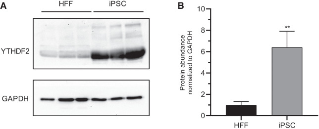
YTHDF2 protein is highly expressed in iPSCs. (A) Western blot to detect YTHDF2 in three independent iPSC and HFF extracts. GAPDH was used as a loading control. (B) Quantification of A. Asterisks indicate significant difference in the relative mean protein expression between iPSC and HFF samples ([**] P-value <0.005).

