

Abstract
The interdisciplinary collaboration of periodontics and orthodontics has allowed teeth to be moved 2–3 times faster, reducing the time required for traditional orthodontic therapy considerably. Periodontally accelerated osteogenic orthodontics (PAOO), also known as Wilckodontics, is a combination of a selective decortication facilitated orthodontics and alveolar augmentation. With this technique, there is no dependence on the pre-existing alveolar volume. This case report describes the treatment of permanent mandibular molar protraction in a 14-year-old patient undergoing orthodontic therapy using PAOO with piezosurgery.
Keywords: Corticotomy, bone grafts, orthodontics, osteogenic, periodontics
Introduction
The twenty- first century is referred to as the century of the biologist, and the regeneration dimension of Periodontics is a front stage player in the script of scientific progress. One of the most salient features in this inevitable progress is the interdisciplinary alliance of orthodontic specialty and periodontal regenerative surgery, which have taken traditional orthodontic tooth movement protocols to a completely new synergism.
In 2001, the Wilcko Brothers combined the refined corticotomy-facilitated orthodontic technique with alveolar augmentation and named the procedure Periodontally accelerated osteogenic orthodontics (PAOO).[1] Treatment of class I malocclusions with moderate to severe crowding, Class II malocclusions requiring expansion or extractions, and mild class III malocclusions can be achieved with PAOO.[2]
Case Report
A 14-year-old male undergoing fixed orthodontic treatment for Class II malocclusion for 1.5 years reported to the department of periodontics at our institution [Figure 1a–c]. The patient had an uneventful extraction of 46 because of the presence of extensive caries, which could not be treated with endodontic intervention 6 months back [Figure 2]. For the successful completion of treatment was to be achieved by orthodontically repositioning 47 in the place of missing 46. As the treatment time for space closure by second molar protraction in adults ranges from 2–3 years, the tooth movement was to be accomplished using the PAOO procedure. The patient had no systemic medical history and had good oral hygiene. As the patient was a minor, the consent of the parents was taken.
Figure 1.

(a): Frontal view (b): Right Lateral view (c): Left Lateral view
Figure 2.

Edentulous space at 46
After administration of local anesthesia, crestal incisions were given in the edentulous area of 46 along with crevicular incisions with respect to 47 and 45 [Figure 3]. A full-thickness mucoperiosteal flap was reflected well beyond the mucogingival junction exposing the buccal and lingual cortical plates [Figure 4]. Vertical corticotomies of approximately 0.5 mm deep were created using the peizosurgical insert no. OT2 [Figure 5] on the buccal and lingual cortical plates, mesial, and distal aspects of 47 [Figures 6 and 7]. Decortication was performed using a round carbide bur no. 8 on the buccal and lingual aspect with respect to 47 and the edentulous area of 46 with a depth of approximately 0.5 mm into the cortical bone [Figure 8]. These corticotomies were deep enough to enter the cancellous aspect of the bone after the corticotomies, and bone grafting was performed using demineralized freeze-dried bone allograft (DFDBA) on the buccal and lingual aspects where the decortication was performed [Figure 9]. Primary closure was achieved using 3–0 silk sutures [Figure 10]. Orthodontic treatment commenced 15 days postsurgery with the application of E chain and lingual buttons and continuous forces were applied to enable the tooth movement [Figure 11].
Figure 3.

Crestal and crevicular incision given
Figure 4.

Full-thickness mucoperiosteal flap reflected
Figure 5.

Completion of vertical corticotomies
Figure 6.

Peizosurgical unit
Figure 7.
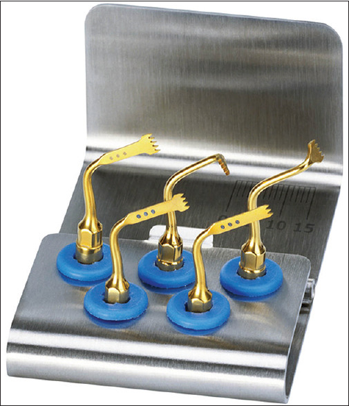
Peizosurgical insert kit
Figure 8.

Decortication of the buccal and lingual cortical plates using micromotor and round carbide bur
Figure 9.

Placement of DFDBA bone graft
Figure 10.

Sutures placed
Figure 11.

Application of E chain with lingual buttons 2 weeks postsurgery
The follow-up at months 3 and 6 showed substantial mesialization of 47 with the closure of the space [Figure 12]. The intraoral periapical radiographs at 3 and 6 months showed signs of active tooth movement [Figure 13]. Complete closure of space was achieved at 10 months [Figure 14a and 14b].
Figure 12.

Reduction of edentulous space 6 months postsurgery
Figure 13.

IOPA after 6 months
Figure 14.

(a): Closure of edentulous space 10 months postsurgery (Right lateral view) (b): Closure of edentulous space 10 months postsurgery (Buccal space closed)
Discussion
The PAOO procedure is based on the principle of regional acceleratory phenomenon (RAP).[3] The duration of RAP depends on the type of tissue and usually lasts about 4 months in human bone.[3] This physical process causes bone healing to occur 10–50 times faster than normal bone turnover.
Köle reported no periodontal pocket formation.[4] Bhattacharya et al. compared the treatment time for the closure of extraction space between conventional orthodontic tooth movement and corticotomy assisted and evaluated the alveolar bone thickness before and after corticotomy procedure using CT scan. They concluded that alveolar corticotomies not only accelerated the orthodontic treatment but also provided the advantage of increased alveolar width to support the teeth as evaluated based on the CT scan analysis.[5] Wilcko T, Wilkco W, Bissada Fconducted an evidence-based analysis of PAOO and stated that this treatment procedure allows conventional orthodontic tooth movement (OTM) 300% to 400% faster, increases the envelope of movement two- to three-fold and alveolar augmentation increases alveolar volume providing an alternative to bicuspid extraction.[6]
Piezosurgery is a relatively modern technique for osteotomy and osteoplasty utilizing ultrasonic vibration. There are various benefits of combining the modified corticotomy-facilitated orthodontics with alveolar augmentation. The most evident is that there is no need to rely on the pre-existing alveolar volume and shape solely.[7]
Dr. Wilcko suggested that analgesics like ibuprofen are not recommended postoperatively as they are nonsteroidal anti-inflammatory drugs (NSAIDs). NSAIDs can actually interfere with the production of prostaglandin hormone in the body and can retard the bone growth process, which is critical to PAOO.[8] Besides, NSAIDs gave during the first 24 h following trauma cause inhibition of clotting. Therefore, ideally, one should not be prescribed NSAIDs before or after undergoing PAOO surgery.[9]
The PAOO procedure does have its advantages and disadvantages.
The advantages include:
Lesser time than traditional orthodontics
Less likelihood of root resorption.
The history of relapse has been meager.
The disadvantages are:
Mildly invasive surgical procedure, and usually like all surgeries, it has a risk of some pain, swelling, and the possibility of infection.[10]
Medically compromised cases on long term intake of NSAIDs cannot be treated with this technique.[11]
Class III malocclusion cases cannot be treated with this approach.[12]
In spite of these drawbacks, the advantages of less treatment time and low relapse rates vastly supersede the disadvantages of this procedure.[13,14]
Conclusion
The fact that the teeth can be moved more rapidly, thus resulting in shortened treatment times, is certainly advantageous in reducing apprehension among the adolescent population regarding the period of the traditional orthodontic treatment. The PAOO technique can correct malocclusion in most patients within 3 to 10 months. This procedure puts orthodontics on a fast track by incorporating changes in the structure of the surrounding bone to accompany the repositioning of the teeth. The PAOO technique requires the application of numerous modified diagnostic and treatment parameters, which once mastered, make it a powerful new treatment option to offer the patients.
Declaration of patient consent
The authors certify that they have obtained all appropriate patient consent forms. In the form, the patient (s) has/have given his/her/their consent for his/her/their images and other clinical information to be reported in the journal. The patients understand that their names and initials will not be published and due efforts will be made to conceal their identity, but anonymity cannot be guaranteed.
Financial support and sponsorship
Nil.
Conflicts of interest
There are no conflicts of interest.
References
- 1.Pathak TS, Kini V, Kanagotagi S, Balasubramanian K, Gupta H. Wilckodontics. J Contemp Dent. 2013;3:15–9. [Google Scholar]
- 2.Wilcko T, Wilcko W. An evidence-based analysis of periodontally accelerated orthodontic and osteogenic techniques:A synthesis of scientific perspectives semin. Orthod. 2008;14:305–16. [Google Scholar]
- 3.Frost HM. The biology of fracture healing:An overview for clinicians. Part II. Clin Orthop Rel Res. 1989;248:283–9. [PubMed] [Google Scholar]
- 4.Köle H. Surgical operations of the alveolar ridge to correct occlusal abnormalities. Oral Surg Oral Med Oral Pathol. 1989;12:515–29. doi: 10.1016/0030-4220(59)90153-7. [DOI] [PubMed] [Google Scholar]
- 5.Bhattacharya P, Bhattacharya H, Anjum A, Bhandari R, Agarwal DK, Gupta A, et al. Assessment of corticotomy facilitated tooth movement and changes in bone thickness –A CT scan study. J Clin Diagn Res. 2014;8:26–30. doi: 10.7860/JCDR/2014/9448.4954. [DOI] [PMC free article] [PubMed] [Google Scholar]
- 6.Sheshan H. Piezosurgery in periodontology and oral implantology. J Indian Soc Periodontol. 2009;13:155–6. doi: 10.4103/0972-124X.60229. [DOI] [PMC free article] [PubMed] [Google Scholar]
- 7.Murphy KG, Wilcko MT, Wilcko WM, Ferguson DJ. Periodontal accelerated osteogenic orthodontics:A description of the surgical technique. J Oral Maxillofac Surg. 2009;67:2160–6. doi: 10.1016/j.joms.2009.04.124. [DOI] [PubMed] [Google Scholar]
- 8.Nazzal AM, Trojan TM, Green M. Uprighting and periodontally accelerated osteogenic orthodontics as an alternative to surgical crown lengthening. J Clin Orthod. 2016;50:507–11. [PubMed] [Google Scholar]
- 9.Chackartchi T, Barkana I, Klinger A. Alveolar bone morphology following periodontally accelerated osteogenic orthodontics:A clinical and radiographic analysis. Int J Periodontics Restorative Dent. 2017;37:203–8. doi: 10.11607/prd.2723. [DOI] [PubMed] [Google Scholar]
- 10.Campbell JH. Uprighting and periodontally accelerated osteogenic orthodontics. J Oral Maxillofac Surg. 2017;75:6. doi: 10.1016/j.joms.2016.10.015. [DOI] [PubMed] [Google Scholar]
- 11.Miyamoto T, Lang M, Khan S, Kumagai K, Nunn ME. The clinical efficacy of deproteinized bovine bone mineral with 10% collagen in conjunction with localized piezosurgical decortication enhanced orthodontics:A prospective observational study. J Periodontol. 2019;90:1106–15. doi: 10.1002/JPER.18-0737. [DOI] [PubMed] [Google Scholar]
- 12.Soltani L, Loomer PM, Chaar EE. A novel approach in periodontally accelerated osteogenic orthodontics (PAOO):A case report. Clin Adv Periodontics. 2019;9:110–4. doi: 10.1002/cap.10045. [DOI] [PubMed] [Google Scholar]
- 13.AlHammadi HA, Wilcho MT, Ferguson DJ. Severe mandibular crowding treated with nonextraction periodontally accelerated osteogenic orthodontics. Int J Periodontics Restorative Dent. 2019;39:e188–94. doi: 10.11607/prd.3926. [DOI] [PubMed] [Google Scholar]
- 14.Jing WD, Xu L, Xu X, Hou JX, Li XT. Association between periodontal biotype and clinical parameters:A cross-sectional study in patients with skeletal class III malocclusion. Chin J Dent Res. 2019;22:9–19. doi: 10.3290/j.cjdr.a41770. [DOI] [PubMed] [Google Scholar]