

Acknowledgements
The patient in this manuscript has given written informed consent to the publication of their case details.
Funding sources
None reported.
Editor
In December 2019, China reported the first group of pneumonia cases associated with a new coronavirus, 2019‐SARS‐CoV‐2. 1 Currently, the novel coronavirus infection has become a pandemic. 2 Significant research efforts are taking place around the world to better understand the transmission dynamics, the spectrum of clinical disease, possible treatment options and prevention measures. Despite this, there are hardly any data or images in the current literature on the cutaneous manifestations produced by COVID‐19 infection. In a series of 1099 patients with confirmed COVID‐19 infection, Guan et al found that only 0.2% of them developed a cutaneous rash. 3
We present the case of a 32‐year‐old female health professional with no significant past medical history whose presenting symptoms were fever, myalgia and asthenia. The following days, she developed cough and diarrhoea. Given the high suspicion of COVID‐19, due to her symptoms and close contact with infected patients, a SARS‐CoV‐2‐PCR of pharyngeal exudate was obtained, which resulted positive. At the moment, the subject remains in home isolation with symptomatic treatment with acetaminophen.
On the sixth day after the onset of symptoms, without a history of taking previous drugs, she presented with a generalized, pruritic morbilliform rash, with a sudden onset, with cephalocaudal progress, associated with low‐grade fever and without accompanying respiratory distress. The cutaneous lesions observed are petechial and maculopapular on an erythematous base (Fig. 1). The distribution of the rash included face, neck, thorax, abdomen, buttocks, extremities, including folds and scalp, respecting the palmo‐plantar region and mucosa. Over the following days, the lesions became itchier, while the erythema intensity decreased (Fig. 2).
Figure 1.
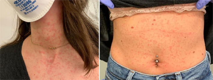
Rash onset.
Figure 2.
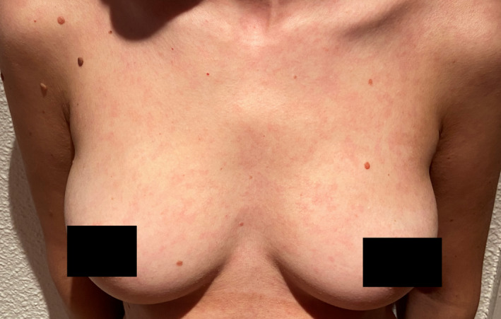
Rash evolution, third day.
A scaly reaction occurred on the fourth day after the rash started and disappeared without leaving visible lesions. As the only therapeutic measure, an intravenous dose of corticosteroid and antihistamines was administered.
In order to accurately define the cause for the rash, it is important to collect as much information as possible about the episode, chronology and characteristics of the injuries. 4 For this purpose, a comprehensive anamnesis, physical examination and, in this case, due to the high suspicion of COVID‐19, a confirmatory RT‐PCR test are necessary for the diagnosis.
Keeping COVID‐19 in the differential diagnosis of a rash is the key because patients may be misdiagnosed by another entity. Joob reports the case of a COVID‐19 positive patient who presented petechial rash, being initially misdiagnosed as dengue, delaying the definitive diagnosis. 5
Recalcati reports in its series a total of 88 COVID‐19 patients, 18 of whom developed skin manifestations (20.4%): erythematous rash, generalized urticaria and varicelliform rash. All of them pruritic in mild intensity, resolving within a few days. There was apparently no correlation with disease severity. 6
It will also be important to determine whether the injuries are caused by COVID‐19, if they are secondary to the use of drugs to treat it and even if they are the consequence of worsening of previous dermatological injuries, either due to emotional stress or by frequent use of disinfectants, hand washing or permanent use of masks. 7 In our case, the patient did not have previous medication intake, nor was she exposed to any disinfectant; therefore, other potential diagnosis was excluded.
Based on our findings and review of current literature, we could consider that cutaneous manifestations, although in a low percentage, are present in COVID19 positive patients without being associated with a worse prognosis. We report the first images of cutaneous manifestation of COVID‐19. The presence of other symptoms, the epidemiological history and the PCR test will be important to establish the diagnosis and to be able to establish early preventive measures. More studies are needed confirm and better understand how COVID‐19 affects the skin.
References
- 1. Zhou P, Yang XL, Wang XG et al. A pneumonia outbreak associated with a new coronavirus of probable bat origin. Nature 2020; 579: 270–273. [DOI] [PMC free article] [PubMed] [Google Scholar]
- 2. World Health Organization . Coronavirus disease (COVID‐19) Situation Dashboard. [WWW document]. URL https://experience.arcgis.com/experience/685d0ace521648f8a5beeeee1b9125cd (last accessed: 4 April 2020).
- 3. Guan W, Ni Z, Hu Y et al. Clinical characteristics of coronavirus disease 2019 in China. N Engl J Med 2020; 382: 1708–1720. [DOI] [PMC free article] [PubMed] [Google Scholar]
- 4. Korman A, Alikhan A, Kaffenberger B. Viral exanthems: An update on laboratory testing of the adult patient. J Am Acad Dermatol J Am Acad Dermatol 2017; 76: 538–550. [DOI] [PubMed] [Google Scholar]
- 5. Joob B, Wiwanitkit V. COVID‐19 can present with a rash and be mistaken for Dengue. J Am Acad Dermatol 2020; 82: e177. [DOI] [PMC free article] [PubMed] [Google Scholar]
- 6. Recalcati S. Cutaneous manifestations in COVID‐19: a first perspective. J Eur Acad Dermatol Venereol 2020. 10.1111/jdv.16387 [DOI] [PubMed] [Google Scholar]
- 7. Zheng Y, Lai W. Dermatology staff participate in fight against Covid‐19 in China. J Eur Acad Dermatol Venereol 2020. 10.1111/jdv.16390 [DOI] [PubMed] [Google Scholar]