

Figure 1.
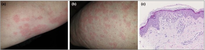
Dear Editor, A previously healthy 57‐year‐old woman presented with fever (39 °C) lasting for 4 days, and dry cough and rash appeared 2 days before. Diffuse fixed erythematous blanching maculopapular lesions were present, asymptomatic over the limbs and trunk, with burning sensation over the palms (a, b). She denied any drug intake, excepting paracetamol for fever. Thorax computed tomography scan was typical of COVID‐19; nasopharyngeal swab polymerase chain reaction (PCR) confirmed SARS‐CoV‐2. Infectious enquiry was otherwise negative. Skin biopsy specimen showed slight spongiosis, basal cell vacuolation and mild perivascular lymphocytic infiltrate (c). PCR on whole‐skin biopsy specimen was negative for SARS‐CoV‐2. Fever and rash resolved within 9 days, dry cough within 2 weeks. Urticarial and chilblain‐like lesions have been reported in patients with COVID‐19, but other phenotypes could be observed.1 2 In our patient an immune reaction to the virus is possible.
Contributor Information
B. Ahouach, Unité de dermatologie Hôpital René Dubos Pontoise France
S. Harent, Unité d'infectiologie Hôpital René Dubos Pontoise France
A. Ullmer, Unité de médecine interne Hôpital René Dubos Pontoise France
P. Martres, Laboratoire de microbiologie Hôpital René Dubos Pontoise France
E. Bégon, Unité de dermatologie Hôpital René Dubos Pontoise France
L. Blum, Unité de dermatologie Hôpital René Dubos Pontoise France
O. Tess, Service d'anatomopathologie Hôpital René Dubos Pontoise France
C. Bachmeyer, Service de médecine interne Hôpital Tenon (AP‐HP) Paris France.
References
- Henry D, Ackerman M, Sancelme E et al. Urticarial eruption in COVID‐19 infection. J Eur Acad Dermatol Venereol 2020; 10.1111/jdv.16472 [DOI] [PMC free article] [PubMed] [Google Scholar]
- Alramthan A, Aldaraji W. A case of COVID‐19 presenting in clinical picture resembling chilblains disease. First report from the Middle East. Clin Exp Dermatol 2020; 10.1111/ced.14243 [DOI] [PMC free article] [PubMed] [Google Scholar]