Abstract
Motivation
Cell-type-specific surface proteins can be exploited as valuable markers for a range of applications including immunophenotyping live cells, targeted drug delivery and in vivo imaging. Despite their utility and relevance, the unique combination of molecules present at the cell surface are not yet described for most cell types. A significant challenge in analyzing ‘omic’ discovery datasets is the selection of candidate markers that are most applicable for downstream applications.
Results
Here, we developed GenieScore, a prioritization metric that integrates a consensus-based prediction of cell surface localization with user-input data to rank-order candidate cell-type-specific surface markers. In this report, we demonstrate the utility of GenieScore for analyzing human and rodent data from proteomic and transcriptomic experiments in the areas of cancer, stem cell and islet biology. We also demonstrate that permutations of GenieScore, termed IsoGenieScore and OmniGenieScore, can efficiently prioritize co-expressed and intracellular cell-type-specific markers, respectively.
Availability and implementation
Calculation of GenieScores and lookup of SPC scores is made freely accessible via the SurfaceGenie web application: www.cellsurfer.net/surfacegenie.
Contact
Rebekah.gundry@unmc.edu
Supplementary information
Supplementary data are available at Bioinformatics online.
1 Introduction
Cell surface proteins play key roles in diverse biological processes and disease pathogenesis through mediation of adhesion and signaling between the extracellular and intracellular space. Owing to their accessible location, cell surface proteins can be exploited as valuable markers for a range of research and clinical applications including immunophenotyping live cells, targeted drug delivery and in vivo imaging. As such, a growing interest in cell-type-specific data has fueled the generation of the Cell Surface Protein Atlas (Bausch-Fluck et al., 2015), Human Protein Atlas (Uhlen et al., 2005), Human Cell Atlas Project (Regev et al., 2017) and related efforts. However, the unique combination of molecules present specifically at the cell surface are not yet described for most cell types or disease states, and thus continued innovation regarding surface protein discovery and annotation efforts are needed.
Specialized chemoproteomic approaches which specifically label and enrich cell surface proteins can provide direct evidence of cell surface localization resulting in empirically derived snapshot views of the cell surface proteome (Kalxdorf et al., 2017; Turtoi et al., 2011; Wollscheid et al., 2009). However, the large sample requirements and technical sophistication required for these experiments preclude their widespread use and application to sample-limited cell types. For these reasons, whole-cell proteomic and transcriptomic-based approaches that can be applied to identify and quantify thousands of molecules from fewer cells will continue to be useful in the search for cell surface proteins that are informative for particular cell types or disease stages.
Independent of the discovery strategy employed, bioinformatic predictions can serve as an important complement to experimental approaches by providing a means to filter data and prioritize the focus on proteins that are predicted to be localized to the cell surface (Bausch-Fluck et al., 2018; da Cunha et al., 2009; Diaz-Ramos et al., 2011; Town et al., 2016). Though transcriptomic and proteomic approaches offer significant advantages over antibody screening with regards to throughput and depth of coverage, candidate markers identified from discovery approaches must be subsequently validated as viable immunodetection or payload-delivery targets. Considering the significant cost and time required for the development of de novo affinity reagents, it is prudent to select candidates in a manner that considers whether a marker is likely to be both accessible and detectable by affinity reagents in a manner that allows cell types of interest (i.e. target cells) to be discriminated from non-target cells. Moreover, these assessments should be objective and suited to the analysis of large datasets such as those generated by proteomic and transcriptomic studies. To address these outstanding needs, we developed GenieScore, a metric to rank and prioritize candidate cell-type-specific surface markers—calculated by integrating a consensus-based prediction of cell surface localization with user-input quantitative data. Here, we describe the development of GenieScore and demonstrate its utility for prioritizing candidate cell surface markers using data obtained from proteomic workflows that specifically identify cell surface proteins [e.g. cell surface capture (CSC)] and more general strategies [e.g. whole-cell lysate (WCL) proteomics and transcriptomics]. We also demonstrate that permutations of GenieScore, termed IsoGenieScore and OmniGenieScore, can efficiently prioritize co-expressed and intracellular cell-type-specific markers, respectively. To facilitate its implementation among users, we developed SurfaceGenie, an easy-to-use web application that calculates GenieScores for user-input data and annotates the data with ontology information particularly relevant for cell surface proteins. SurfaceGenie is freely available at www.cellsurfer.net/surfacegenie.
2 Materials and methods
All experimental details are provided in Supporting Material.
2.1 Cell culture
Human lymphocyte cell lines (Ramos, HG-3, RCH-ACV, Jurkat) were cultured and passaged as described previously (Haverland et al., 2017). α-TC1 clone 6 (CRL-2934; ATCC, Manassas, VA) and β-TC-6 (CRL-11506; ATCC) cells were maintained at 37˚C and 5% CO2, cultured in Dulbecco’s modified Eagle’s medium supplemented with 10% heat-inactivated fetal bovine serum containing 16.6 or 5.5 mM glucose, respectively.
2.2 Cell lysis, protein digestion and peptide cleanup
For WCL analysis of lymphocytes, cell pellets were lysed in 100 mM ammonium bicarbonate containing 20% acetonitrile and 40% Invitrosol (Thermo Fisher Scientific, Waltham, MA), digested with trypsin (Promega, Madison, WI) overnight, and cleaned by SP2 (Waas et al., 2019). Peptides were quantified using Pierce Quantitative Fluorometric Peptide Assay (Thermo Fisher Scientific) according to manufacturer’s instructions on a Varioskan LUX Multimode Microplate Reader and SkanIt 5.0 software (Thermo Fisher Scientific). For CSC analysis of mouse islet cell lines, samples were prepared as previously described (Boheler et al., 2014; Gundry et al., 2012; Haverland et al., 2017).
2.3 Mass spectrometry acquisition and analysis
Lymphocyte peptides and CSC samples of mouse islet cell types were analyzed by LC-MS/MS using a Dionex UltiMate 3000 RSLCnano system (Thermo Fisher Scientific) in line with a Q Exactive (Thermo Fisher Scientific). Lymphocyte samples were prepared as 50 ng/µl total sample peptide concentration with Pierce Peptide Retention Time Calibration Mixture (PRTC, Thermo Fisher Scientific) spiked in at a final concentration of 2 fmol/µl and queued in blocked and randomized order with two technical replicates analyzed per sample. CSC samples of mouse islet cell types were analyzed as described previously (Mallanna et al., 2016, 2017). MS data were analyzed using Proteome Discoverer 2.2 (Thermo Fisher Scientific) and SkylineDaily (v4.2.1.19095) (Schilling et al., 2012). One-way ANOVA of CSC and WCL peptide-spectrum matches (PSMs) were performed using R (R Core Team, 2018).
2.4 Construction of a consensus dataset of predicted surface proteins
Four published surfaceome datasets (Bausch-Fluck et al., 2018; da Cunha et al., 2009; Diaz-Ramos et al., 2011; Town et al., 2016), each of which used a distinct methodology to bioinformatically predict the subset of the proteome which can be surface localized, were concatenated into a single consensus dataset. The ‘retrieve/mapping ID’ function within UniProt (www.uniprot.org) was used to convert the gene names provided in the published datasets to UniProt Accession numbers. Ambiguous matches were clarified by any supplementary information provided in the datasets in addition to gene name (e.g. alternate name, molecule name and chromosome).
2.5 GenieScore: a mathematical representation of surface marker potential
An equation was developed to mathematically represent key features deemed relevant when considering whether a protein has high potential to qualify as a cell surface marker for distinguishing between cell types or experimental groups. The equation [Equation (1)], which returns a metric termed GenieScore, is the product of (i) the SPC scores (described above); (ii) signal dispersion, a measure of the disparity in observations among investigated samples that is mathematically equivalent to the square of the normalized Gini coefficient (Gini, 1912) and (iii) signal strength, a logarithmic transformation of the experimental data (e.g. number of PSMs, MS1 peak area, FKPM or RKPM).
| (1) |
A thorough definition and rationalization of the individual equation terms are provided in Supporting Material.
2.6 Application of GenieScore
Details for the strategies applied to calculate GenieScores for each study are provided in Supporting Material.
2.7 SurfaceGenie web application
A web application for accessing SurfaceGenie was developed as an interactive Shiny app written in R (https://www.R-project.org/) and is available for use at www.cellsurfer.net/surfacegenie. Source code is available at www.github.com\GundryLab\SurfaceGenie.
3 Results
3.1 Generation of a surface prediction consensus dataset for predictive localization
Four previous bioinformatics-based constructions of the human cell surface proteome were compiled into a single, surface prediction consensus (SPC) dataset containing 5407 protein accession numbers (Supplementary Dataset S1.1). The strategies used to generate these predicted human surface protein datasets varied markedly, from manual curation to machine learning and resulted in dataset sizes ranging from 1090 to 4393 surface proteins (Fig. 1A). Despite these differences, there was considerable overlap among these predictions, with 69% and 41% of proteins in the SPC dataset occurring in ≥2 or ≥3 individual prediction sets, respectively. The number of proteins exclusive to a prediction strategy is positively correlated to the original dataset size, albeit not linearly, comprising 1.7%, 4.4%, 9.6% and 26.5% for the Diaz-Ramos, Bausch-Fluck, Town and Cunha datasets, respectively (Fig. 1B). To reflect the difference in the consensus of surface localization, each protein was assigned one point for each of the individual predicted datasets in which that protein appeared, termed SPC score (Fig. 1B and Supplementary Dataset S1.1). The distribution of SPC scores is shown in Figure 1B where 1671, 1507, 1497 and 732 proteins are assigned a score of 1, 2, 3 and 4, respectively. To enable more widespread applicability, mouse and rat SPC score databases were generated by mapping the human proteins to mouse and rat homologs using the Mouse Genome Informatics database (http://www.informatics.jax.org, Supplementary Dataset S1.2–3).
Fig. 1.
Generation and benchmarking of a SPC score. (A) The four previously published human surfaceome databases used, designated by first author of the corresponding publication, with details about how the databases were generated and the number of Uniprot Accessions within each database. (B) An UpSet plot (Conway et al., 2017; Lex et al., 2014) depicting the intersections between the individual surfaceome databases. The proteins were stratified by the number of individual datasets they appeared in, termed SPC. The number of proteins with each SPC score is shown. The full dataset is provided in the Supplementary Dataset S1.4.1. (C) The distribution of Gene Ontology Cellular Component Ontology (GO-CCO) annotations across different SPC scores depicted as a bubble chart, where the size of the bubble represents the number of proteins in the intersection between the particular SPC score and GO-CCO annotations
3.2 Benchmarking the SPC dataset against other annotations
The SPC dataset was compared to three established resources for obtaining cell surface localization annotations: (i) Gene Ontology Cellular Component Ontology (GO-CCO) (Ashburner et al., 2000; The Gene Ontology Consortium, 2019), (ii) annotations within the Cell Surface Protein Atlas (CSPA) (Bausch-Fluck et al., 2015) and (iii) annotations generated through application of HyperLOPIT (Christoforou et al., 2016). Comparisons to GO-CCO were consistent with expectations as ‘nucleus’ and ‘cytoplasm’ were the two most common terms for proteins with SPC scores of 0, ‘integral component of membrane’ and ‘membrane’ for SPC scores of 1, and ‘integral component of membrane’ and ‘plasma membrane’ for SPC scores of 2–4 (Fig. 1C). The ‘confidence’ assignment to proteins in the CSPA agreed with SPC scores for both human and mouse, with the notable exception of ∼17% of proteins assigned ‘high confidence’ having an SPC score of 0 (Supplementary Fig. S1A). However, upon closer inspection, 95% of these proteins are predicted to be secreted or extracellular matrix proteins (Secretome P, Bendtsen et al., 2005), which can be captured in CSC experiments but are not integral membrane proteins. The most common HyperLOPIT annotation in proteins with SPC scores of 3 or 4 was ‘plasma membrane’; however, ‘ER/Golgi apparatus’ was the most common annotation in proteins with SPC scores of 1 or 2 (Supplementary Fig. S1B). Though these comparisons demonstrated agreement overall, the SPC dataset provides unique and specific information in addition to assigning the predictions in a non-binary manner. Furthermore, as the SPC score is not dependent on experimental observation, it is more comprehensive in coverage than the CSPA and HyperLOPIT. These differences offer significant advantages for mathematically assigning the likelihood that a protein is present at the cell surface in a predictive manner. Moreover, the calculation of SPC score is straightforward and flexible to allow easy integration of results from future efforts of cell surface localization prediction.
3.3 Defining features of a cell surface protein marker based on first principle
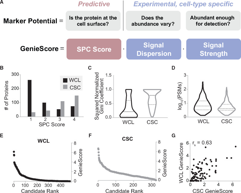
By defining the term ‘marker’ to designate a cell surface protein which is capable of distinguishing between cell types of interest on the basis of signal obtained by immunodetection, there are three features that can be used to evaluate the capacity of a protein to serve as a marker (Fig. 2A). These include (i) SPC score—presence at the cell surface, (ii) signal dispersion—difference in abundance among cell types and (iii) signal strength—scaling factor to account for the aim of obtaining specific antibody-based detection. The product of these three terms, which we define as GenieScore, is a metric that can be used to rank proteins from experimental data for their capacity to serve as a marker. Importantly, prioritization of cell surface proteins that are likely capable of serving as informative markers should consider experimental data from relevant cell types, including the target and non-target cell types that are to be discriminated. Hence, although a consensus-based predictive approach can be adopted to represent whether a protein is capable of being present at the cell surface (SPC score), the signal dispersion and signal strength must be determined empirically, as these will differ among cell types. Additional descriptive details and rationalization for the GenieScore components are provided in Supporting Material along with examples of GenieScore calculations for different experimental observations (Supplementary Fig. S2B). As the two mathematical terms chosen to represent signal dispersion and signal strength are agnostic to the data type, we investigated the effects of different sources of input data to these terms with respect to the calculated GenieScores
Fig. 2.
GenieScore components and application to two proteomic analyses of four lymphocyte lines. (A) The features of a protein that were hypothesized to predominate the capacity of a protein to serve as a cell surface marker are shown with the names of the mathematical terms derived to represent them. The marker potential features are annotated by the applied approach (i.e. predictive or experimental) to answer the relevant questions. The remaining panels depict the distribution of the individual components and GenieScores calculated from the data acquired from application of whole-cell lysate (WCL) or Cell Surface Capture (CSC) to four lymphocyte cell lines (n = 3 per cell line, N = 485 data points for WCL, N = 325 data points for CSC). (B) A histogram depicting the distribution of SPC scores within predicted surface proteins (SPC score >0) identified by application of WCL and CSC. (C) A violin plot depicting the distribution of signal dispersion for the predicted surface proteins identified by WCL and CSC. (D) A violin plot depicting the distribution of signal strength for the predicted surface proteins identified by WCL and CSC. (E) Plot of GenieScore against rank-order of candidate cell surface markers for predicted surface proteins identified by WCL. (F) Plot of GenieScore against rank-order of candidate cell surface markers for predicted surface proteins identified by CSC. (G) GenieScores calculated using either WCL or CSC data are plotted against each other the 91 proteins identified by both approaches along with the Spearman’s Correlation for those scores
3.4 Testing GenieScore by comparing two proteomic approaches for marker discovery
We previously demonstrated that CSC applied to four human lymphocyte cell lines resulted in sufficient depth of surface protein identification to allow discrimination among the lines (Haverland et al., 2017). Here, we performed WCL digestion of these same cell lines to determine whether a generic proteomic approach would be sufficient to detect divergent cell surface proteins to do the same. Notably, the WCL approach only required a peptide amount equivalent to ∼1000 cells compared to CSC which used ∼100 million cells/experiment. While the majority, 75% (325), of the CSC-identified proteins are predicted to be cell surface localized (i.e. SPC scores of 1–4), only 13% (485) of the WCL proteins met this criterion. Although these datasets were collected on the same cell lines, only 91 proteins with SPC scores 1–4 were observed in both datasets, which represent 28% and 19% of the CSC and WCL predicted surface proteins, respectively. Despite these differences, applying hierarchical clustering to the subset of proteins in each dataset with SPC scores of 1–4 recapitulated the clustering predicted based on the entire dataset for both proteomic approaches (Supplementary Fig. S3). These data highlight the utility of applying the SPC metric as a strategy to filter and compare between datasets and demonstrate that a generic proteomic strategy can provide sufficient predicted surface protein detection to differentiate among cell types using 0.1% of the cellular material required for CSC, albeit without the empirical evidence of localization for the detected set predicted surface proteins provided by application of CSC.
GenieScores were calculated for each protein in the CSC and WCL datasets using PSMs as input for signal dispersion and signal strength calculations (Supplementary Dataset S.1–2). Though predicted surface proteins were identified by both proteomic approaches, the distributions of SPC scores, signal dispersion and signal strength were markedly different between CSC and WCL (Fig. 2B–D). These observations are expected due to the highly selective nature of CSC which primarily captures N-glycosylated peptides resulting in higher specificity for bona fide surface proteins and fewer peptides identified per protein (Boheler et al., 2014; Gundry et al., 2012; Haverland et al., 2017; Wollscheid et al., 2009). GenieScores were plotted against the rank for CSC and WCL data resulting in a rectangular hyberbola-like shape, namely a subset of higher-scoring proteins that trail off into a majority of lower-scoring proteins (Fig. 2E and F) with a similar range (6.59 and 6.16 for CSC and WCL, respectively) but significant difference in the distribution. GenieScores for the 91 proteins identified in both proteomic approaches were strongly correlated (rs = 0.63) (Supplementary Dataset S2.3 and Fig. 2G).
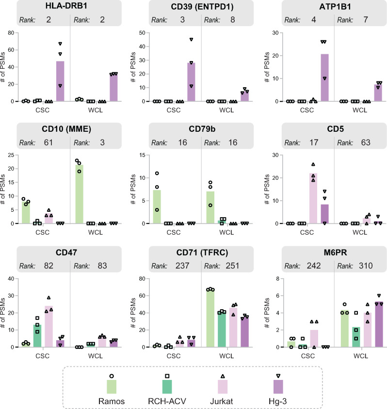
The top-scoring candidate markers in both the CSC and WCL datasets are proteins for which the majority (if not the totality) of PSMs are in a single cell line. The numbers of PSMs per cell line for selected proteins are shown for CSC and WCL in Figure 3 along with the ranks determined by application of GenieScore (plotted in Fig. 2E and F). Many of the high ranking candidates have previously been reported as markers for cancer types modeled by the cell lines used here—including ATP1B1, CD39 and HLA-DR for chronic lymphocytic leukemia (CLL) (Damle et al., 2002; Johnston et al., 2018; Pulte et al., 2007) (Hg-3 cell line); CD10 and CD79b for Burkitt Lymphoma [He et al., 2018; WHO Classification: Tumours of the Haematopoietic and Lymphoid Tissues (2008), 2016] (Ramos cell line) (Fig. 3). Proteins with moderate ranks often had PSMs spread evenly among two or more of the cell lines. The examples here include CD5, a known T cell marker that is often expressed in CLL (Damle et al., 2002) (accounting for its observation in Jurkat and Hg-3 cell lines, respectively), and CD47, a protein reported to be upregulated in many cancer subtypes (Takimoto et al., 2019). Proteins with low ranks are equally spread among all the cell lines, often ‘housekeeping’-type proteins such as transferrin receptor 1 and mannose-6-phosphate receptor (Fig. 3).
Fig. 3.
Distributions of observed abundance for selected proteins in the lymphocyte data with a range of GenieScores. The number of peptide-spectrum matches (PSMs) assigned to selected proteins for both Cell Surface Capture (CSC) and whole-cell lysate (WCL) experiments. Biological replicates (n = 3) are shown as data points and averages are shown as columns. The ranks assigned to each protein, according to the set of calculated GenieScores, are shown for both CSC and WCL datasets
As the calculation of GenieScore relies on averages (as opposed to individual replicate measurements), the relationship between the product of the experimental terms (signal dispersion and signal strength) used to calculate GenieScore and the statistical difference (which considers variability in measurement) between cell lines was investigated. A positive relationship was observed, with Spearman’s correlations (rs) of 0.66 and 0.64 for WCL and CSC, respectively, suggesting that the equation for GenieScore is likely to be prioritizing proteins for which there is a statistical difference (Supplementary Fig. S4A). Finally, recognizing the limitations of relying on PSMs for quantitative comparisons, GenieScores were calculated using MS1 peak areas for selected proteins. Results from this strategy had a very strong correlation to GenieScores using PSMs; rs = 0.88 and 0.80 for WCL and CSC, respectively (Supplementary Fig. S4B). Altogether, application of GenieScore to the data collected from diverse lymphocyte cell lines produced similar ranking of candidate markers independent of the type of input data, including several surface proteins previously linked to relevant cancer subtypes, which indicates GenieScore is a robust and valid prioritization metric.
3.5 Benchmarking GenieScore against two published surface protein marker studies
A major application of GenieScore is to prioritize candidate markers for immunophenotyping. Hence, we sought to benchmark the performance of GenieScore ranking against two published studies that performed flow cytometry analyses to orthogonally validate putative markers for cell types of interest which were originally identified from proteomics and/or transcriptomic data. In the first study, Martinko et al. performed CSC and RNA-Seq on MCF10A KRASG12V cells (comparing the results to empty vector control MCF10A cells) to identify surface proteins indicative of RAS-driven cancer phenotype (Martinko et al., 2018). Antibodies were subsequently developed against seven candidate markers, all of which demonstrated positive signal on the MCF10A KRASG12V cells. Using GenieScore as a prioritization metric, we investigated the relative ranks of these validated markers among the CSC and RNA-Seq datasets.
As the goal of the original study was to identify surface proteins which were upregulated in cancer, GenieScores were only calculated for predicted surface proteins (SPC score > 0) which met this additional criterion—resulting in 122 candidates from CSC and 330 candidates from RNA-Seq (Fig. 4A and Supplementary Dataset S2.1–2). The validated proteins were among the highest scoring candidates in both the CSC and RNA-Seq datasets (Fig. 4A). GenieScores calculated from the CSC and RNA-Seq data had a moderate correlation (rs = 0.41) with most of the validated markers scoring highly for both CSC and RNA-Seq (Fig. 4B). The rank of the putative markers by GenieScore was compared to the rank by log2 fold changes (a metric denoted as selection criteria in the original manuscript) (Fig. 4C). In all but one case, the candidates rank higher by GenieScore than by log2-fold ratio. These results support GenieScore as a useful, single metric that enables selection of cell surface proteins which can serve as markers for immunodetection. Though the validated markers were among the top-ranking candidates, other proteins with high GenieScores emerge as potential targets, highlighting the utility of GenieScore to reveal new biological insights or targets from previously published data. Two such targets that scored well by both CSC and RNA-Seq are THBD and NRP1, which have been previously implicated in a KRAS-driven myeloid malignancy and KRAS-driven tumorigenesis (Meyerson et al., 2017; Vivekanandhan et al., 2017). Additionally, several proteins score well in one dataset (i.e. CSC or RNA-Seq) but are completely absent from the other. SAT-1, ranked 5 within the RNA-Seq data but not observed by CSC, plays a role in a polyamine synthesis pathway that is upregulated by KRAS-driven cancers (Arruabarrena-Aristorena et al., 2018; Linsalata et al., 2004). Inspection of the primary sequence of SAT-1 reveals that the N-glycosite is located within a peptide that would make it unlikely to be detected by mass spectrometry. Conversely, there are proteins within the CSC dataset for which there were no matching transcripts such as LRP1, a protein associated with tumorigenesis and tumor progression (Chen et al., 2010; Xing et al., 2016). Altogether, these results present the complementary nature of CSC and RNA-Seq as discovery techniques and demonstrate that GenieScore-based analyses of these data, either independently or together, provide a rapid strategy for prioritizing candidates for immunophenotyping.
Fig. 4.
Benchmarking GenieScore against two published cell surface marker studies which validated candidate markers by flow cytometry. (A–C) Data from application of GenieScore to data from Martinko et al. (D) Data from application of GenieScore to data from Boheler et al. (A) The subset of proteins for which GenieScores were calculated is the intersection of the set of proteins with SPC scores >0 with the set of proteins that were increased in the KRAS mutant—shown by the shaded overlap. Plots of GenieScores against candidate rank are shown for the Cell Surface Capture (CSC) and RNA-Seq datasets. Proteins selected in the original manuscript for antibody development and subsequently validated as surface markers by flow cytometry are shown as black diamonds and labeled with gene names. (B) The GenieScores calculated using either CSC or RNA-Seq data are plotted against each other for the 211 surface proteins identified by both approaches along with the Spearman’s correlation of those scores. The flow cytometry-validated markers are shown as black diamonds. (C) A table containing the ranks assigned, according to either GenieScores or log2-fold, for each protein. The change in rank, calculated as GenieScore rank minus log2-fold rank, is shown for each flow cytometry-validated marker. (D) GenieScores for the 495 proteins identified by CSC in human fibroblast and stem cells are plotted against the log2-fold ratio. The reference stem cell markers, as well as the negative and positive markers for pluripotency selected for validation by flow cytometry are highlighted in their own plots
In the second study, Boheler et al. performed CSC on human fibroblasts, embryonic stem cells and induced pluripotent stem cells to identify surface markers for stem cells (Boheler et al., 2014). Candidate pluripotency markers were selected by comparing the set of proteins observed on stem cells to CSC data from the CSPA, specifically, requiring that a protein was not detected in fibroblasts and detected in fewer than four other somatic cell types (excluding cancer cell types). Negative markers of pluripotency were selected in a similar manner, specifically, not detected in stem cells and detected in six or more non-diseased cell types in the CSPA. Flow cytometry analysis of human fibroblasts and stem cells was used to orthogonally validate 17 putative positive and 3 putative negative pluripotency markers and included 3 previously reported positive pluripotency markers as controls. The GenieScores for proteins observed in the human fibroblast and stem cell CSC experiments were plotted against the log2-fold ratio of PSMs between the cell types providing a visual depiction of capacity to serve as a marker segregated by cell type wherein the reference, putative negative and putative positive pluripotency markers are denoted in individual plots (Fig. 4D and Supplementary Dataset S3.1). The reference stem cell markers are among the top-scoring (with ranks of 2 and 10) candidate pluripotency markers from the CSC dataset, except for Thy1, a protein for which both CSC and flow cytometry results provided evidence for its presence in fibroblasts and stem cells. The GenieScores for the putative negative and positive markers were spread over a greater range in this dataset compared to the distribution observed for the Martinko et al. study. This difference is likely attributable to the notably divergent strategies employed for candidate selection. Specifically, the Boheler study relied on qualitative (presence/absence) rather than quantitative comparisons, considered data from cell types outside those included in the study, and restricted validation to candidates for which commercially available monoclonal antibodies were available. Notably, several of the validated markers were identified by relatively few PSMs in the original dataset (IL27RA, EFNA3). While the number of PSMs is sometimes used as a filter to eliminate proteins from consideration, in this case, comparisons to 50 other cell types suggested that these candidates are putatively restricted to stem cells. Thus, despite being identified by relatively few PSMs in CSC analyses, proteins that are uniquely observed in a single cell type can be valuable immunophenotyping markers provided there are data of a similar type and quality on other cell types for comparison. Altogether, these data highlight the importance of context during marker selection and the value of considering additional datasets. Specifically, if additional datasets are integrated prior to calculation of GenieScore, candidates with a lower signal strength (few PSMs) would rank more highly because they would have a higher signal dispersion (all PSMs coming from a single cell type). Overall, these evaluations of previously validated datasets illustrate how GenieScore is a useful strategy to prioritize candidate cell surface markers using both proteomic and transcriptomic datasets.
3.6 Integrating GenieScores of proteomic and transcriptomic data to reveal candidate markers for mouse islet cell types
As GenieScore provided a useful rank ordering of potential protein markers from both RNA-Seq and CSC data that was consistent with published results, we sought to evaluate its utility for integrating data from disparate studies for marker discovery. To this end, we performed CSC on mouse α and β cell lines and compared the results to published RNA-Seq data acquired on primary α and β cells from dissociated mouse islets (Benner et al., 2014). The datasets shared 321 predicted surface proteins (Fig. 5A andSupplementary Dataset S4.1), and GenieScores from the CSC data were plotted against GenieScores from the RNA-Seq data (Fig. 5B). A possible explanation for the weak correlation (rs = 0.26) between GenieScores is that the CSC dataset was acquired on cell lines and the RNA-Seq dataset was acquired on primary cells. However, in the context of marker discovery, each of these approaches offers advantages, namely, the CSC data provides experimental evidence regarding protein abundance at the cell surface and the RNA-Seq analysis of primary cells avoids possible artifacts introduced by culturing cells ex vivo. Recognizing the complementary benefits of these approaches, the data were combined in a manner that weights them equally, namely, the signal dispersion was calculated using the average of the normalized CSC and normalized RNA-Seq data. The combined GenieScores were distributed similarly to scores calculated using CSC or RNA-Seq individually and when plotted against the log2-fold ratio between α and β cells allow for visual discrimination of the candidate markers for each cell type (Fig. 5C and Supplementary Dataset S4.2). Among the top candidate markers for α and β cells revealed by this combined approach are proteins with well-established roles in islet biology including GLP1, GABBR2, GALR1, KCNK3 and SLC7A2—proteins highlighted in a recent review of the β cell literature (Rorsman and Ashcroft, 2018). ALCAM (CD166), CHR1 and CEACAM1 are proteins that have been studied in the context of the islet biology, though have less defined roles (DeAngelis et al., 2008; Fujiwara et al., 2014; Schmid et al., 2011). Altogether, GenieScore provided a useful framework for integrating proteomic and transcriptomic data for surface marker prioritization.
Fig. 5.

Application of GenieScore and its permutations to islet cell types. (A–C) Data from application of GenieScore to Cell Surface Capture (CSC) data from mouse α and β cell lines collected as part of this study integrated with RNA-Seq on mouse primary α and β cells from Benner et al. (D) Data from application of GenieScore and its permutations to human islet single-cell RNA-Seq data from Lawlor et al. (A) The subset of proteins for which GenieScores were calculated is the set of proteins with SPC scores >0 that were identified by both CSC and RNA-Seq shown as the shaded overlap. (B) The GenieScores calculated using either CSC or RNA-Seq data are plotted against each other for the 321 proteins identified by both approaches along with the Spearman’s correlation of those scores. (C) GenieScores calculated using the combined CSC and RNA-Seq data are plotted against candidate rank and against the log2-fold ratio (N = 321 proteins). Selected candidate markers which have previously been associated with islet cell biology are labeled with gene names. (D) The top-scoring proteins from application of the different permutations of GenieScore are shown grouped either by cell type or by biological function
To extend the analysis beyond the identification of proteins which might be capable of distinguishing α and β cells to finding cell-type-specific markers within the context of the islet, we applied GenieScore to a single-cell RNA-Seq dataset that was collected on cells from dissociated human islets (Lawlor et al., 2017) (Supplementary Dataset S5.1). Lawlor et al. partitioned the data on single cells into seven different cell types—α, β, γ, δ, acinar, ductal and stellate—based on a subset of genes that were determined to be representative of each of the clusters. Top-ranking markers for each of the seven cell types are listed in Figure 5D. Many of the proteins identified as capable of distinguishing between α and β cells in the analysis of CSC and RNA-Seq data were not cell-type specific when data from other cell types found within the islet were considered. For example, NRCAM and SLC4A10 are proteins more abundant in β than α cells, but the levels expressed in β cells are equivalent to γ or δ cells, respectively. PTPRK is expressed at a higher level in α than β cells in all studies, but the level of expression is 26-fold lower than acinar cells and 35-fold lower than ductal cells. Altogether, while the cell-type specificity that is ultimately required will depend on the desired downstream application, these observations highlight that consideration of a larger cellular context is important for the identification of cell-type-specific markers.
Recognizing the utility of the GenieScore approach for prioritizing cell-type-specific surface proteins, the equation was further adapted to enable prioritization of other classes of proteins using the same input data. First, removal of the SPC score component from GenieScore, a permutation termed OmniGenieScore, allowed for the identification of proteins which can be used as cell-type-specific markers without considering their surface localization. Application of OmniGenieScore to the islet cell single-cell RNA-seq data revealed many known cell-type-specific markers, such as glucagon (GCG) for α cells, insulin (INS) for β cells, pancreatic polypeptide (PPY) for γ cells and somatostatin (SST) for δ cells (Fig. 5D). By inverting the signal dispersion term [i.e. 1 − (G/Gmax)2], a permutation termed IsoGenieScore, the set of cell surface proteins which are relatively abundant and similar in signal among all cell types in the analysis were prioritized. The classes of proteins (e.g. adhesion, cell growth and insulin signaling) which were at the top of this ranking system were largely involved in generic processes that are not specific to any cell type (Fig. 5D). Reversing the signal dispersion and ignoring the SPC score, termed IsoOmniGenieScore, resulted in prioritization of proteins typically selected to be loading controls for western blotting or reference genes for PCR (e.g. GAPDH, ACTB and B2M) in addition to many of the proteins involved in mitochondrial oxidation (Fig. 5D). Altogether, these four permutations of the GenieScore enabled the prioritization of candidate markers for a broad range of applications, including cell surface and intracellular markers that distinguish cell types as well as those that are co-expressed among cell types.
3.7 SurfaceGenie: a web-based application for GenieScore calculation and ontological annotation
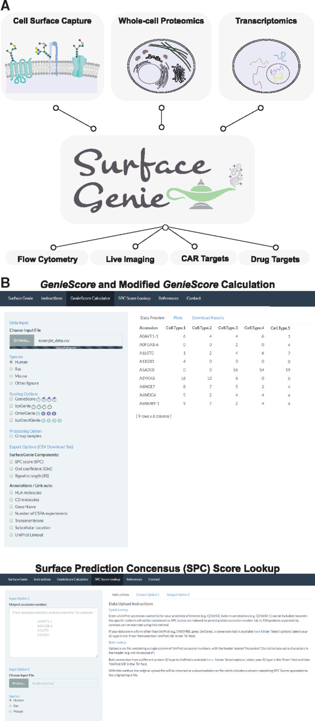
To enable calculation of GenieScores for user-input data, a shinyApp, SurfaceGenie, was developed in R. In this interface, users upload data from proteomic or transcriptomic experiments as a .csv file and can view the distribution of GenieScores and SPC scores for the proteins contained in their data (Fig. 6A). SurfaceGenie is compatible with human, mouse and rat data. As part of the analysis, input proteins are annotated with ontological information including Cluster of Differentiation (CD) and Human Leukocyte Antigen (HLA) molecule designations. In addition, proteins are annotated with the number of cell types within the CSPA in which the protein has been observed—a factor found to be relevant for marker prioritization in the Boheler et al. data. The plots and data generated are available for download, including the results for individual terms used to calculate GenieScore. The permutations of GenieScore applied in Figure 5D are also available. Additional functionality includes the ability to query accession numbers in single or batch mode, independent of data type, to obtain SPC scores. SurfaceGenie is freely available at http://www.cellsurfer.net/surfacegenie (screen captures are shown in Fig. 6B). A User Guide with step-by-step tutorials is provided in the Supplementary Material; it can be alternatively accessed through the web application or the Github repository.
Fig. 6.

Overview of the utility of SurfaceGenie and screen captures from the web application. (A) A schematic depicting the tested inputs and potential applications of SurfaceGenie, a web-based application which calculates GenieScore permutations from user-input data. (B) Screen captures of the different modes of use for the SurfaceGenie web application
4 Discussion
Despite the central role cell surface proteins play in maintaining cellular structure and function, the cell surface is not well documented for most human cell types. There is currently no comprehensive reference repository of experimentally determined cell surface proteins cataloged by individual human cell types that can be used as a baseline for comparison to experimentally perturbed or diseased phenotypes. Although specialized proteomic approaches allow for probing the occupancy of the cell surface, the sample requirements and technical sophistication often preclude widespread application, and quantitation is challenging. To overcome these challenges, predictions of surface localization can enable insights from more easily implemented proteomic and transcriptomic approaches, which can be performed on smaller sample sizes. However, with technologies that allow for ‘omic’ evaluation of individual cell types, there is a need to develop methodologies to prioritize the value contained within these studies in order to extract useful knowledge from acquired data.
Here, we describe the development of GenieScore, a prioritization approach that integrates a predictive metric regarding surface localization with experimental data to rank-order proteins which may be useful as cell surface markers. We demonstrate that GenieScore is compatible with quantitative data from CSC, WCL and RNA-Seq experiments and is a useful strategy by which to integrate multiple sources of data for candidate marker prioritization. The design of GenieScore was intended for comparing data collected for various cell types or conditions within the same batch. Comparisons involving data from multiple sources may benefit from utilization of batch correction (Espin-Perez et al., 2018) or signal harmonization methods (Pino et al., 2018) for transcriptomic or proteomic data, respectively. SurfaceGenie, a web-based application, was developed to enable the calculation of SPC scores and GenieScores, and the various permutations thereof, from user-input data. SurfaceGenie also supplements the data with annotations relevant for marker selection.
Beyond immunophenotyping, SurfaceGenie is expected to facilitate the identification of valuable drug targets as the features of cell surface markers (e.g. surface localization and cell-type specificity) are also advantageous when designing efficient and specific therapies. Independent of GenieScore, the ability to query SPC scores within SurfaceGenie can deliver value in-and-of-itself, providing users with an additional resource to interrogate surface localization for proteins which are not yet characterized experimentally. However, whether an expressed protein is localized to the cell surface on a specific cell type in a specific experimental or biological condition remains difficult to predict. This is especially true for proteins that (i) lack traditional sequence motifs (e.g. signal peptides), (ii) are only trafficked to the cell surface upon ligand binding (e.g. glucose transporter 3, GLUT3) or (iii) have proteoforms that exhibit different subcellular localization than the canonical version of a protein for which predictions are typically based upon. For these reasons, experimental workflows that provide capabilities for discovery (i.e. not limited to available affinity reagents) while providing experimental evidence of cell surface localization on a particular cell type of interest with a specific context (e.g. experimental condition, disease state) will remain invaluable.
In conclusion, we anticipate that SurfaceGenie will enable effective prioritization of informative candidate cell surface markers to support a broad range of research questions, from mechanistic to disease-related studies. The candidates prioritized using SurfaceGenie are expected to be of use to a range of applications including immunophenotyping, immunotherapy and drug targeting.
Supplementary Material
Acknowledgements
We specially thank Dr. Christopher Ashwood and Linda Berg Luecke for critical review of the manuscript and insightful discussions.
Funding
This work was supported by the National Institutes of Health [R01-HL126785 and R01-HL134010 to R.L.G., F31-HL140914 to M.W., DK-052194 and AI-44458 to J.A.C.] and Juvenile Diabetes Research Foundation [2-SRA-2019-829-S-B to R.L.G. and J.A.C.]. S.S. is a member of the MCW-MSTP, which was partially supported by a T32 grant from National Institute of General Medical Sciences [GM080202].
Conflict of Interest: none declared.
References
- Arruabarrena-Aristorena A. et al. (2018) Oil for the cancer engine: the cross-talk between oncogenic signaling and polyamine metabolism. Sci. Adv., 4, eaar2606. [DOI] [PMC free article] [PubMed] [Google Scholar]
- Ashburner M. et al. (2000) Gene ontology: tool for the unification of biology. The Gene Ontology Consortium. Nat. Genet., 25, 25–29. [DOI] [PMC free article] [PubMed] [Google Scholar]
- Bausch-Fluck D. et al. (2015) A mass spectrometric-derived cell surface protein atlas. PLoS One, 10, e0121314. [DOI] [PMC free article] [PubMed] [Google Scholar]
- Bausch-Fluck D. et al. (2018) The in silico human surfaceome. Proc. Natl. Acad. Sci. USA, 115, E10988–E10997. [DOI] [PMC free article] [PubMed] [Google Scholar]
- Bendtsen J.D. et al. (2005) Non-classical protein secretion in bacteria. BMC Microbiol., 5, 58. [DOI] [PMC free article] [PubMed] [Google Scholar]
- Benner C. et al. (2014) The transcriptional landscape of mouse beta cells compared to human beta cells reveals notable species differences in long non-coding RNA and protein-coding gene expression. BMC Genomics, 15, 620. [DOI] [PMC free article] [PubMed] [Google Scholar]
- Boheler K.R. et al. (2014) A human pluripotent stem cell surface N-glycoproteome resource reveals markers, extracellular epitopes, and drug targets. Stem Cell Rep., 3, 185–203. [DOI] [PMC free article] [PubMed] [Google Scholar]
- Chen J.S. et al. (2010) Secreted heat shock protein 90alpha induces colorectal cancer cell invasion through CD91/LRP-1 and NF-kappaB-mediated integrin alphaV expression. J. Biol. Chem., 285, 25458–25466. [DOI] [PMC free article] [PubMed] [Google Scholar]
- Christoforou A. et al. (2016) A draft map of the mouse pluripotent stem cell spatial proteome. Nat. Commun., 7, 8992. [DOI] [PMC free article] [PubMed] [Google Scholar]
- Conway J.R. et al. (2017) UpSetR: an R package for the visualization of intersecting sets and their properties. Bioinformatics, 33, 2938–2940. [DOI] [PMC free article] [PubMed] [Google Scholar]
- da Cunha J.P. et al. (2009) Bioinformatics construction of the human cell surfaceome. Proc. Natl. Acad. Sci. USA, 106, 16752–16757. [DOI] [PMC free article] [PubMed] [Google Scholar]
- Damle R.N. et al. (2002) B-cell chronic lymphocytic leukemia cells express a surface membrane phenotype of activated, antigen-experienced B lymphocytes. Blood, 99, 4087–4093. [DOI] [PubMed] [Google Scholar]
- DeAngelis A.M. et al. (2008) Carcinoembryonic antigen-related cell adhesion molecule 1: a link between insulin and lipid metabolism. Diabetes, 57, 2296–2303. [DOI] [PMC free article] [PubMed] [Google Scholar]
- Diaz-Ramos M.C. et al. (2011) Towards a comprehensive human cell-surface immunome database. Immunol. Lett., 134, 183–187. [DOI] [PubMed] [Google Scholar]
- Espin-Perez A. et al. (2018) Comparison of statistical methods and the use of quality control samples for batch effect correction in human transcriptome data. PLoS One, 13, e0202947. [DOI] [PMC free article] [PubMed] [Google Scholar]
- Fujiwara K. et al. (2014) CD166/ALCAM expression is characteristic of tumorigenicity and invasive and migratory activities of pancreatic cancer cells. PLoS One, 9, e107247. [DOI] [PMC free article] [PubMed] [Google Scholar]
- Gini, C. (1912) Variabilità e mutabilità. In: Pizetti,E. and Salvemini,T. (eds.) Reprinted in Memorie di metodologica statistica. Libreria Eredi Virgilio Veschi, Rome.
- Gundry R.L. et al. (2012) A cell surfaceome map for immunophenotyping and sorting pluripotent stem cells. Mol. Cell. Proteomics, 11, 303–316. [DOI] [PMC free article] [PubMed] [Google Scholar]
- Haverland N.A. et al. (2017) Cell surface proteomics of N-linked glycoproteins for typing of human lymphocytes. Proteomics, 17. doi: 10.1002/pmic.201700156. [DOI] [PMC free article] [PubMed] [Google Scholar]
- He X. et al. (2018) Continuous signaling of CD79b and CD19 is required for the fitness of Burkitt lymphoma B cells. Embo J., 37, e97980. [DOI] [PMC free article] [PubMed] [Google Scholar]
- Johnston H.E. et al. (2018) Proteomics profiling of CLL versus healthy B-cells identifies putative therapeutic targets and a subtype-independent signature of spliceosome dysregulation. Mol. Cell. Proteomics, 17, 776–791. [DOI] [PMC free article] [PubMed] [Google Scholar]
- Kalxdorf M. et al. (2017) Monitoring cell-surface N-glycoproteome dynamics by quantitative proteomics reveals mechanistic insights into macrophage differentiation. Mol. Cell. Proteomics, 16, 770–785. [DOI] [PMC free article] [PubMed] [Google Scholar]
- Lawlor N. et al. (2017) Single-cell transcriptomes identify human islet cell signatures and reveal cell-type-specific expression changes in type 2 diabetes. Genome Res., 27, 208–222. [DOI] [PMC free article] [PubMed] [Google Scholar]
- Lex A. et al. (2014) UpSet: visualization of intersecting sets. IEEE Trans. Vis. Comput. Graph., 20, 1983–1992. [DOI] [PMC free article] [PubMed] [Google Scholar]
- Linsalata M. et al. (2004) Polyamine biosynthesis in relation to K-ras and p-53 mutations in colorectal carcinoma. Scand. J. Gastroenterol., 39, 470–477. [DOI] [PubMed] [Google Scholar]
- Mallanna S.K. et al. (2016) Mapping the cell-surface N-glycoproteome of human hepatocytes reveals markers for selecting a homogeneous population of iPSC-derived hepatocytes. Stem Cell Rep., 7, 543–556. [DOI] [PMC free article] [PubMed] [Google Scholar]
- Mallanna S.K. et al. (2017) N-glycoprotein surfaceome of human induced pluripotent stem cell derived hepatic endoderm. Proteomics. 17, 1600397. doi: 10.1002/pmic.201600397. [DOI] [PMC free article] [PubMed] [Google Scholar]
- Martinko A.J. et al. (2018) Targeting RAS-driven human cancer cells with antibodies to upregulated and essential cell-surface proteins. Elife, 7, e31098. [DOI] [PMC free article] [PubMed] [Google Scholar]
- Meyerson H. et al. (2017) Juvenile myelomonocytic leukemia with prominent CD141+ myeloid dendritic cell differentiation. Hum. Pathol., 68, 147–153. [DOI] [PubMed] [Google Scholar]
- Pino L.K. et al. (2018) Calibration using a single-point external reference material harmonizes quantitative mass spectrometry proteomics data between platforms and laboratories. Anal. Chem., 90, 13112–13117. [DOI] [PMC free article] [PubMed] [Google Scholar]
- Pulte D. et al. (2007) CD39 activity correlates with stage and inhibits platelet reactivity in chronic lymphocytic leukemia. J. Transl. Med., 5, 23. [DOI] [PMC free article] [PubMed] [Google Scholar]
- R Core Team (2018) R: a language and environment for statistical computing. R Foundation for Statistical Computing, Vienna, Austria.
- Regev A. et al. (2017) The Human Cell Atlas. Elife, 6, e27041. [DOI] [PMC free article] [PubMed] [Google Scholar]
- Rorsman P., Ashcroft F.M. (2018) Pancreatic beta-cell electrical activity and insulin secretion: of mice and men. Physiol. Rev., 98, 117–214. [DOI] [PMC free article] [PubMed] [Google Scholar]
- Schilling B. et al. (2012) Platform-independent and label-free quantitation of proteomic data using MS1 extracted ion chromatograms in skyline: application to protein acetylation and phosphorylation. Mol. Cell. Proteomics, 11, 202–214. [DOI] [PMC free article] [PubMed] [Google Scholar]
- Schmid J. et al. (2011) Modulation of pancreatic islets-stress axis by hypothalamic releasing hormones and 11beta-hydroxysteroid dehydrogenase. Proc. Natl. Acad. Sci. USA, 108, 13722–13727. [DOI] [PMC free article] [PubMed] [Google Scholar]
- Takimoto C.H. et al. (2019) The macrophage ‘do not eat me’ signal, CD47, is a clinically validated cancer immunotherapy target. Ann. Oncol., 30, 486–489. [DOI] [PubMed] [Google Scholar]
- The Gene Ontology Consortium (2019) The Gene Ontology Resource: 20 years and still GOing strong. Nucleic Acids Res., 47, D330–D338. [DOI] [PMC free article] [PubMed] [Google Scholar]
- Town J. et al. (2016) Exploring the surfaceome of Ewing sarcoma identifies a new and unique therapeutic target. Proc. Natl. Acad. Sci. USA, 113, 3603–3608. [DOI] [PMC free article] [PubMed] [Google Scholar]
- Turtoi A. et al. (2011) Novel comprehensive approach for accessible biomarker identification and absolute quantification from precious human tissues. J. Proteome Res., 10, 3160–3182. [DOI] [PubMed] [Google Scholar]
- Uhlen M. et al. (2005) A human protein atlas for normal and cancer tissues based on antibody proteomics. Mol. Cell. Proteomics, 4, 1920–1932. [DOI] [PubMed] [Google Scholar]
- Vivekanandhan S. et al. (2017) Genetic status of KRAS modulates the role of Neuropilin-1 in tumorigenesis. Sci. Rep., 7, 12877. [DOI] [PMC free article] [PubMed] [Google Scholar]
- Waas M. et al. (2019) SP2: Rapid and automatable contaminant removal from peptide samples for proteomic analyses. J Proteome Res., 18, 1644–1656. [DOI] [PMC free article] [PubMed] [Google Scholar]
- WHO Classification: Tumours of the Haematopoietic and Lymphoid Tissues (2008) Hoffbrand,A.V. et al (2016) Postgraduate Haematology, 7th edn., John Wiley & Sons. pp. 885–887. [Google Scholar]
- Wollscheid B. et al. (2009) Mass-spectrometric identification and relative quantification of N-linked cell surface glycoproteins. Nat. Biotechnol., 27, 378–386. [DOI] [PMC free article] [PubMed] [Google Scholar]
- Xing P. et al. (2016) Roles of low-density lipoprotein receptor-related protein 1 in tumors. Chin. J. Cancer, 35, 6. [DOI] [PMC free article] [PubMed] [Google Scholar]
Associated Data
This section collects any data citations, data availability statements, or supplementary materials included in this article.


