Abstract
Neurological symptoms and gastrointestinal symptoms were rare at onset in COVID-19. Here we report a 37-year-old man with vertigo, fever, and diarrhea symptoms as the first manifestation. 18F-FDG PET/CT spotted multiple ground glass opacity (GGO) lesions in the lungs, with increased tracer uptake in both lung GGOs and the whole colon. Serial CT examinations showed the emersion and dissipation of lung GGOs. We illustrate the symptoms initiation, the laboratory test results, the imaging examination, and the treatment strategy in the duration of COVID-19 with a timeline chart.
Key Words: COVID-19, SARS-CoV-2, 18F-FDG, PET/CT
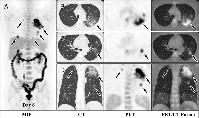
FIGURE 1.

A 37-year-old man was admitted to hospital on January 2, 2020, because of vertigo for 2 days. On the first day of hospital admission, his brain MRI indicated no abnormalities (not shown), yet the chest CT spotted a 3.6-cm ground glass opacity (GGO) in the upper lobe of the left lung. On day 6, 18F-FDG PET/CT was performed to make differentiation of benign and malignant of the GGO. MIP (A) image revealed multiple 18F-FDG uptake in bilateral lungs (arrows) and along the whole colon. The GGO lesions at the apical segment of the left upper lobe, the posterior segment of the left upper lobe, and the apical segment of the right upper lobe were demonstrated in the axial images (B and C) and in the coronal images (D). The SUVmax of lung GGOs range from 2.4 to 12.4. The patient’s respiratory virus (influenza A, influenza B, parainfluenza, respiratory syncytial virus, adenovirus) antigen tests were negative. Considering the epidemiological history of Wuhan residence, fever symptom, and multiple GGOs in lungs, COVID-19 infection was clinically diagnosed.
FIGURE 2.

The patient received treatment against COVID-19 immediately after the diagnosis. Serial CT demonstrated how lung lesions developed in the duration of COVID-19. Chest CT on day 9 showed more GGO lesions, although the GGO lesion in the upper lobe of the left lung dissipated. New GGO lesions emerged on day 14, whereas some old lesions dissipated or developed into consolidation. Most of the lesions disappeared on day 19, leaving slight remnant consolidation.
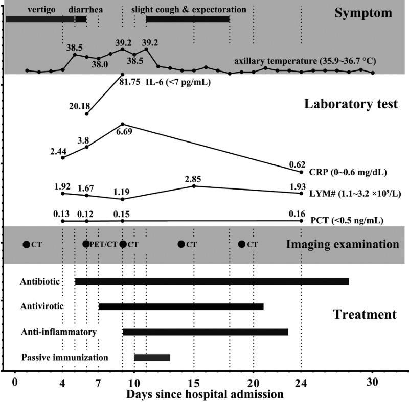
FIGURE 3.

The timeline chart showed how the disease developed and how the treatment was managed during the COVID-19 course. The patient’s fever emerged on day 5, along with 5 times of diarrhea for 1 day. Interleukin 6 and C-reactive protein increased quickly during the fever stage, whereas lymphocyte counts decreased a little but were still within normal reference range. Procalcitonin never got elevated in the duration of the disease. Antibiotics on day 5 and antivirotics on day 7 were used to treat against COVID-19. However, his fever persisted until the treatment of anti-inflammatory and passive immunization. The patient was symptom-free since day 18 and was discharged from hospital on day 30. The successful treatment corroborated the initial diagnosis of COVID-19 infection. COVID-19 shares the same respiratory symptoms and lung pathological characteristics with severe acute respiratory syndrome and Middle East respiratory syndrome1,2; however, this case and other reports indicate neurological symptoms, and gastrointestinal symptoms may also suggest suspected COVID-19.3,4 Due to the unsatisfactory positive rate of SARS-CoV-2 nucleic acid test,5 chest imaging examination serves as an important diagnostic method.6–8 18F-FDG PET/CT plays a complementary role for COVID-19 diagnosis.9 It demonstrates inflammatory lesions of the whole body, which may offer value for treatment strategy in the duration of COVID-19.
Footnotes
Conflicts of interest and sources of funding: This work was supported by the research program of Wuhan Municipal Health Commission (no. WX18Q32) and the Natural Science Foundation of Hubei Province (no. 2019CFB287). None declared to all authors.
REFERENCES
- 1.Zu ZY, Jiang MD, Xu PP, et al. Coronavirus disease 2019 (COVID-19): a perspective from China. Radiology. 2020;200490. [DOI] [PMC free article] [PubMed] [Google Scholar]
- 2.Xu Z, Shi L, Wang Y, et al. Pathological findings of COVID-19 associated with acute respiratory distress syndrome. Lancet Respir Med. 2020. Epub ahead of print. [DOI] [PMC free article] [PubMed] [Google Scholar]
- 3.Shi H, Han X, Jiang N, et al. Radiological findings from 81 patients with COVID-19 pneumonia in Wuhan, China: a descriptive study. Lancet Infect Dis. 2020. Epub ahead of print. [DOI] [PMC free article] [PubMed] [Google Scholar]
- 4.Li YC, Bai WZ, Hashikawa T. The neuroinvasive potential of SARS-CoV2 may be at least partially responsible for the respiratory failure of COVID-19 patients. J Med Virol. 2020. Epub ahead of print. [DOI] [PMC free article] [PubMed] [Google Scholar]
- 5.Xie C, Jiang L, Huang G, et al. Comparison of different samples for 2019 novel coronavirus detection by nucleic acid amplification tests. Int J Infect Dis. 2020. Epub ahead of print. [DOI] [PMC free article] [PubMed] [Google Scholar]
- 6.Guan W, Ni Z, Hu Y, et al. Clinical characteristics of coronavirus disease 2019 in China. N Engl J Med. 2020. Epub ahead of print. [DOI] [PMC free article] [PubMed] [Google Scholar]
- 7.Ai T, Yang Z, Hou H, et al. Correlation of chest CT and RT-PCR testing in coronavirus disease 2019 (COVID-19) in China: a report of 1014 cases. Radiology. 2020;200642 Epub ahead of print. [DOI] [PMC free article] [PubMed] [Google Scholar]
- 8.Pan F, Ye T, Sun P, et al. Time course of lung changes on chest CT during recovery from 2019 novel coronavirus (COVID-19) pneumonia. Radiology. 2020;200370 Epub ahead of print. [DOI] [PMC free article] [PubMed] [Google Scholar]
- 9.Qin C, Liu F, Yen TC, et al. 18F-FDG PET/CT findings of COVID-19: a series of four highly suspected cases. Eur J Nucl Med Mol Imaging. 2020. Epub ahead of print. [DOI] [PMC free article] [PubMed] [Google Scholar]


